|
|
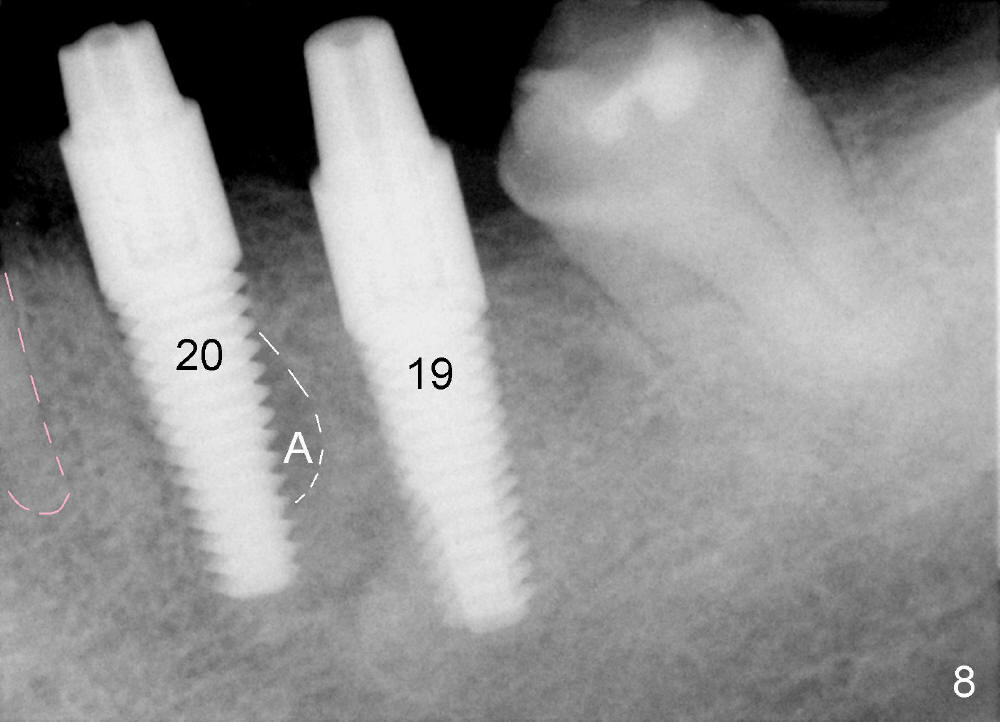
Fig.8: Two of 5x17 mm implants are placed at the sites of #19 and #20; 4x5 mm and 4x3 mm abutments are placed, respectively.
A: apex of the original socket of #20 (no bone graft being placed). Instead, the coronal portion of the mesiobuccal socket is filled with autogenous bone (harvested from the osteotomy at the site of #19, mixed with Osteogen).
Pink outline: the root of the tooth #21.
Return to Trajectory
Xin Wei, DDS, PhD, MS 1st edition 07/22/2014, last revision 01/19/2018