
|
|
|
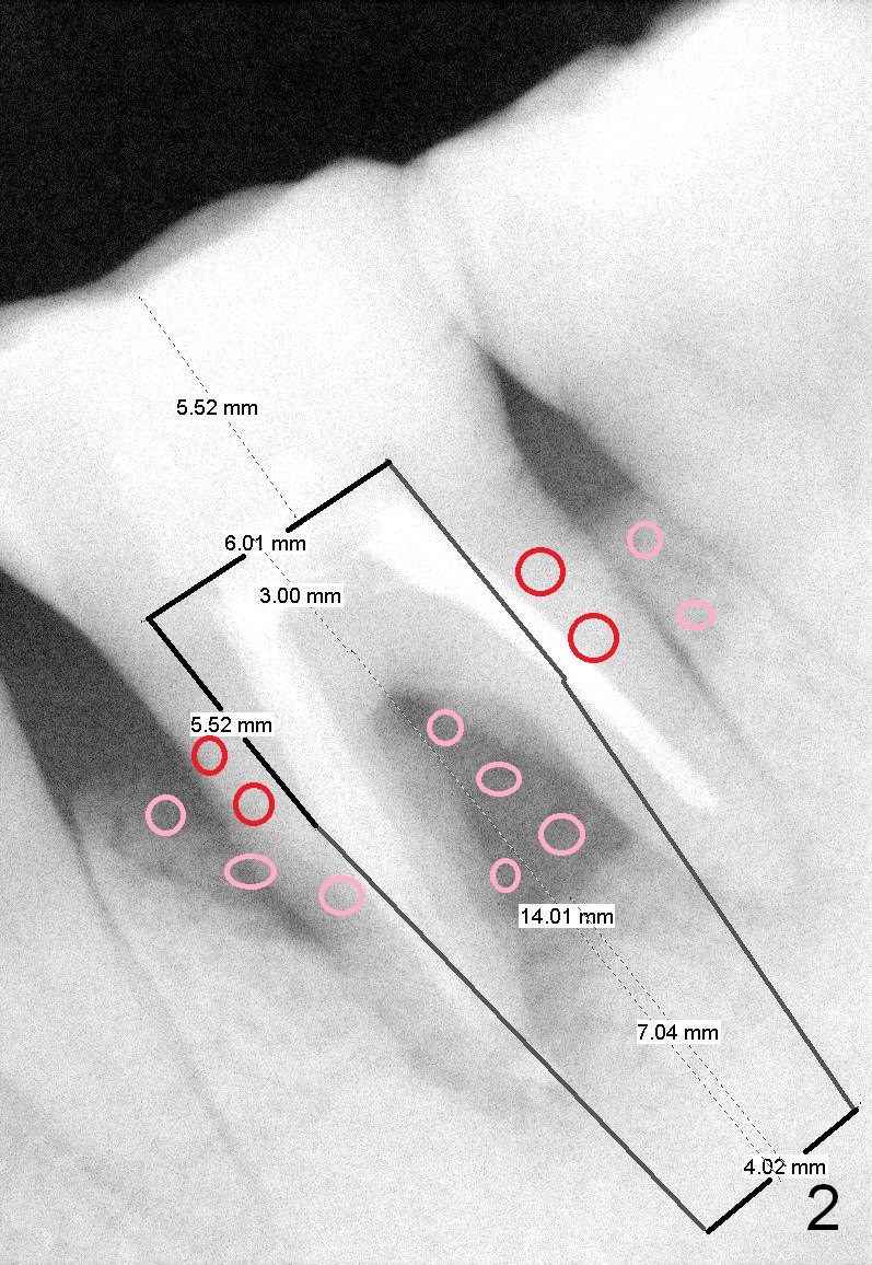
Tentative size of the implant is 6x14 mm with ~ 7 mm in new bone (septum; Fig.2). The septum is low, and flat (Fig.3 red line, S), while the mesial and distal sockets are shallow (brown (M) and pink (D) outlines).
Bone graft is placed twice: one (pink circles) before and the other (red circles) after implantation. The first one is placed to area predictably difficult to reach post implantation. In this case, the lingual plate is defective. When osteotomy is finished, a piece of Osteotape (collagen membrane saturated with a type of Hydroxyapaptite (Impladent) is placed against the lingual defect, followed by bone graft (mixture of autogenous bone (from reamers), allograft and Osteogen) lingually. More bone graft is placed in the peri-implant gaps (Fig.8,9 *).
Xin Wei, DDS, PhD, MS 1st edition 02/19/2015, last revision 01/19/2018