|
|
|
|
|
|
|
|
|
Severe Vertical Bone Loss
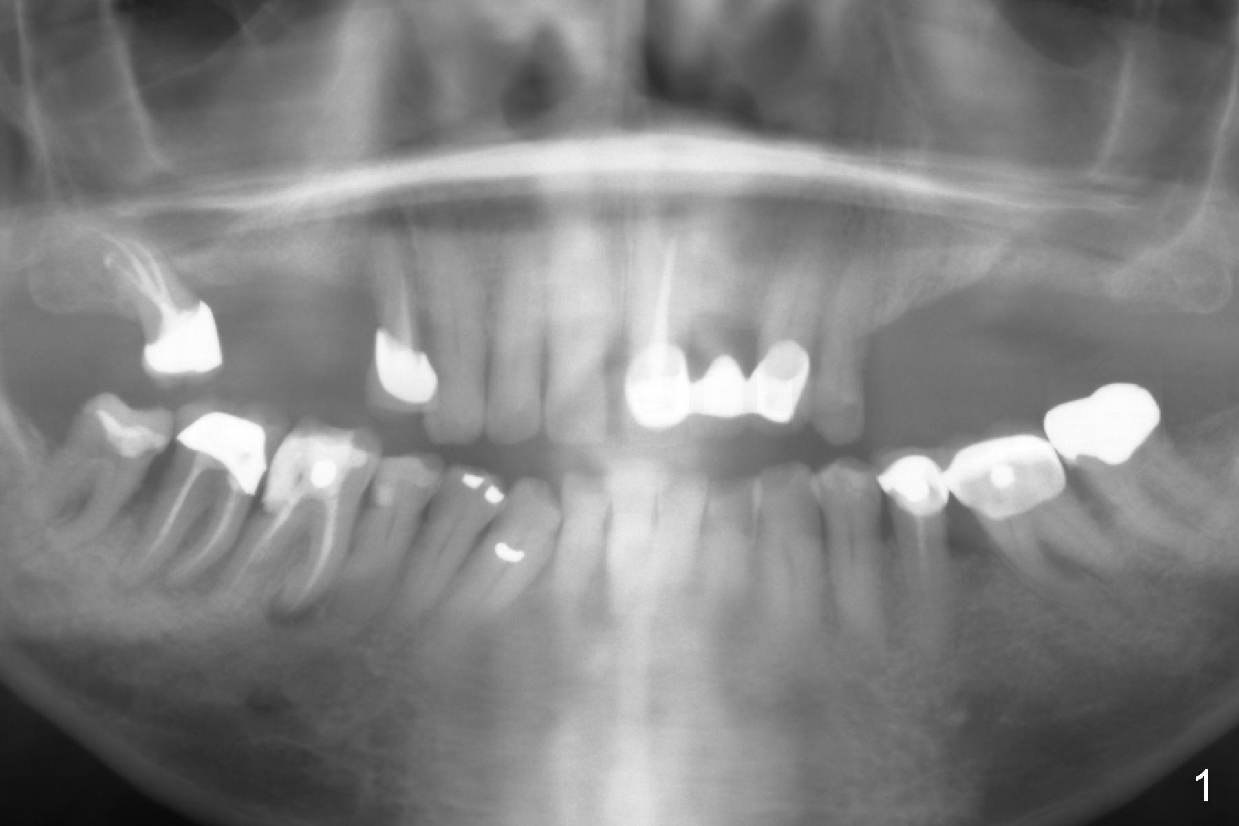
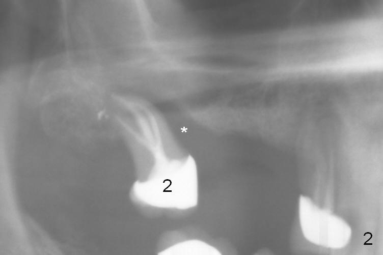
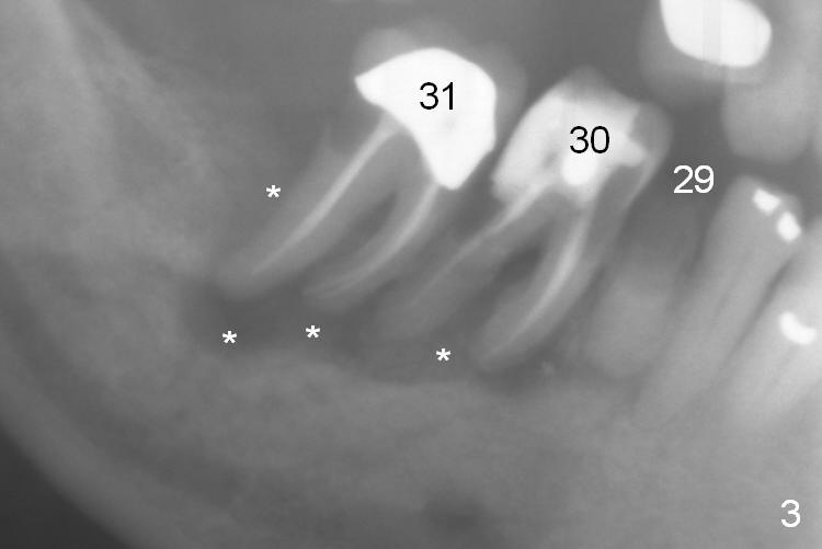
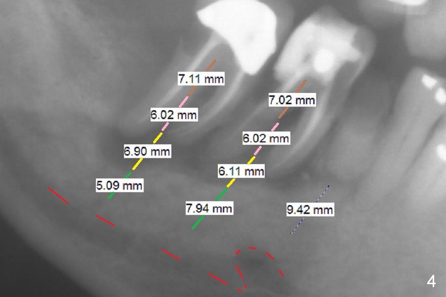
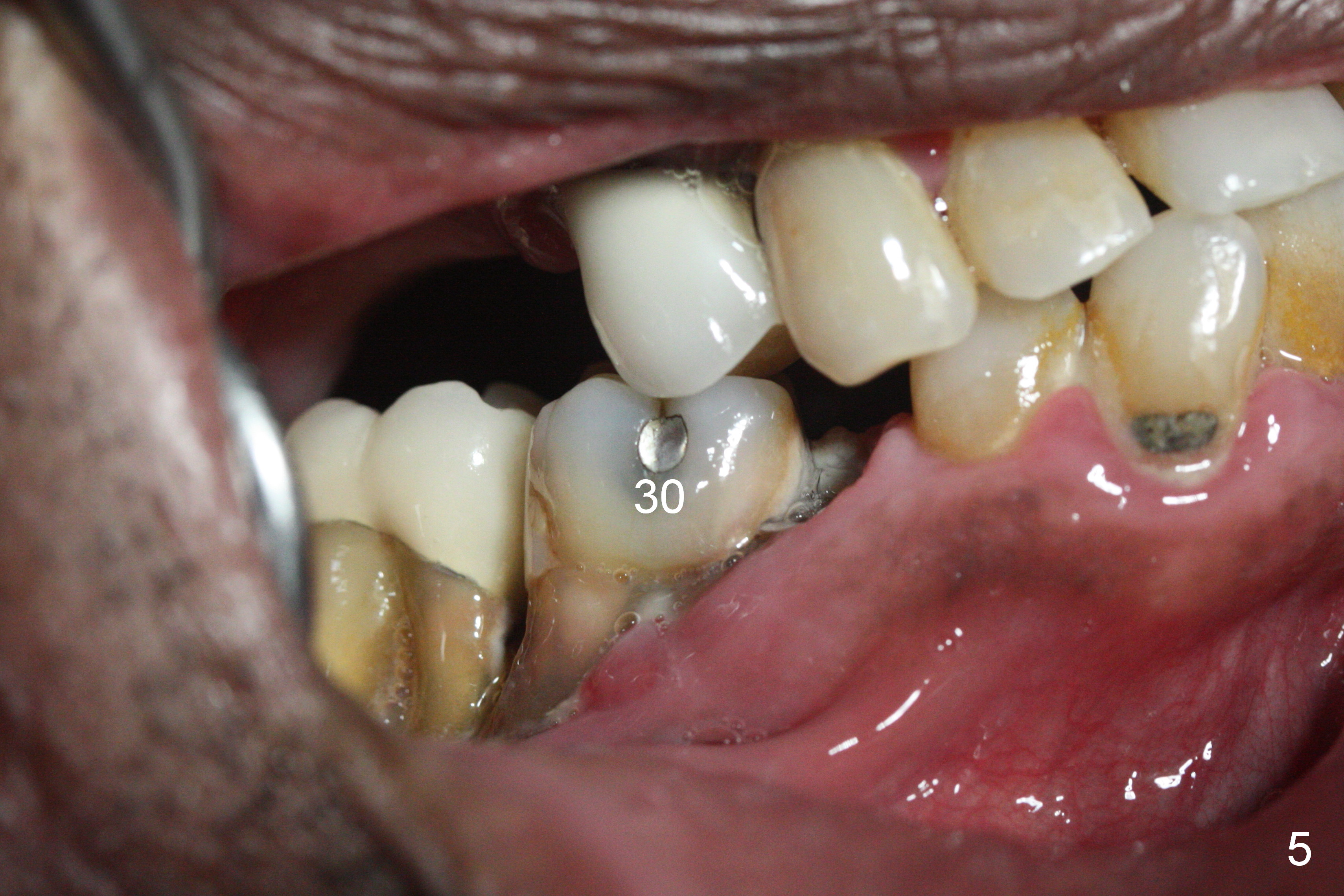
A 71-year-old man had poor dentition 4 years ago (Fig.1). Since then severe bone loss has occurred at the teeth #2 (Fig.2 *), 30 and 31 (Fig.3 *). An original plan is to place implants at #29 and 31 and fabricate a 3-unit bridge. Since the bone height at #31 is limited, primary stability may be questionable. It is advantageous to place 3 implants at #29-31. As the bone loss is severe and the sockets at #30 and 31 are long (Clindamycin), a fairly long portion of the implants at #30 and 31 will be not covered by the native bone (Fig.4 yellow line: 6-7 mm, bare) in spite of using 6 mm cuff (pink) of 7 mm abutments (brown). Use initial drill from DIO Sinus Master Kit with 5 mm stopper at #31 (green line), followed by insertion of a marked parallel pin. Use regular drills with stoppers of 10 and 8 mm at #29 and 30 (green line). Continue osteotomy at #31 with round drills (2.8 and 3.6 mm) with shorter stopper to avoid injury to the underlying Inferior Alveolar Canal (red dashed line). Pack allograft well around the implants at #30 and 31 before placing abutments to reduce periimplantitis and insert collagen plug around the abutments to prevent loss of the bone graft underneath. The last method to secure the bone graft is a retentive, splinted provisional.
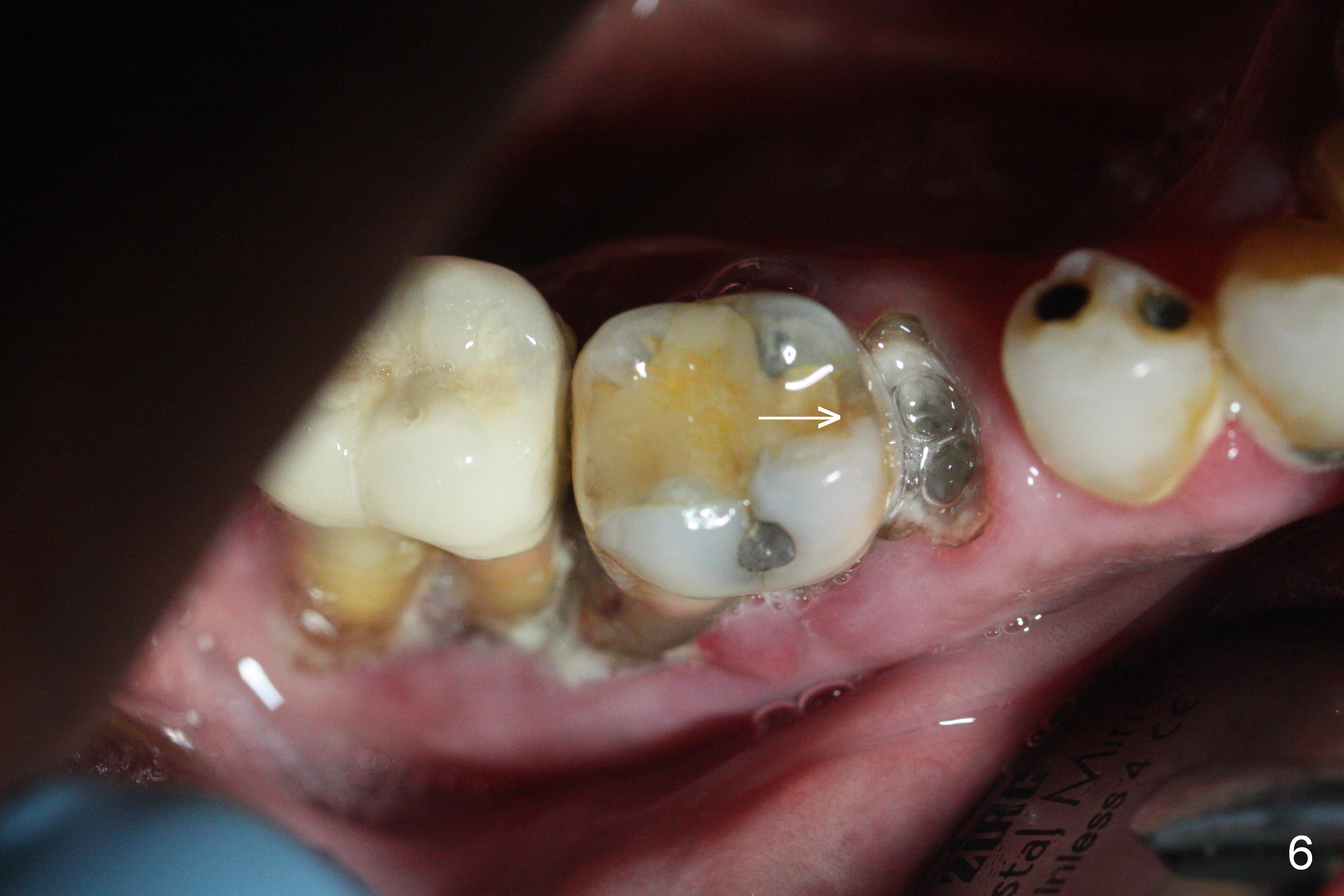
It appears that the endo perio disease is more severe than 6 months ago (Fig.5,6). Prepare PRF and Extra Wide implants as well.
Return to
Lower
Molar Immediate Implant, Prevent
Molar Periimplantitis (Protocols,
Table), IBS,
Xin Wei, DDS, PhD, MS 1st edition 03/18/2017, last revision 06/16/2019