|
|
|
|
|
|
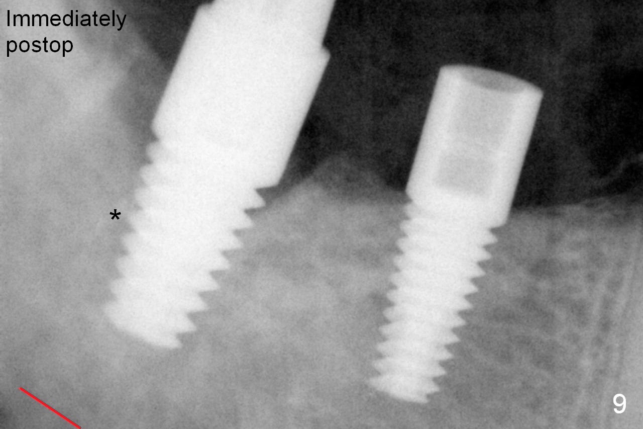
The insertion torques of the implant at the sites of #30 and 31 (4.5x14 and 6x14 mm) are both more than 60 Ncm (Fig.9). The septum (*) is pushed lower (wears down) as the osteotomy increases. Red dashed line: the upper border of the Inferior Alveolar Canal.
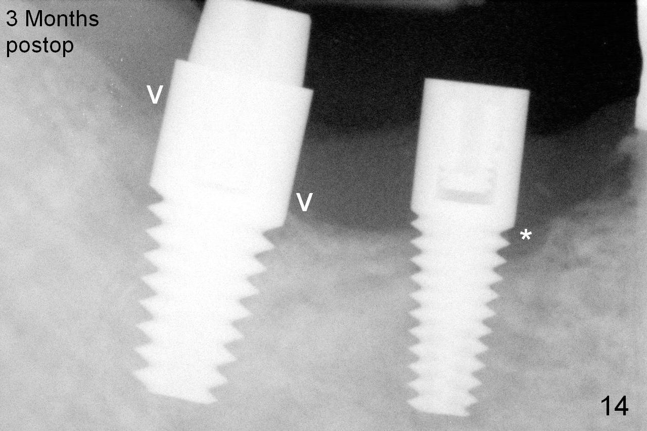
Three months postop, the mesial and distal gaps appear to have closed around the implant at the site of #31 (Fig.14 arrowheads), while there is bone resoprtion mesial to the implant at #30 (*). This suggests that lack of bone width is associated with bone loss. In contrast, there is ample bone around the immediate implant when it is placed, which is related to bone deposit (with bone graft).
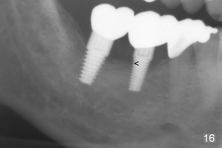
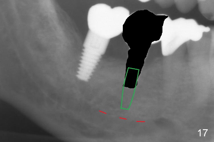
A few of the coronal threads at #30 appear not to be covered by the bone 6 months post cementation (15 months postop, Fig.16). Clinically the lingual gingiva recession is moderate. If peri-implantitis develops, either remove the exposed threads or place a narrower implant deeper (Fig.17).
Xin Wei, DDS, PhD, MS 1st edition 05/14/2015, last revision 08/10/2016