
|
|
|
Fig.4: the coronal view of the extracted tooth, with buccal on the bottom of the image, showing C shaped canal.
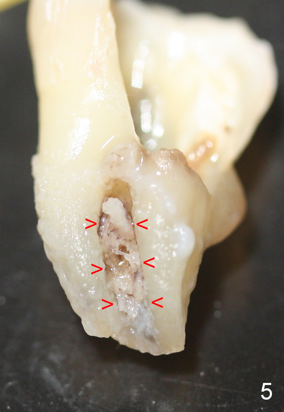
Fig.5: the lingual view of the extracted tooth. The septal bone (arrowheads) is embedded between incompletely fused roots. The distolingual wall is fractured during extraction.
In brief, this type of the lower 2nd molar root resembles a half cup, a common finding in Chinese population.
Xin Wei, DDS, PhD, MS 1st edition 06/25/2015, last revision 06/25/2015