|
|
|
|
||
|
|
|
|
||
|
|
|
|
|
|
|
|
|
|
|
|
|
|
|
|||
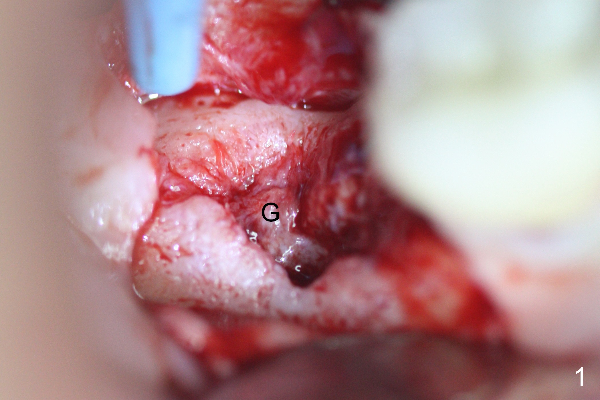
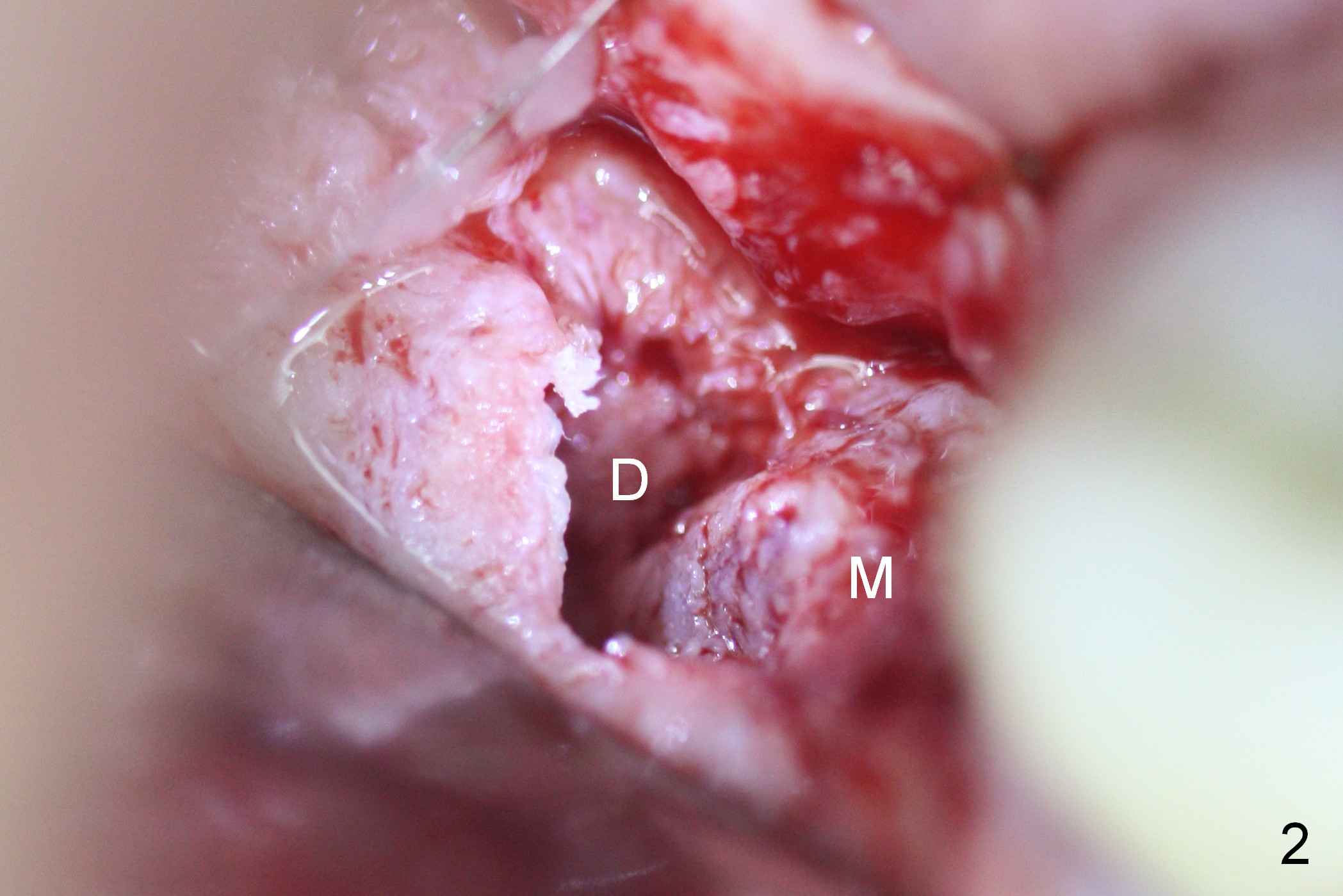
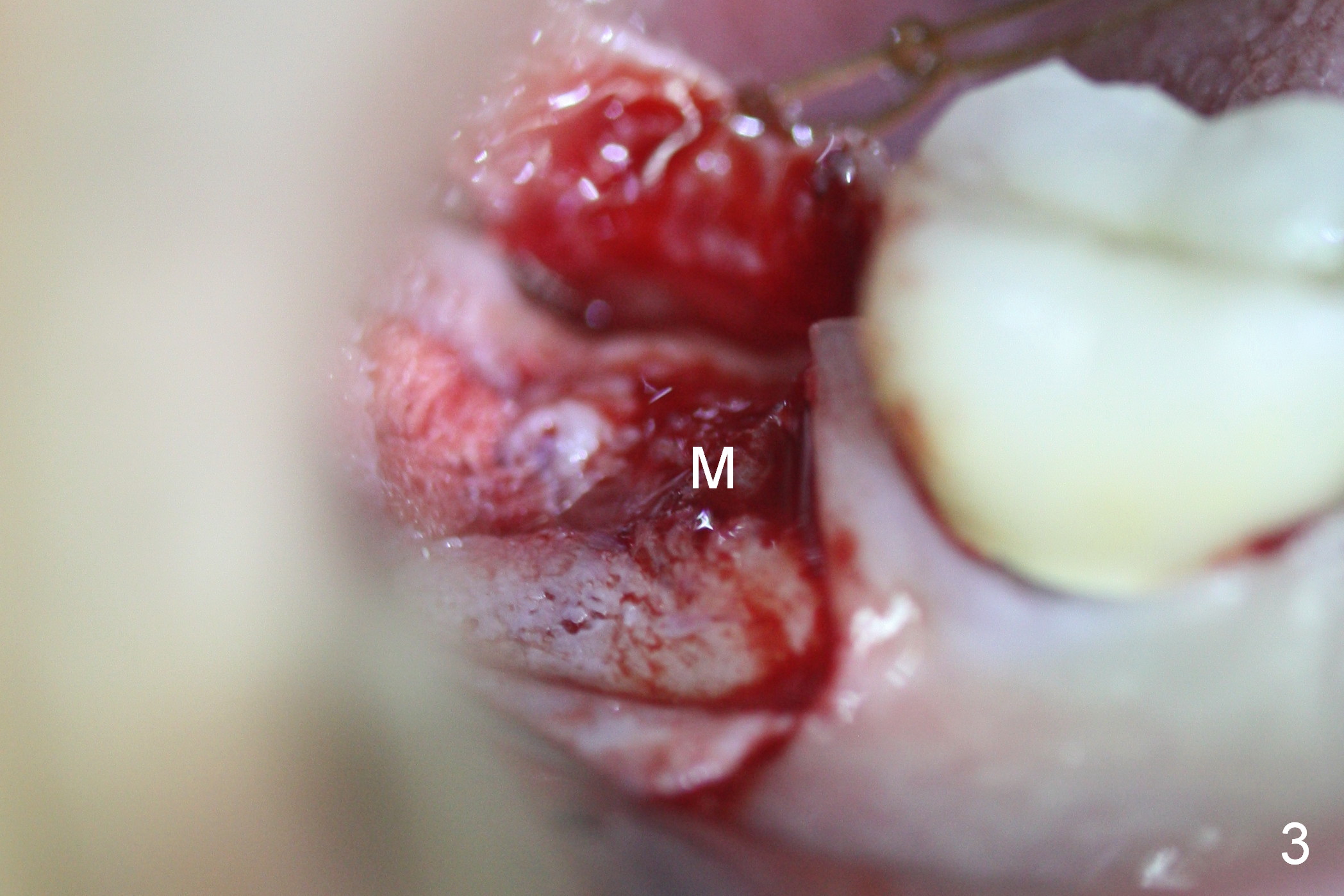
Mesial Socket Placement
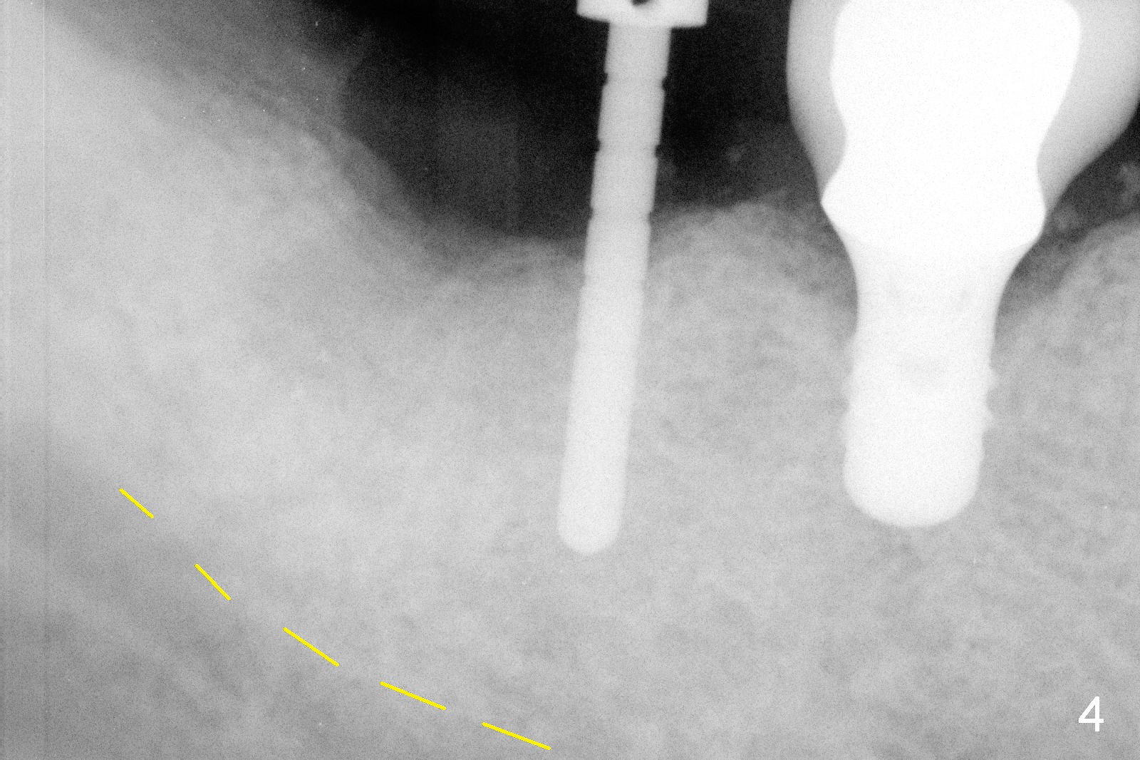
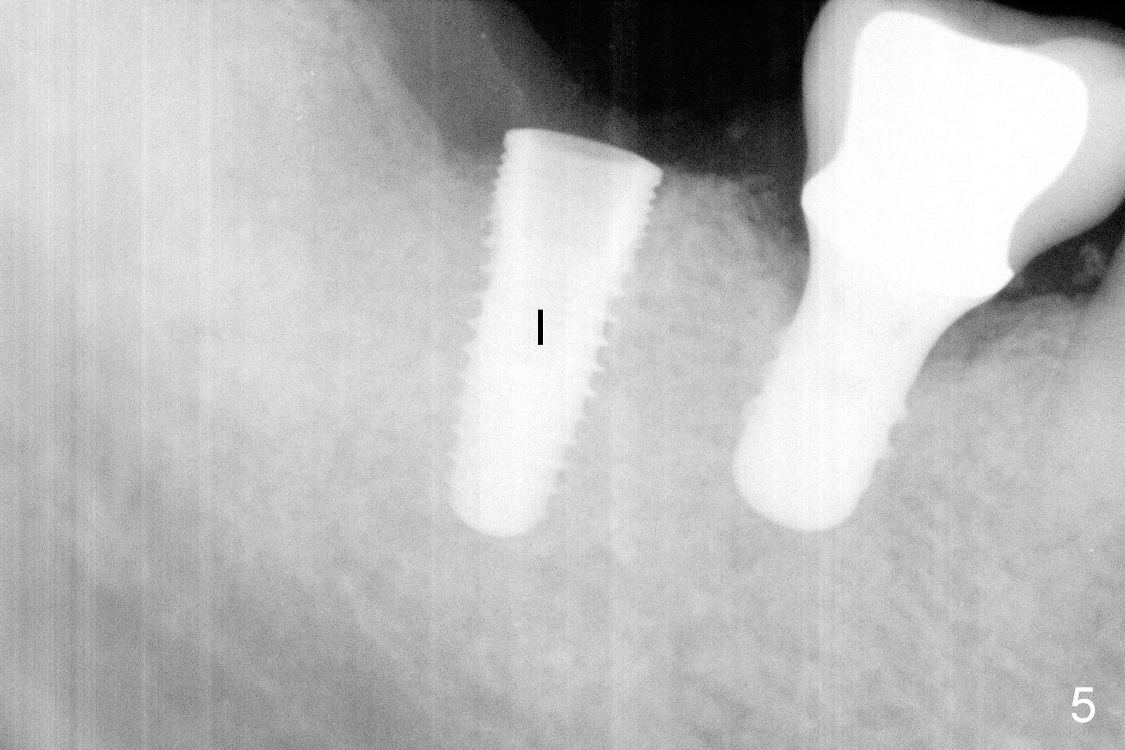
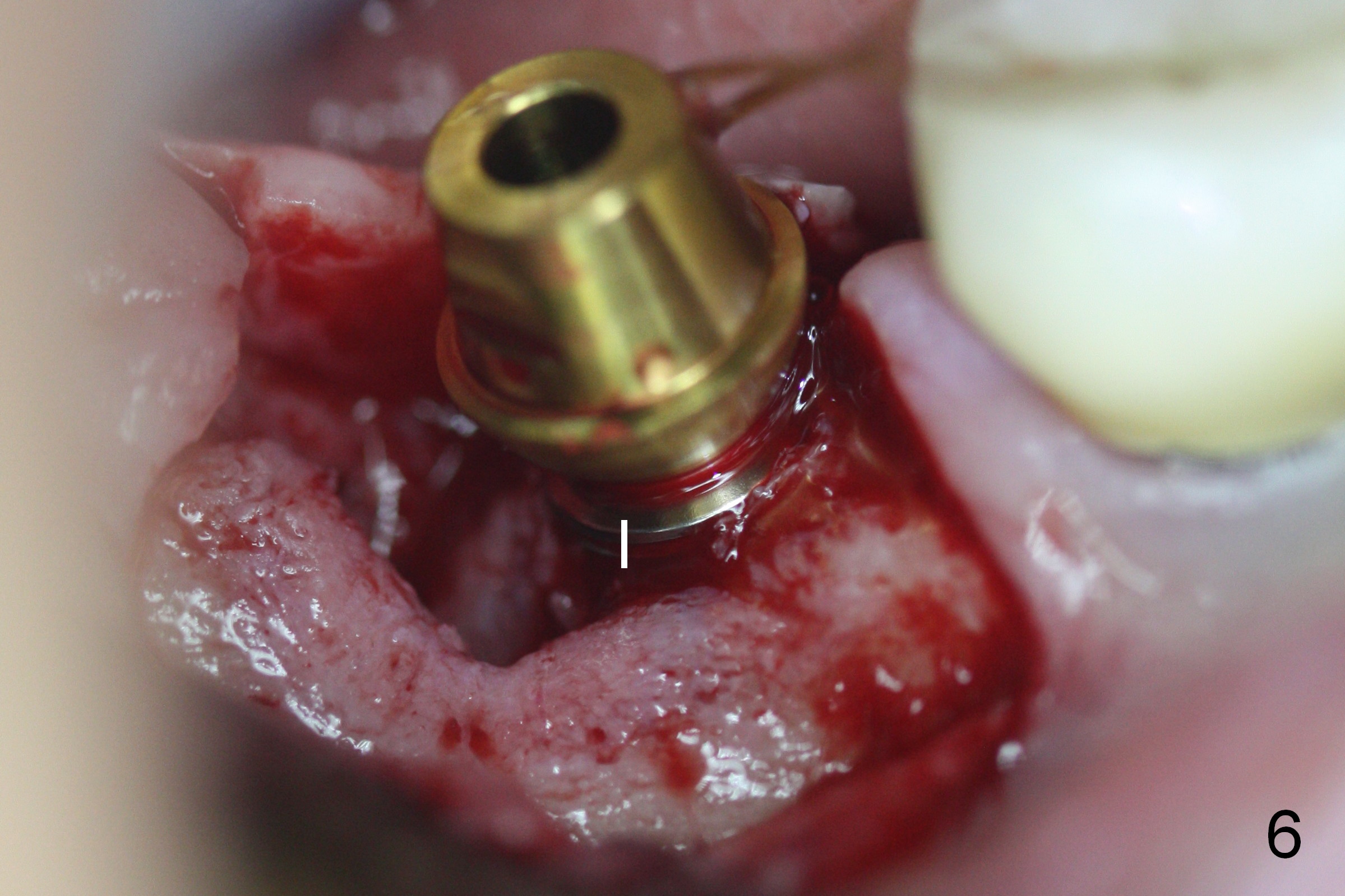
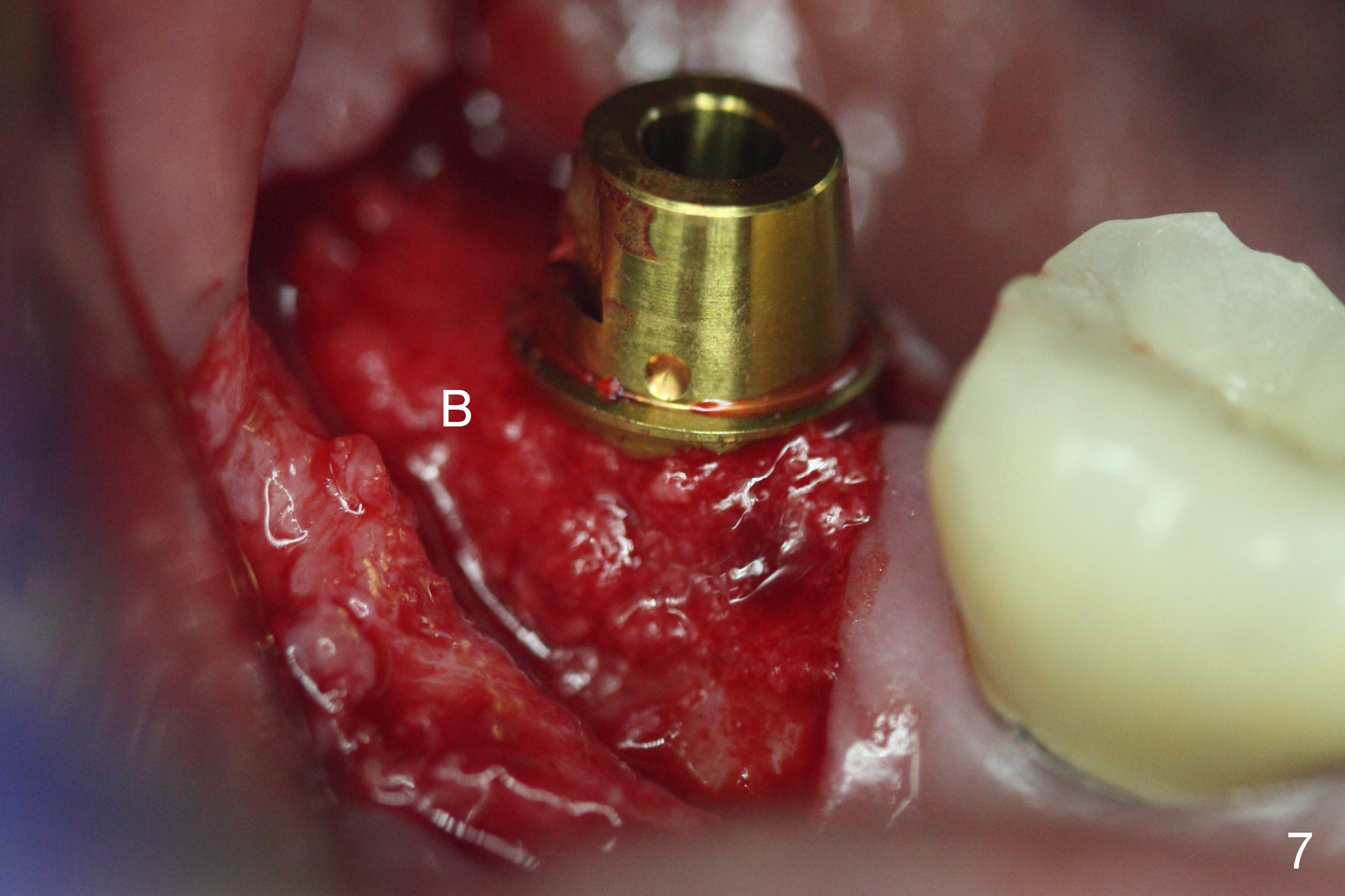
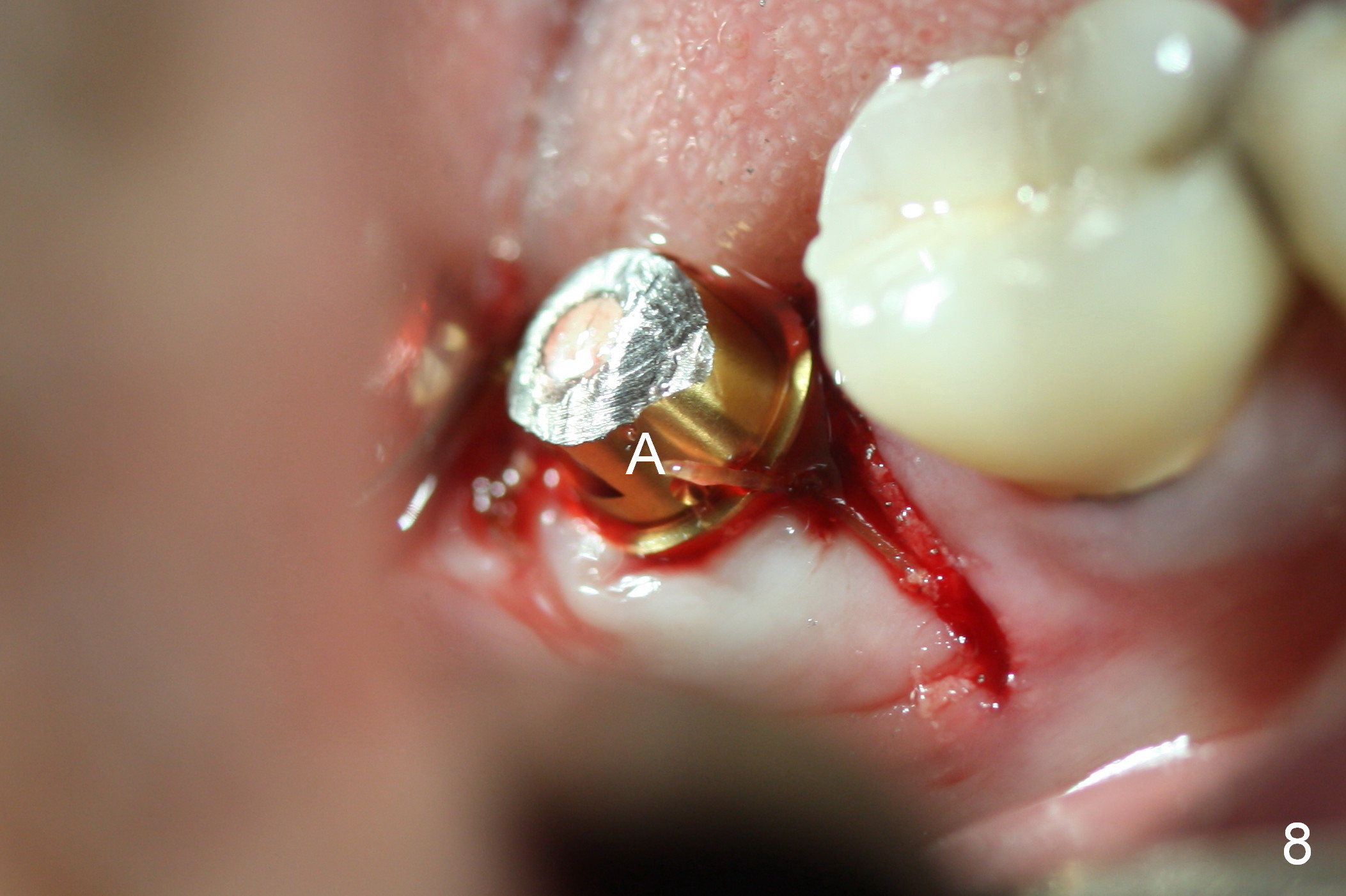
Three months post extraction, the site of #31 is surgical exposed with granulation tissue in the distal socket (Fig.1 G), corresponding to the distal root fracture. After debridement of the distal socket (Fig.2 D), the mesial socket is found to have healed with solid bone and sufficient width (Fig.2.3 M). Initial osteotomy (more or less lingual) depth is 10 mm; there is ~ 6 mm clearance from the Inferior Alveolar Canal (Fig.4 yellow dashed line). A 5x12 mm implant (Fig.5 I) is surrounded by bone except distobuccally (Fig.6). Autogenous bone (harvested from osteotomy, Fig.7 B) is placed in the distal socket. After suturing the flaps, the height of the abutment (Fig.8 A, 6.8x4(3) mm) is adjusted for an immediate provisional. The mesiobuccal incision is not completely closed by suture. Perio dressing is applied for wound protection.
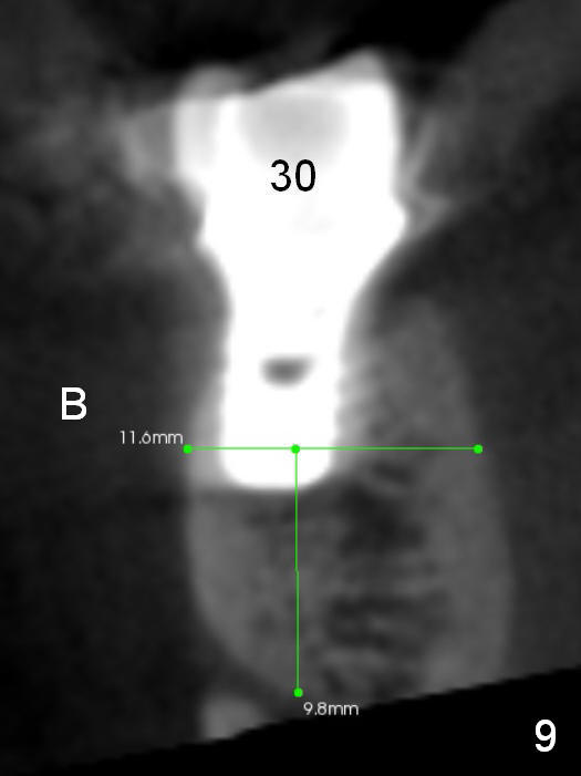
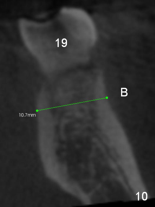
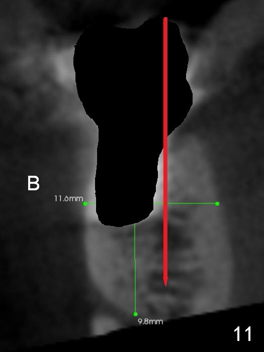
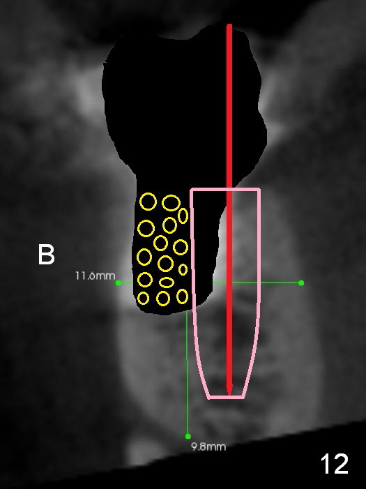
The implant at the site of #30 has sign of peri-implantitis (Fig.4,5). It may be associated with its buccal placement (Fig.9 B, as compared to the contralateral side (Fig.10)). If the implant needs to be removed (Fig.11 black area), osteotomy is initiated immediately and as lingual as possible (red line). As the osteotomy increases and a new implant is placed (Fig.12 pink), the latter may be deviated buccally. The buccal defect will be bone grafted (yellow circles).
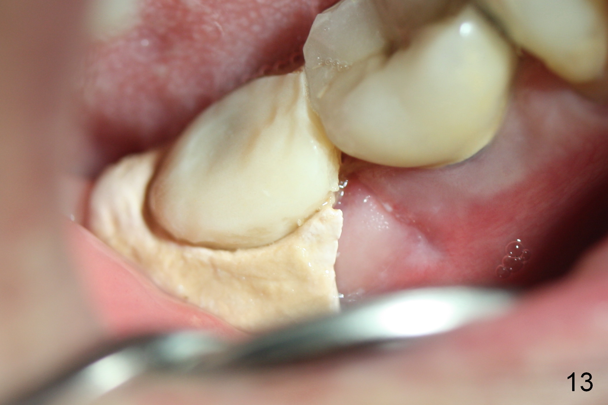
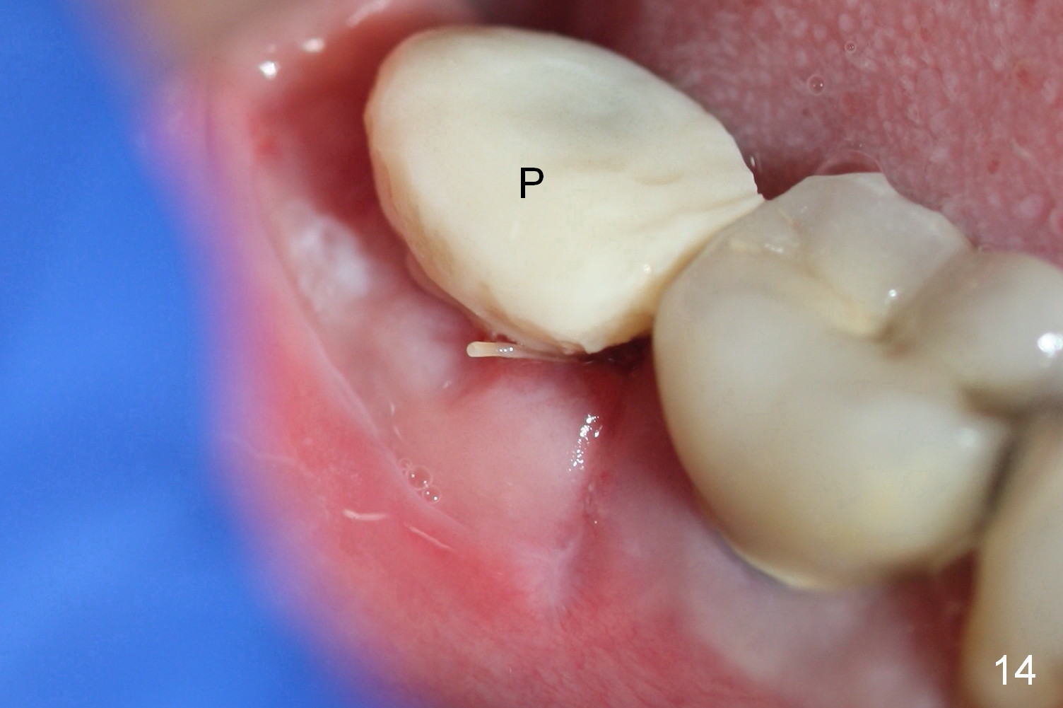
Perio dressing is partially dislodged 11 days postop (Fig.13). The mesiobuccal wound is healing, as compared to Fig.8. It appears that the immediate provisional (Fig.14 P) helps keep perio dressing in place, which in turn protects the wound.
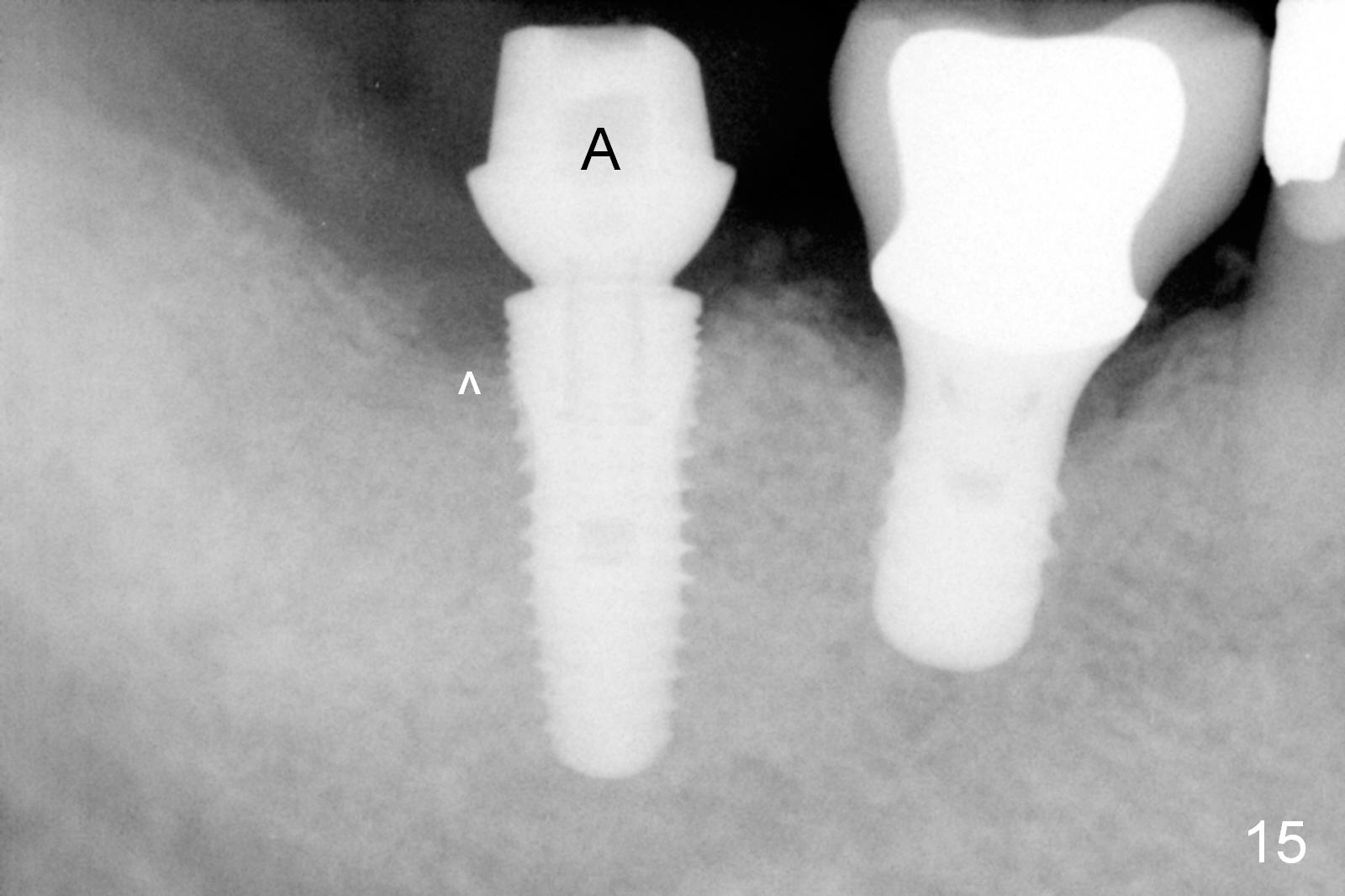
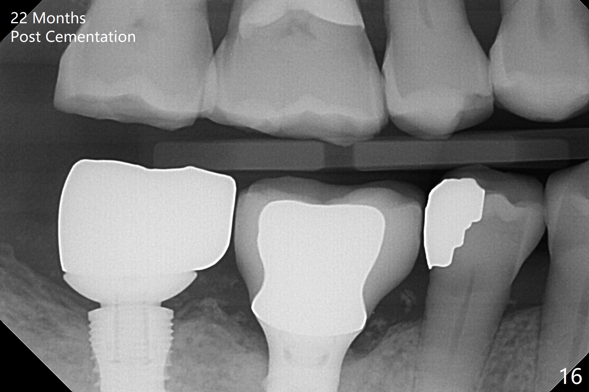
The patient returns for impression 2 months postop (Fig.15) and for follow-up 22 months post cementation (Fig.16). There is no bone loss around the implant at #31. Food impaction occurs mesial and distal to the implant at #30. The patient will return for #29 DO onlay and #31 mesial porcelain addition 4 year post cementation.
Return to Lower Molar Immediate Implant,
Localized Use of Antibiotic for Healed Socket
Trajectory II
Xin Wei, DDS, PhD, MS 1st edition 07/23/2015, last revision 10/27/2019