|
|
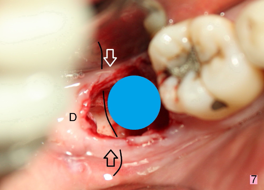
Fig.7 is an illustration (from Fig.4) to show how the socket is closed.. A suture is placed (black line) to approximate buccal (black arrow) and lingual (white arrow) gingiva distal to the implant (blue circle). D: distal portion of the gingiva. Sometimes collagen dressing is placed above the bone graft without suture. The socket can shrink.
Return to No Drill Implantation
Xin Wei, DDS, PhD, MS 1st edition 10/10/2014, last revision 11/02/2014