
|
|
|
|
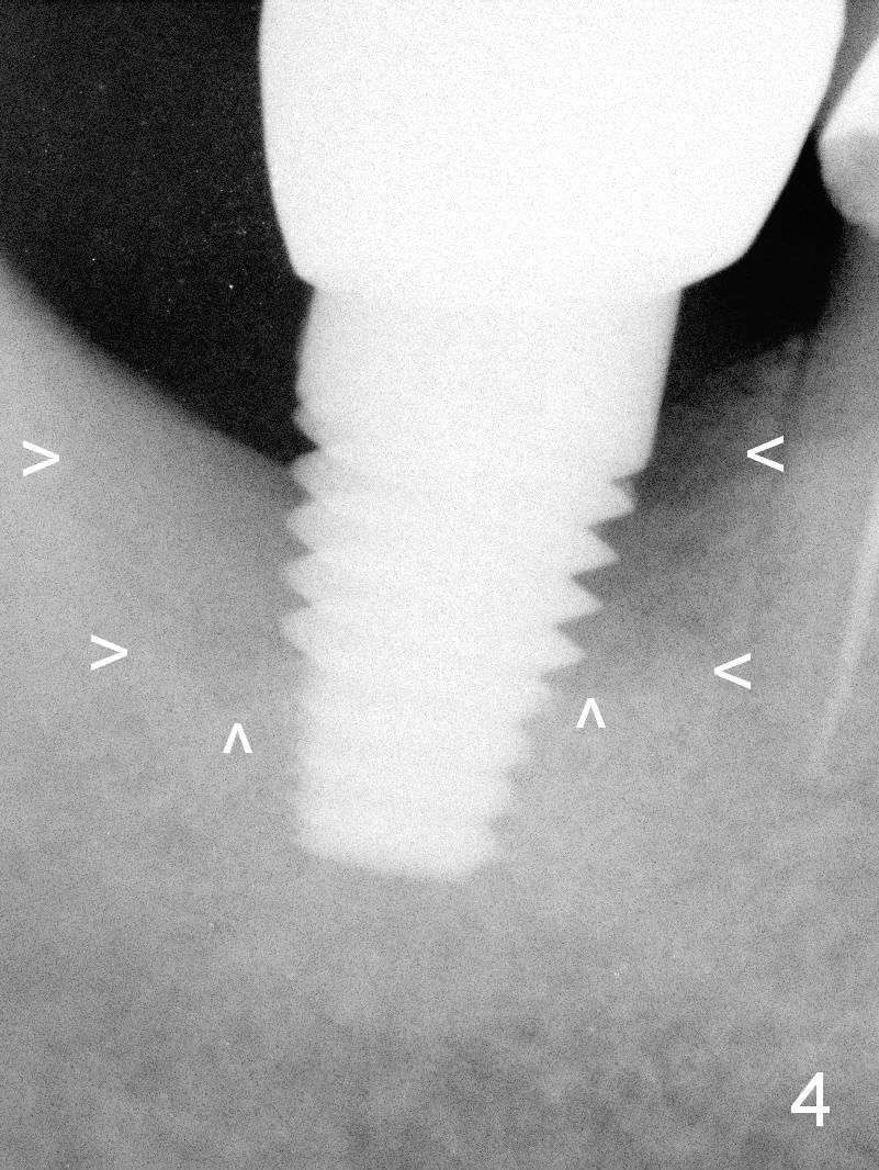
PA taken 1 month preop (bone graft, 2 years 4 months post implant placement) shows bony defect around the implant at #31 (Fig.4 arrowheads).
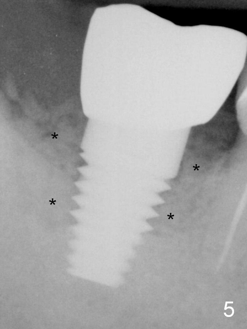
Immediately postop PA demonstrates that the defect is filled with .5-2 mm mineralized cortical and cancellous allograft (Fig.5 *).
There is purulent discharge in the buccal vestibule with unknown origin 6 months postop. The gingiva looks healthy. There is bone loss (Fig.6).
Bone Graft for Periimplantitis
Xin Wei, DDS, PhD, MS 1st edition 03/12/2016, last revision 03/04/2018