|
|
|
|
|
|
|
|
|
Try Again: No Drill Osteotomy
Once the anterior implant-supported immediate provisionals are stabilized, the tooth #29 is planned to be extracted and have an immediate implant as the second stage of lower arch reconstruction. At the first stage, left inferior alveolar nerve paresthesia occurs probably due to complete block of mental nerve with Articaine. The patient is not cooperative in intraop X-ray.
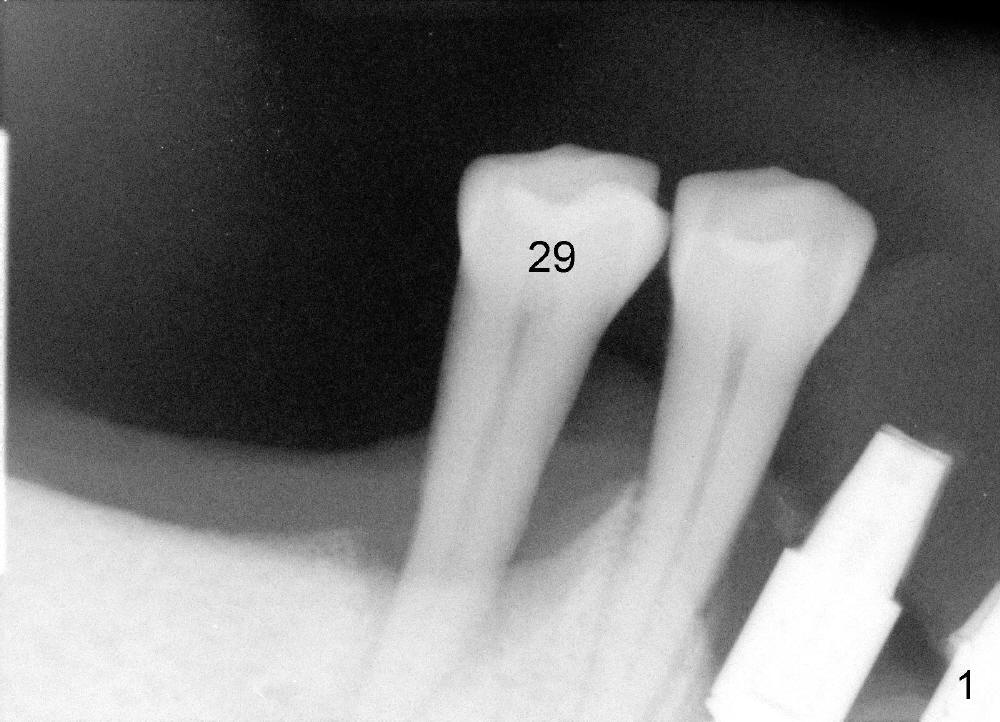
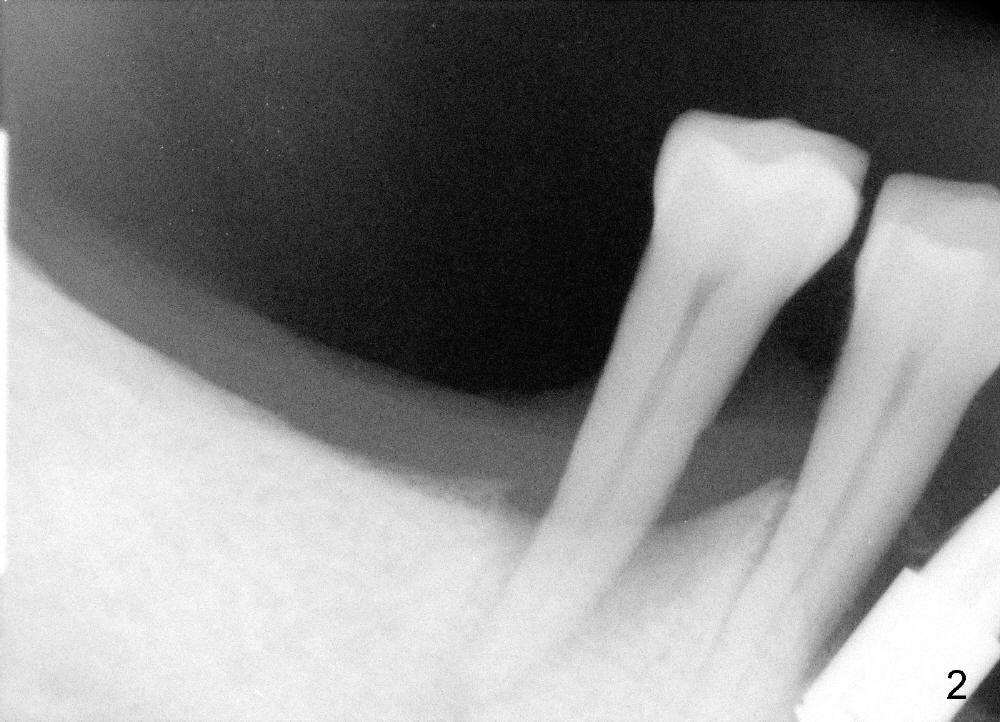
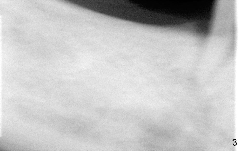
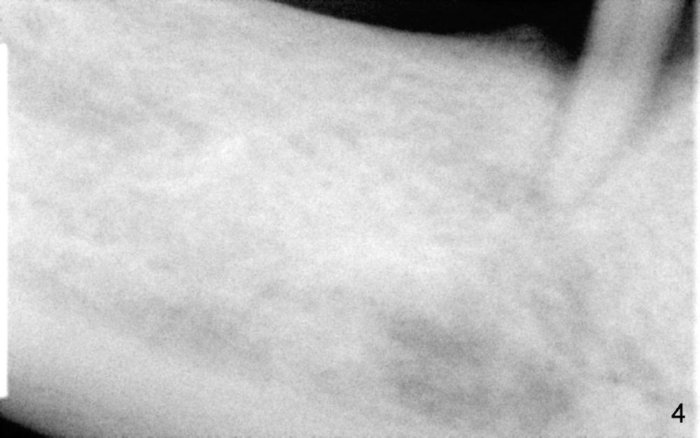
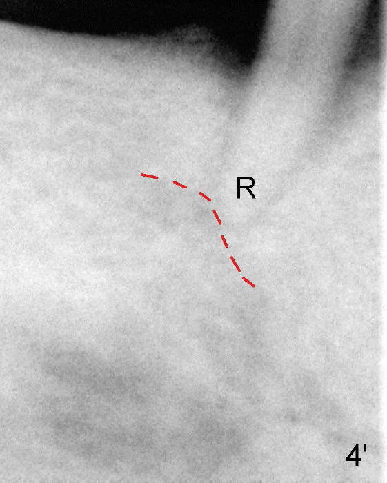
To avoid paresthesia on the right side, several preop PAs are taken (Fig.1-4) so that we know how to take good X-ray for this gagging patient. At first, #2 Sensor is used to take the first PA (Fig.1). It does not show the apex of the tooth #29. The second PA is a little better (Fig.2), but the mental nerve is out of view. Then #1 sensor is used. It allows us to position the sensor lower (Fig.3), but the image is blurred. When it is retaken, the image is clear (Fig.4), showing the proximity of the mental nerve loop (Fig.4': red dashed line) to the root tip of the tooth #29 (R).
The second step to avoid paresthesia for this case is local anesthetic. Do not to use Articaine, since it is the most potent. No injection is given around the mental foramen. The anesthetic may retrograde block the mental nerve and loop. There will be no intraligamental injection. High injection pressure may force the anesthetic to infiltrate the major trunk of the mental loop.
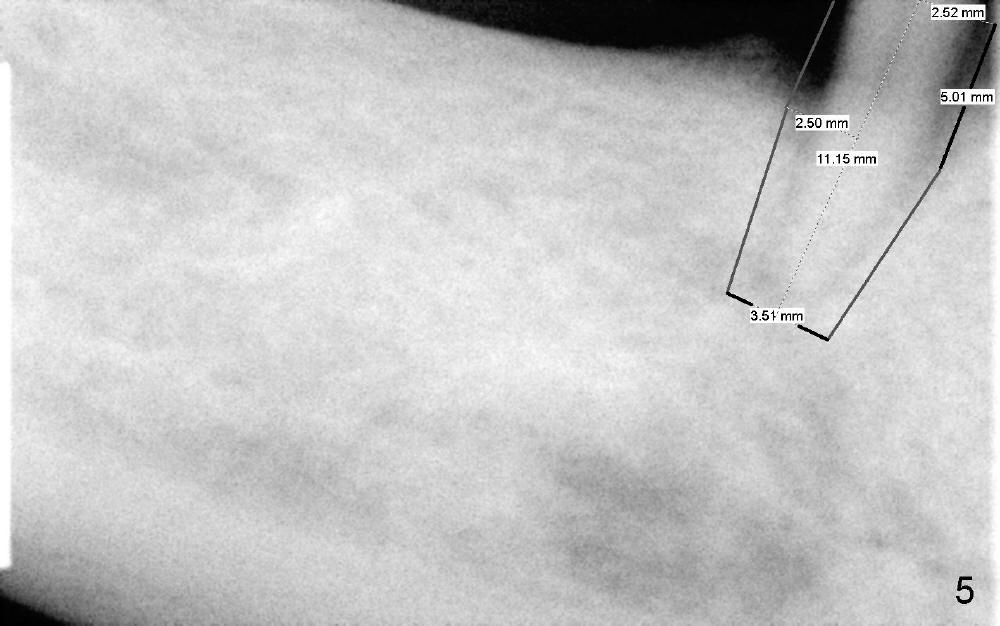
The third step to avoid paresthesia is called no drill osteotomy. When the tooth is extracted, the depth of the socket is measured. Tap(s) will be used to create the osteotomy (to form threads); the depth is controlled with precision. How to decide the diameter of the implant? It should be large enough to obtain primary stability, but an oversized implant may also injury the loop. It is estimated that a 5x11 mm tissue-level tapered implant will be used (Fig.5) or 4.5x11 mm. Mesiodistally the implant should be a little larger than the root, since buccolingual width of the socket is larger. The osteotomy will be as lingual as possible, since the mental loop is buccally located. A photo is going to be taken to show the oval shape of the socket. If visibility is poor, a flap may be raised.
Return to Lower Arch Reconstruction
Xin Wei, DDS, PhD, MS 1st edition 05/31/2014, last revision 06/01/2014