,%204m%20postop.jpg)
|
|
|
|
|
|
|
|
|
|
|
|
|
|
|
|
|
|
Flap Guided Surgery (I)
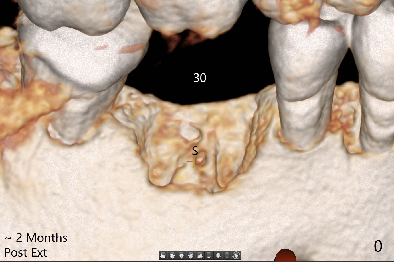
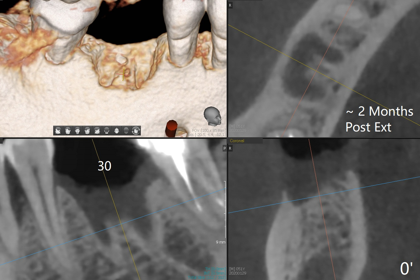
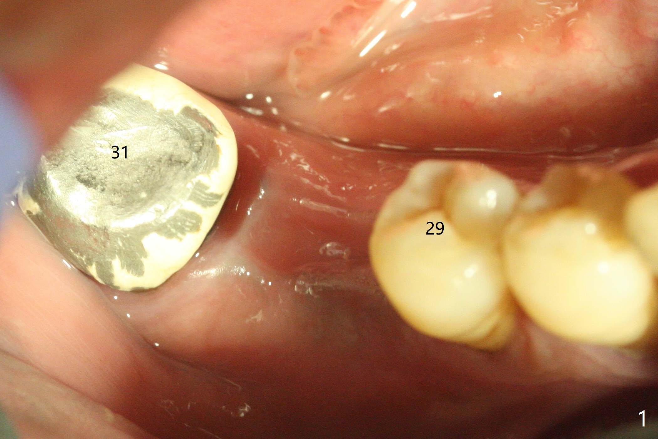
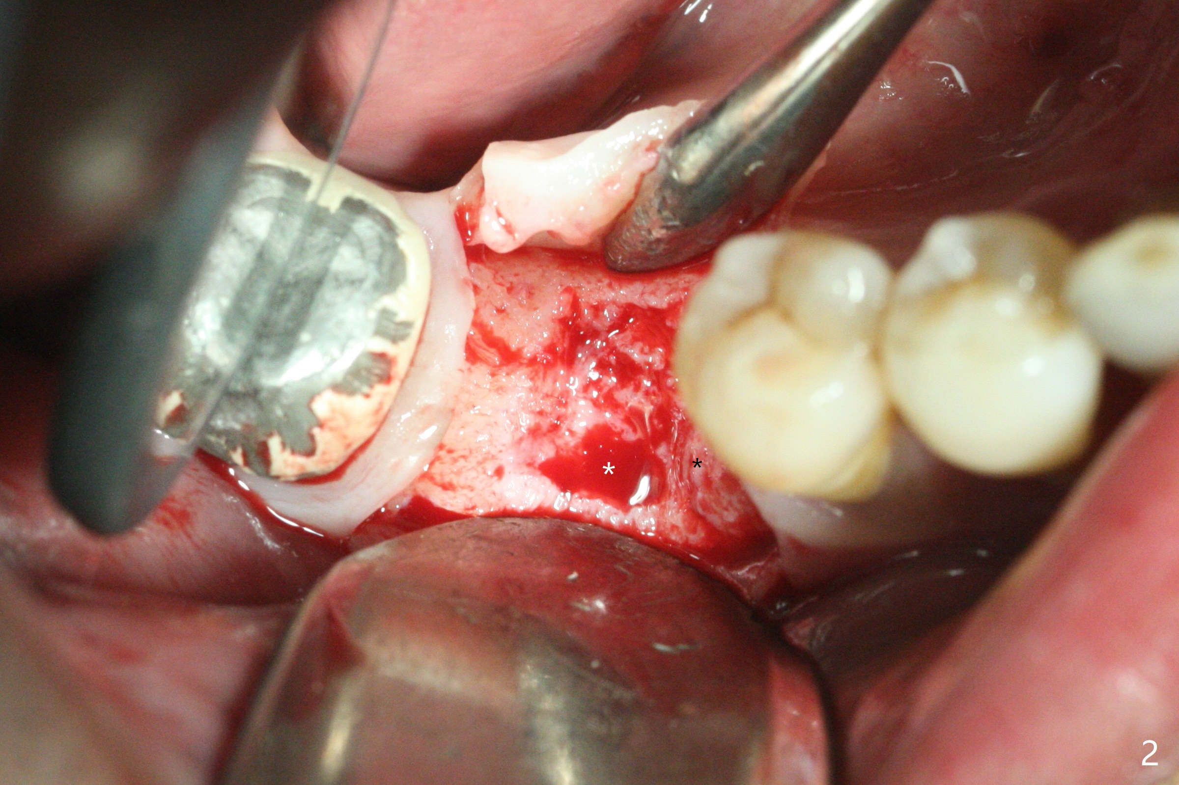
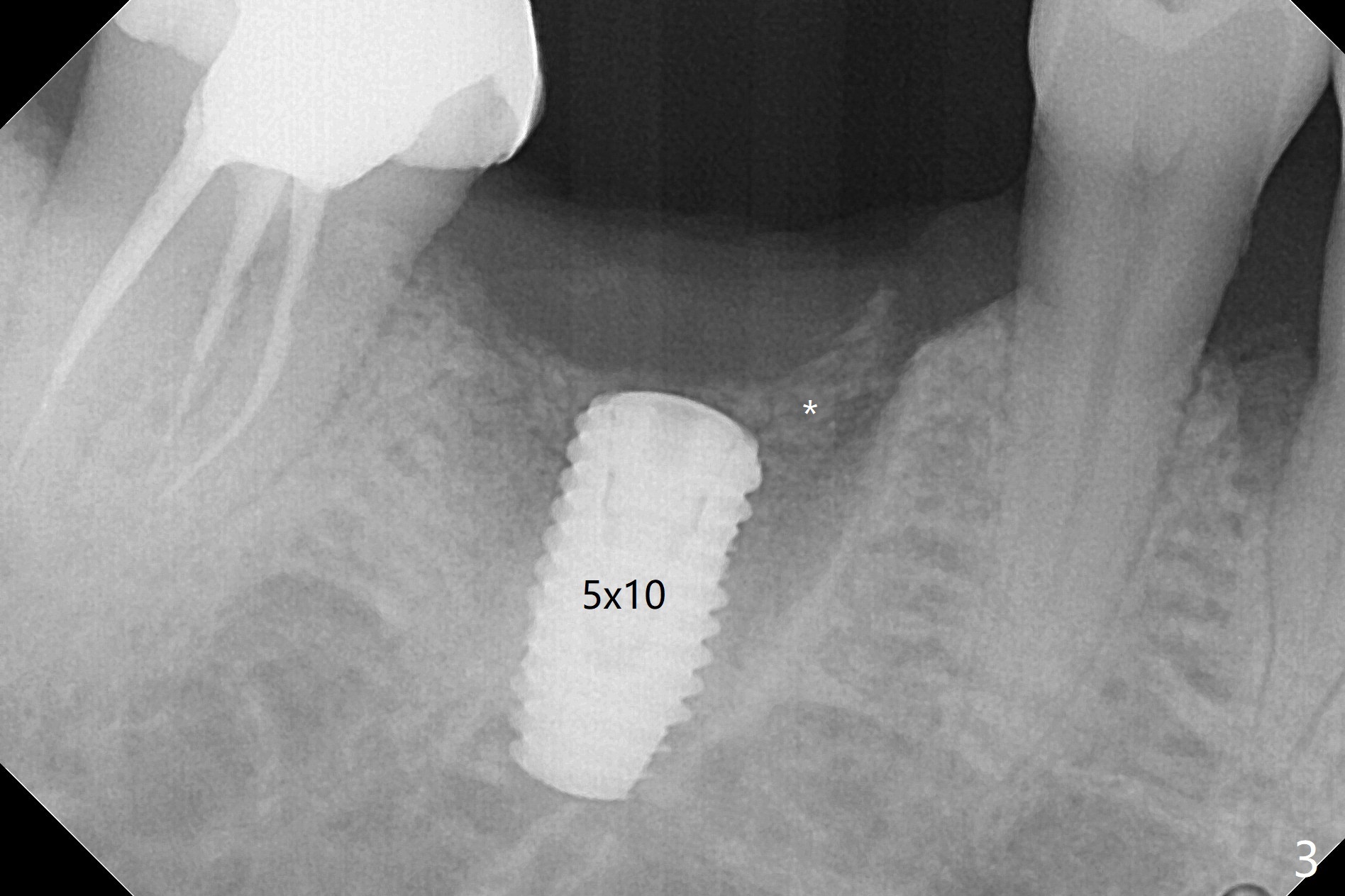
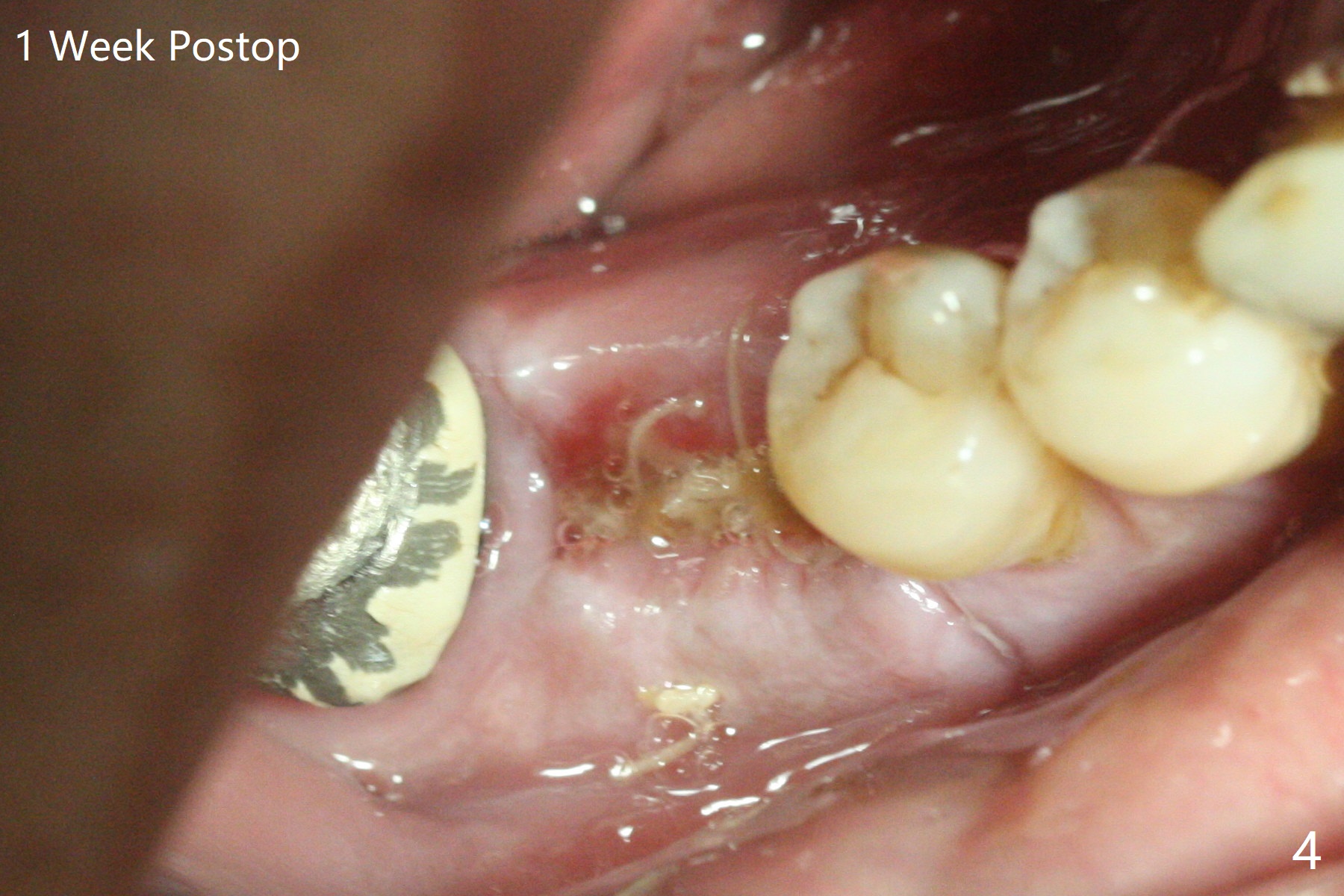
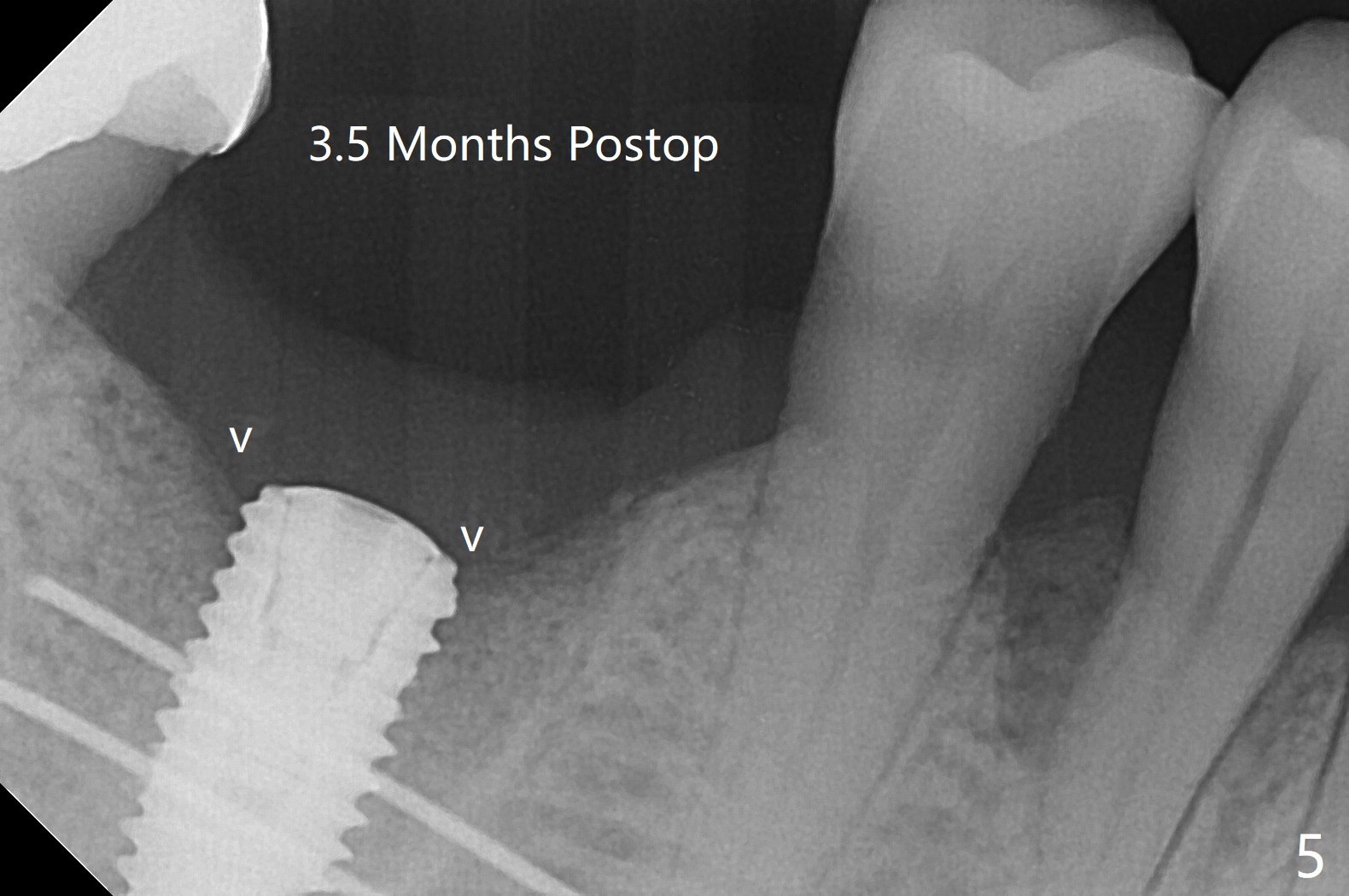
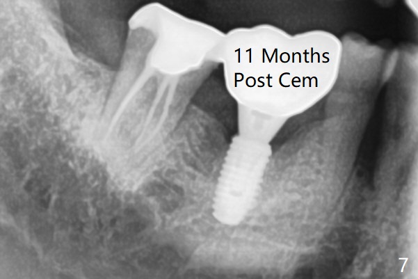
The sockets of #30 (M) of a smoker (M) are apparently not healing ~ 2 months post extraction (Fig.0, 0'). The ridge looks wide 3 months post extraction without bone graft in other office before (Fig.1) and after (Fig.2) flap surgery. The socket appears to have healed perfectly consi-dering buccal defect (M) revealed by CT ~ 1 month earlier. When a 5x10 mm implant is placed with guide and high torque (in spite of overprep), granu-lation tissue is found mesio-buccal (MB, Fig.2 *). After curettage, bone graft is placed around the implant, especially MB (Fig.3 *), followed by PRF. Periodontal dressing dislodged a few days postop because of mastication on the right side (Fig.4), whereas the anterior one (24/26) remains in place. There is a small gap around the implant when it is uncovered (Fig.5 arrowheads). It appears that some of bone graft gets lost from the incision (smoker). A 6.5x5.5(4) mm cemented abutment is seated and torqued at 30 Ncm before impression (Fig.6). There is no bone loss 11 months post cementation (Fig.7).
Return to
No Deviation
7 Density
3
Xin Wei, DDS, PhD, MS 1st edition
03/05/2020, last revision
06/17/2021