,%20then%20Vanilla,%20Osteogen%20plug,%20perio%20dressing.jpg)
%20occlusal%20view.jpg)
|
|
|
|
|
|
|
|
|
|
|
|
|
|
|
|
|
|
||
Smallest 1-Piece Implant to Treat Periimplantitis
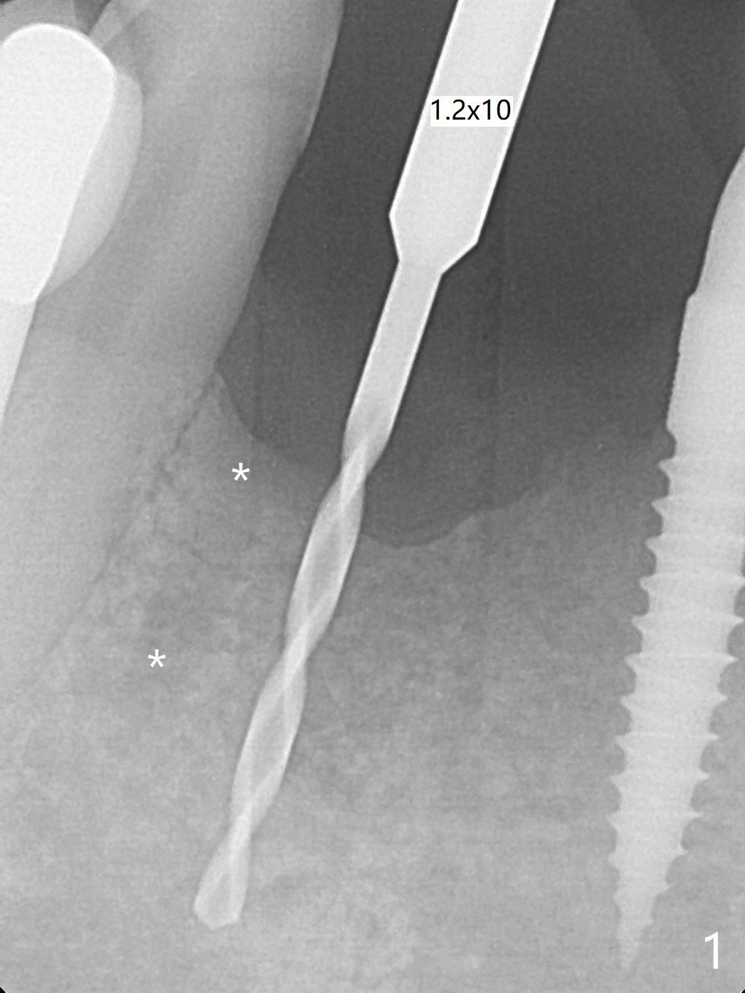
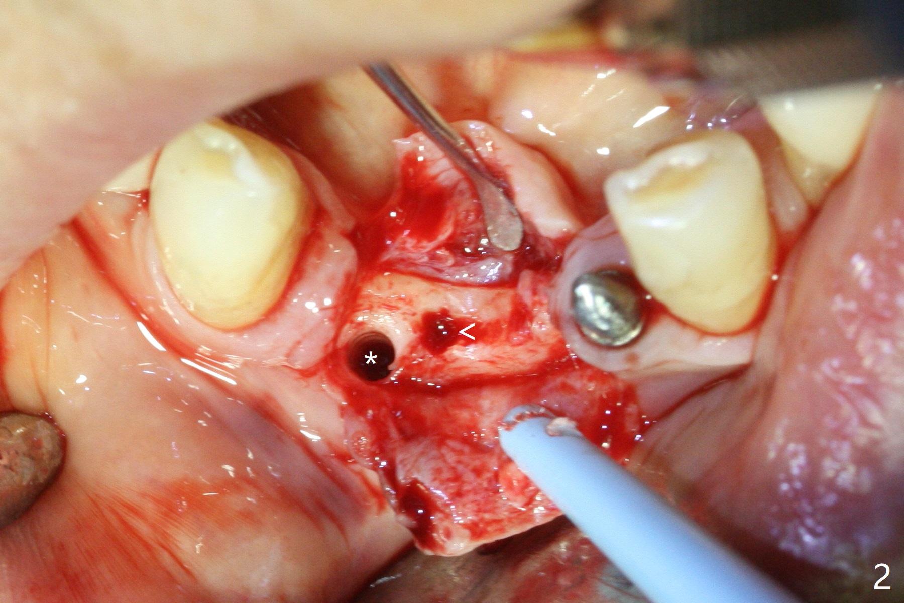
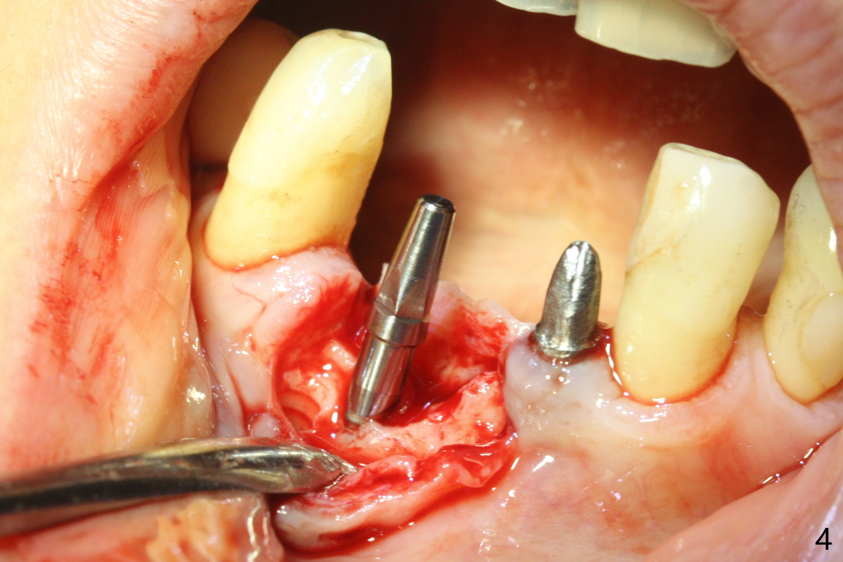
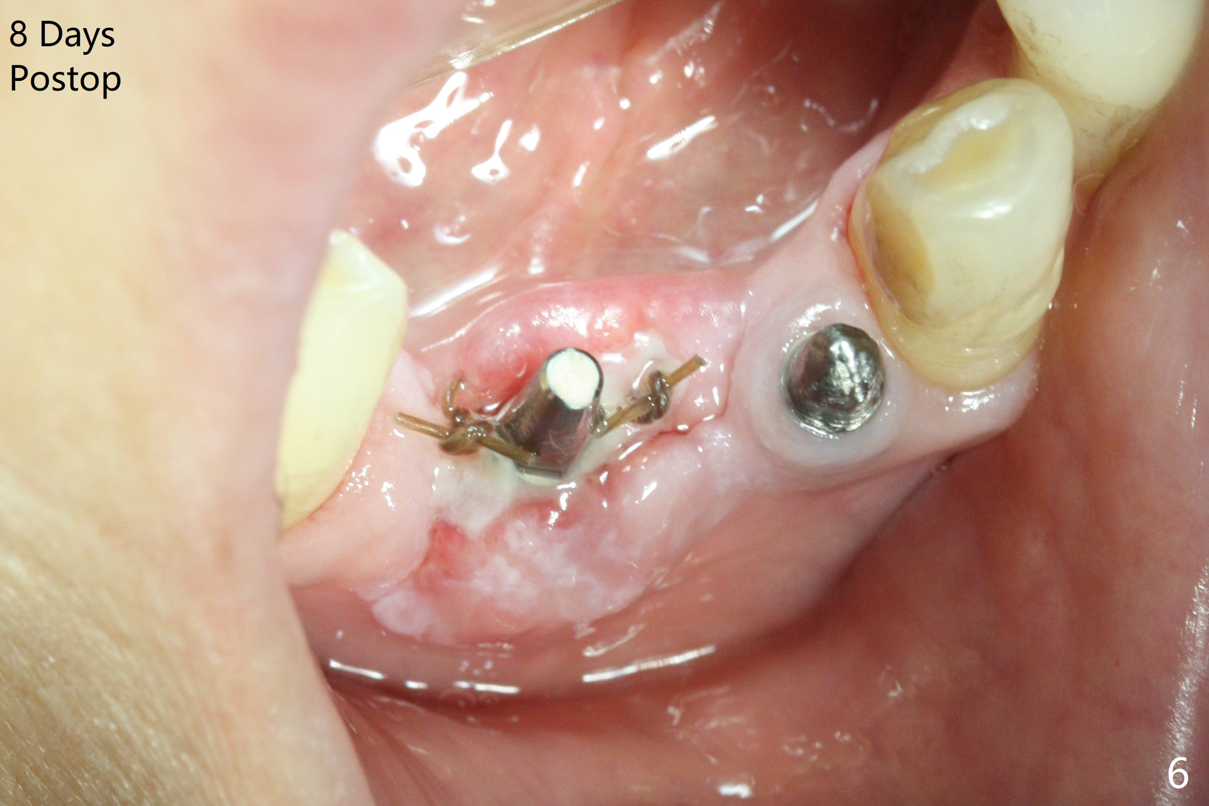
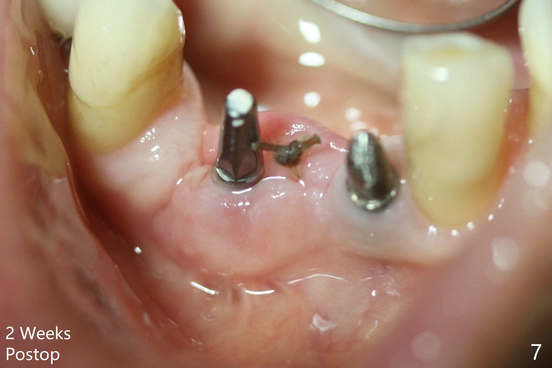
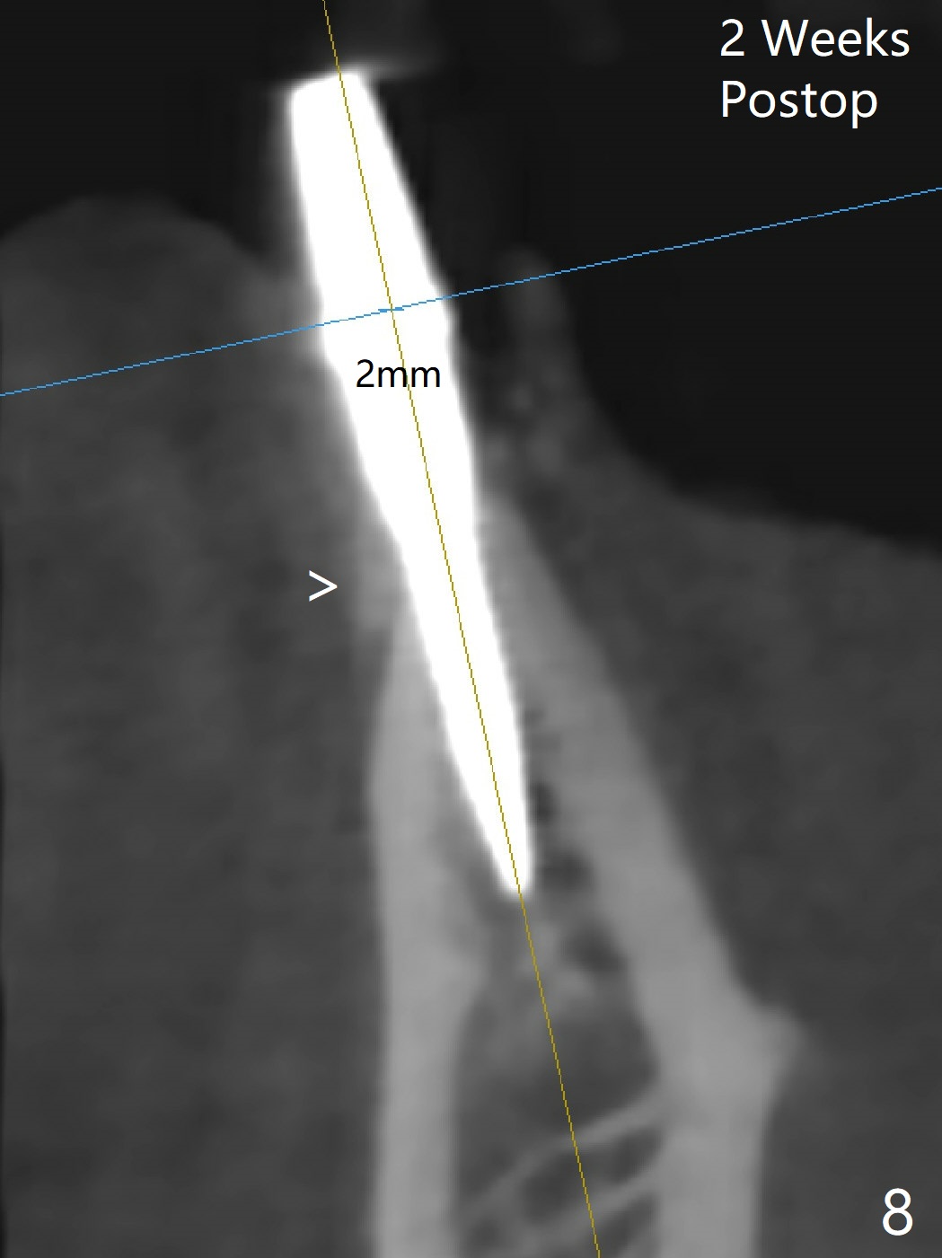
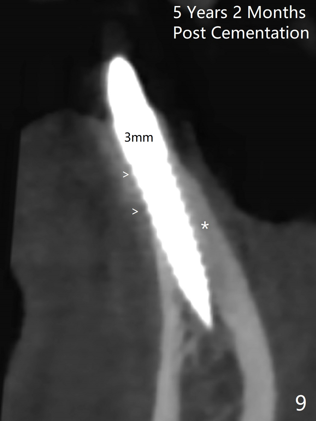
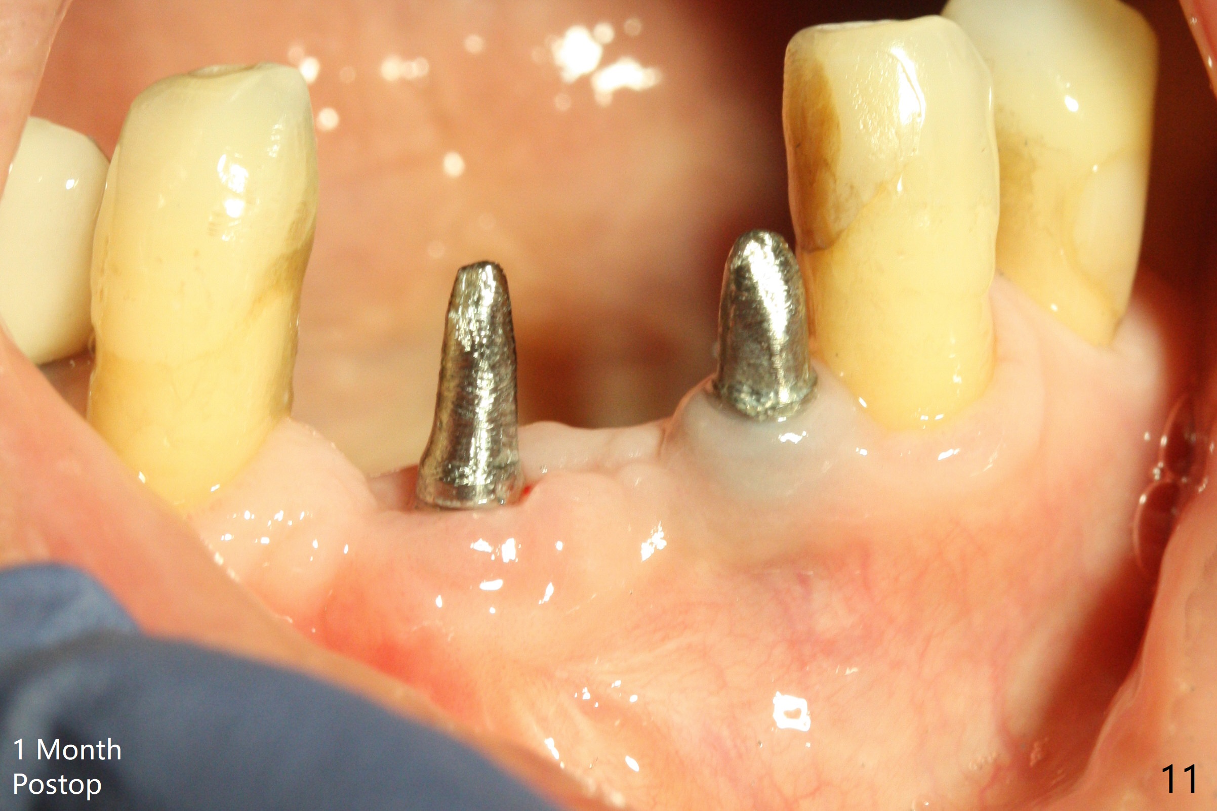
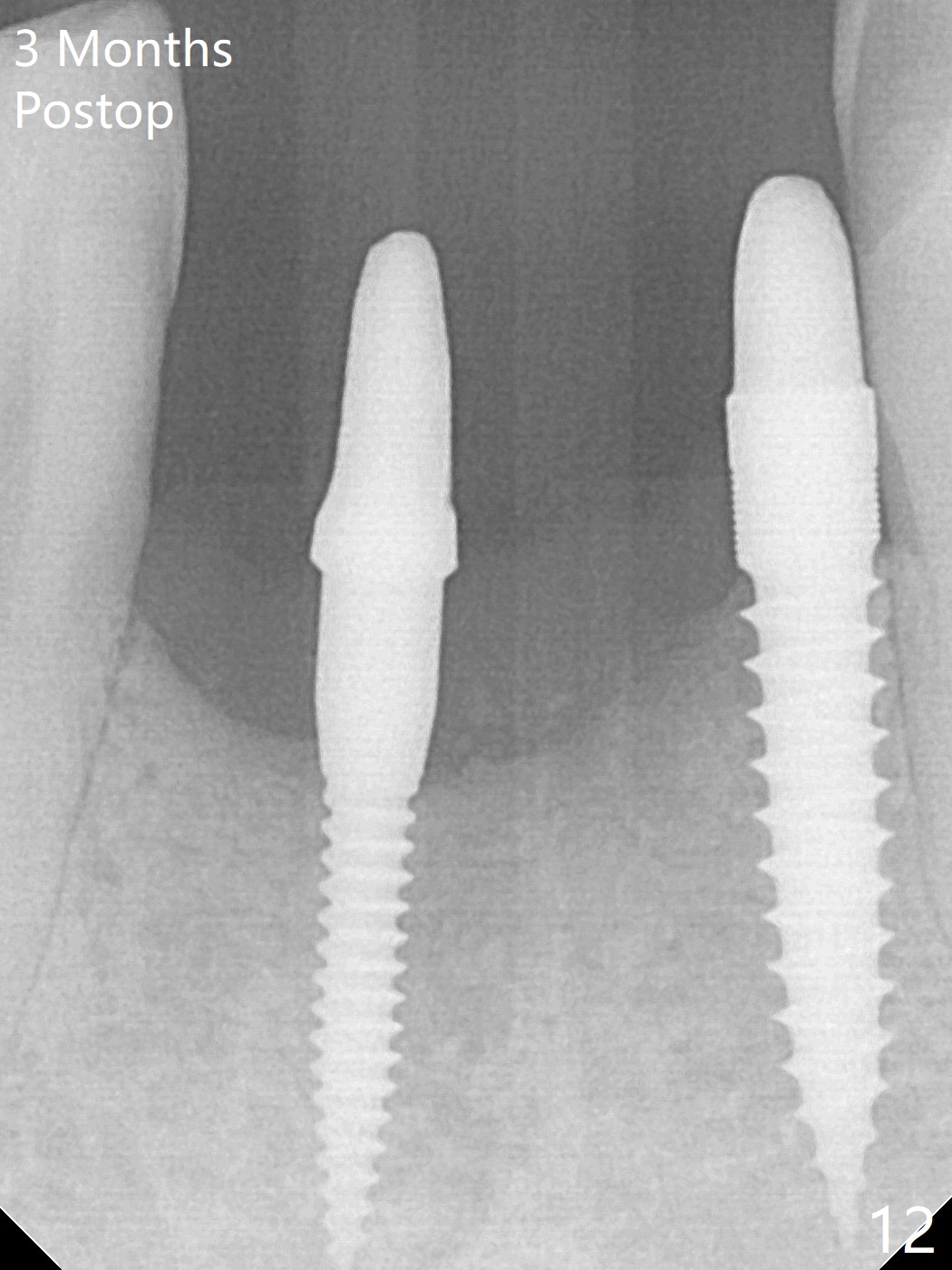
After removing #23-26 FPD and the 3x14 mm 1-piece implant at #26 (Fig.1,2 *), a new osteotomy is initiated in the narrow ridge (after ridge top reduction) approximately at #25 (Fig.2 <). Following placement a 2x10(4) mm implant at #25 (Fig.3-5) and Osteogen plug in the osteotomy at #26, Vanilla graft is placed around the implant, especially buccal. Periodontal dressing is applied after suturing. The buccal and lingual flaps are erythermatous and edematous without pain 8 days postop (Fig.6). The wound seems to be healing 2 weeks postop (Fig.7), no sign of osteonecrosis. With placement of a 2 mm implant at #25 (Fig.8 (>: bone graft buccally)), the buccal plate remains normal in thickness. When a 3 mm implant is placed at #23 (Fig.9), approximately 4 threads appear to be exposed (between arrowheads), partially due to the thick lingual plate (*).
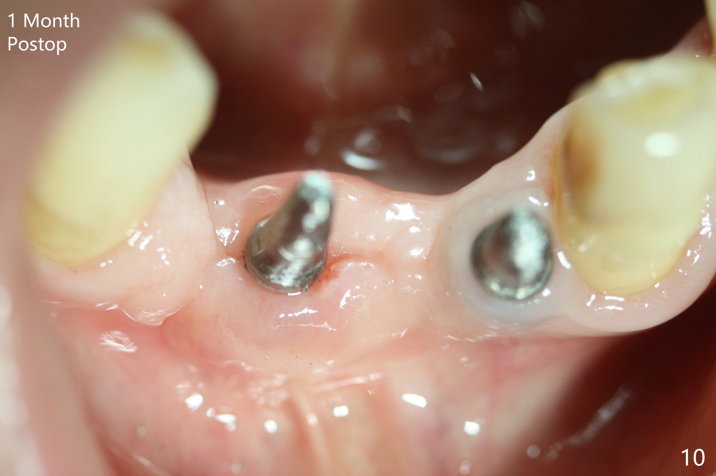
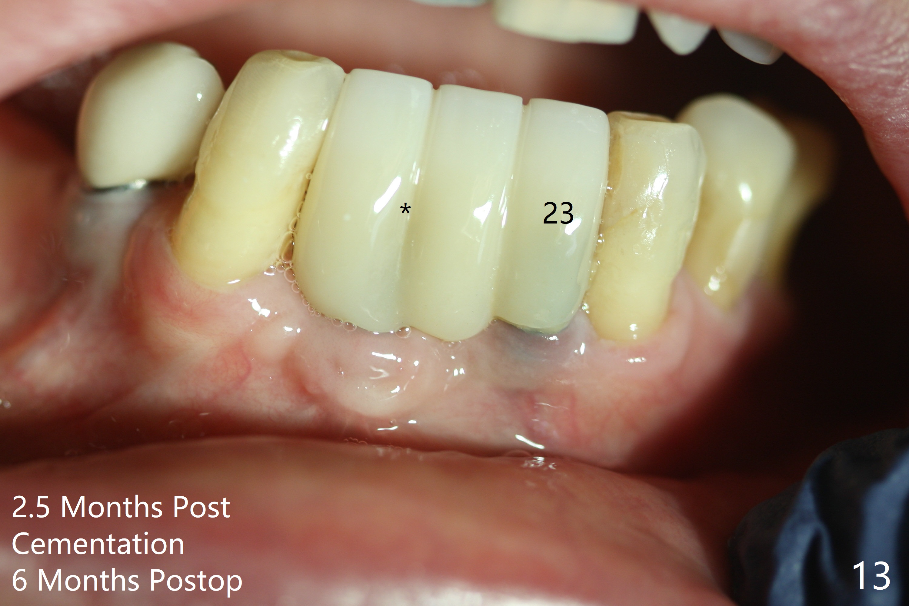
The wound at #25 seems to heal 1 month postop; after prep (Fig.10,11), a provisional FPD is fabricated. Impression is taken for surgical guides of UL and LL implants. The patient requests early final restoration (Fig.12). The small implant placed lingually (Fig.12 *, 2 mm) is associated with the pleasing gingiva in color, as compared to the 3 mm one at #23 with the metal shaded gingiva.
Return to
Lower Incisor
Immediate Implant,
Armaments
1-Piece
Xin Wei, DDS, PhD, MS 1st edition 07/10/2018, last revision 12/26/2020