
|
|
|
|
|
|
|
|
|
|
|
|
|
|
|
|
||
Control Length of Implant In Graft Bone
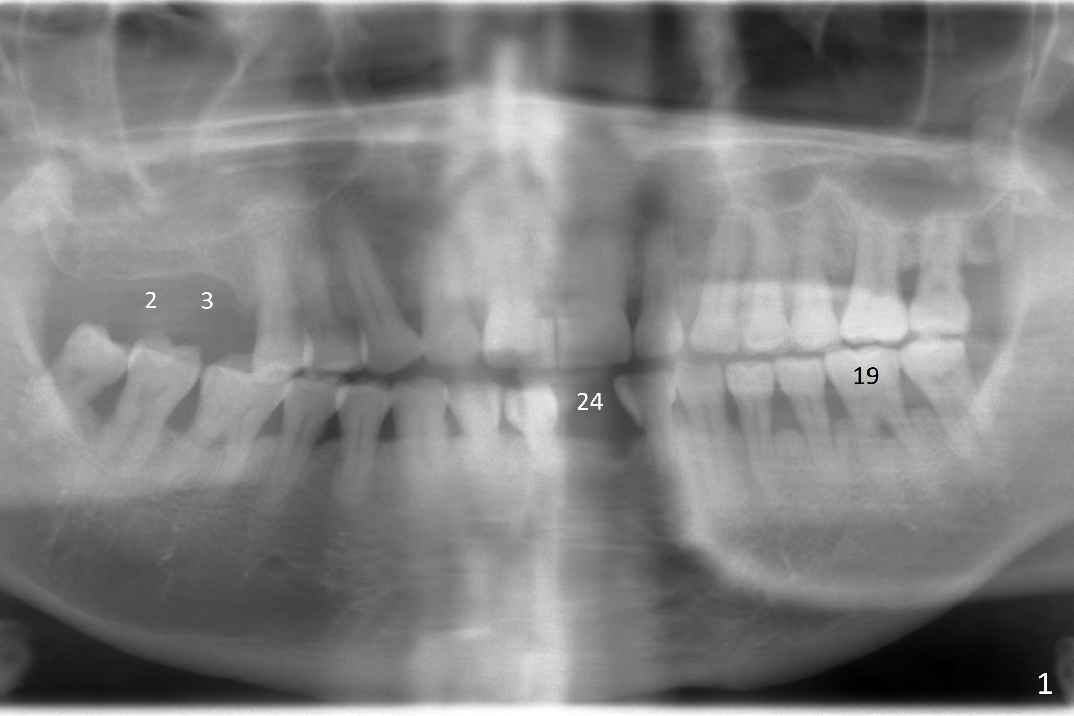
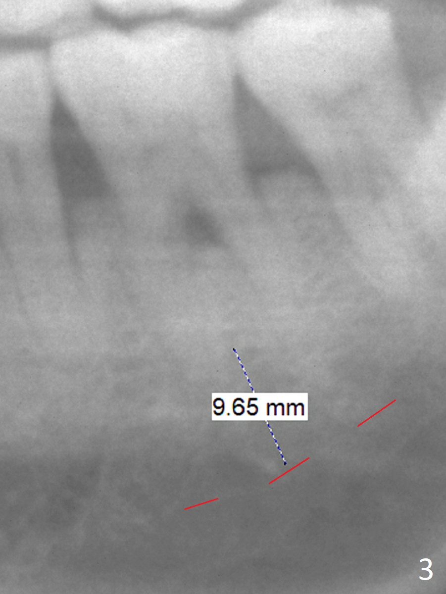
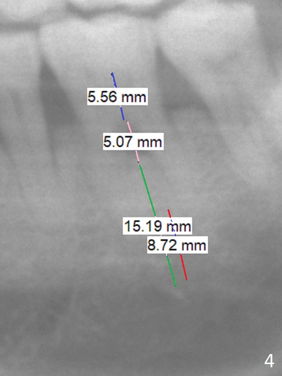
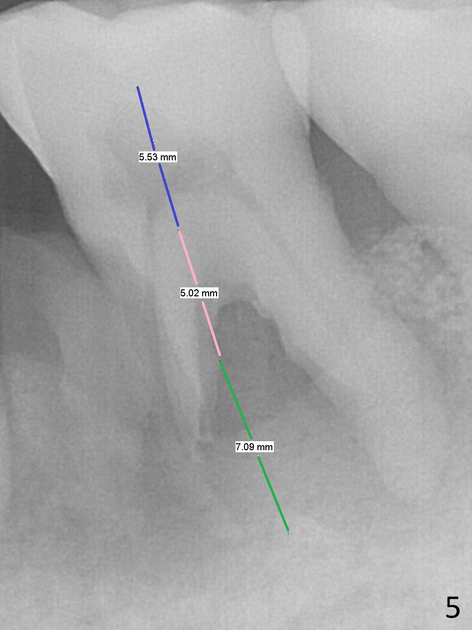
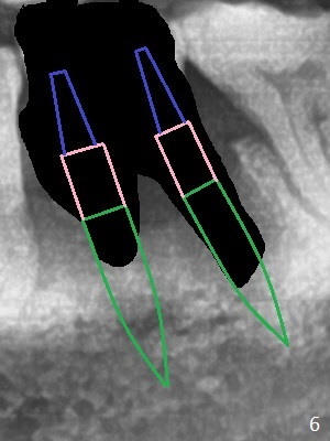
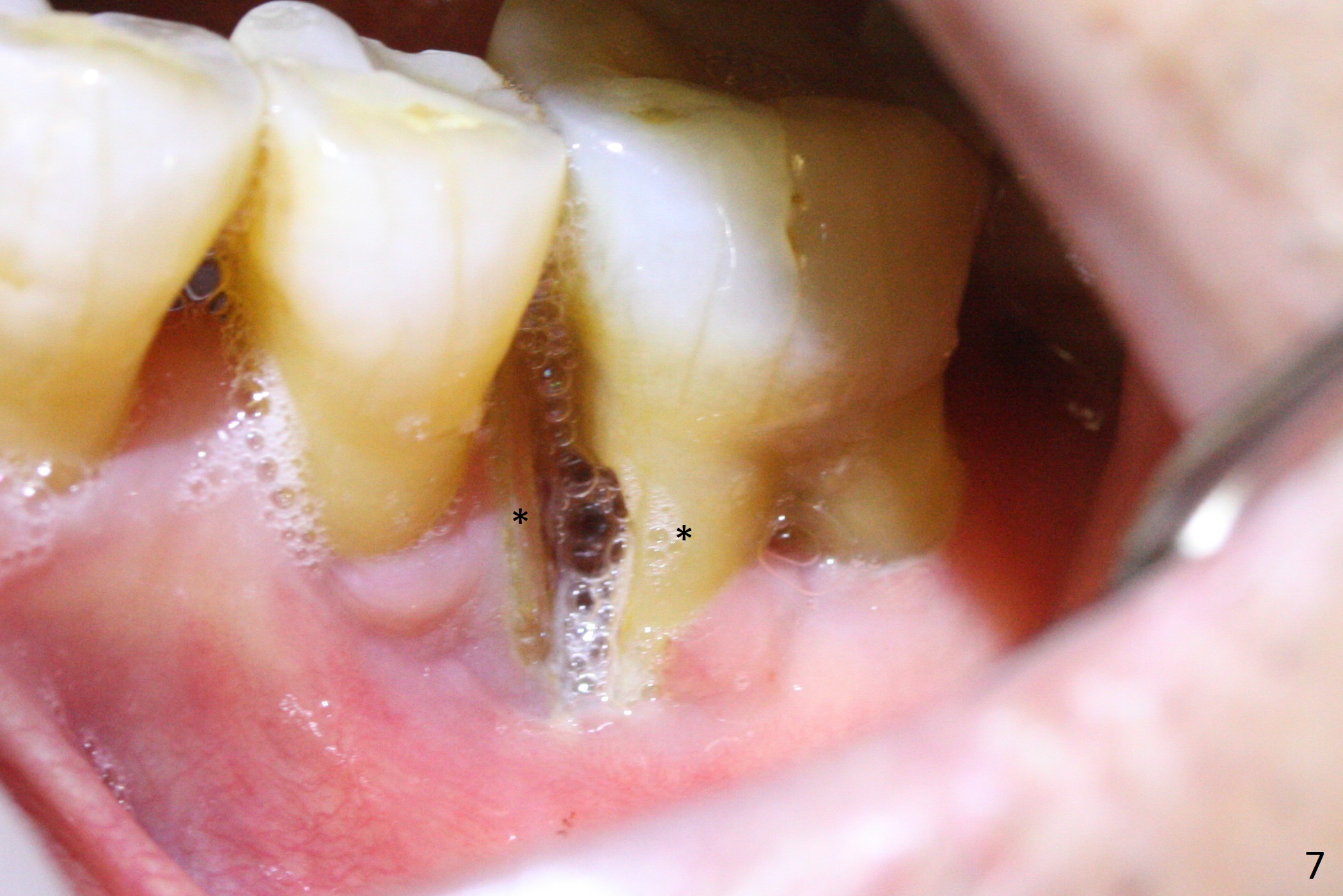
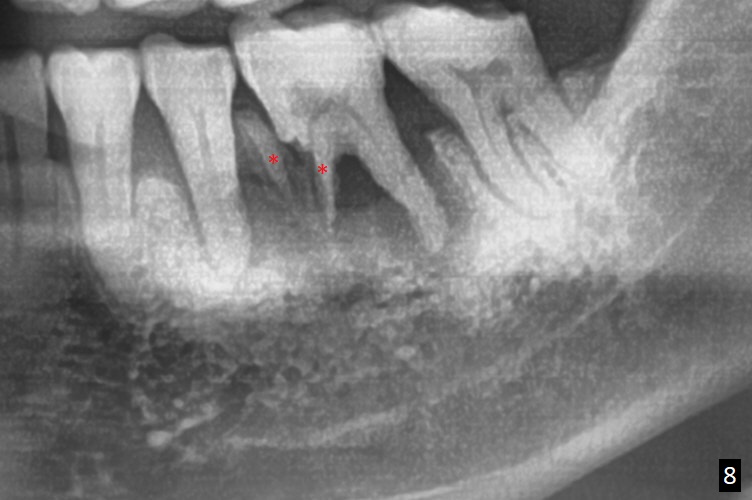
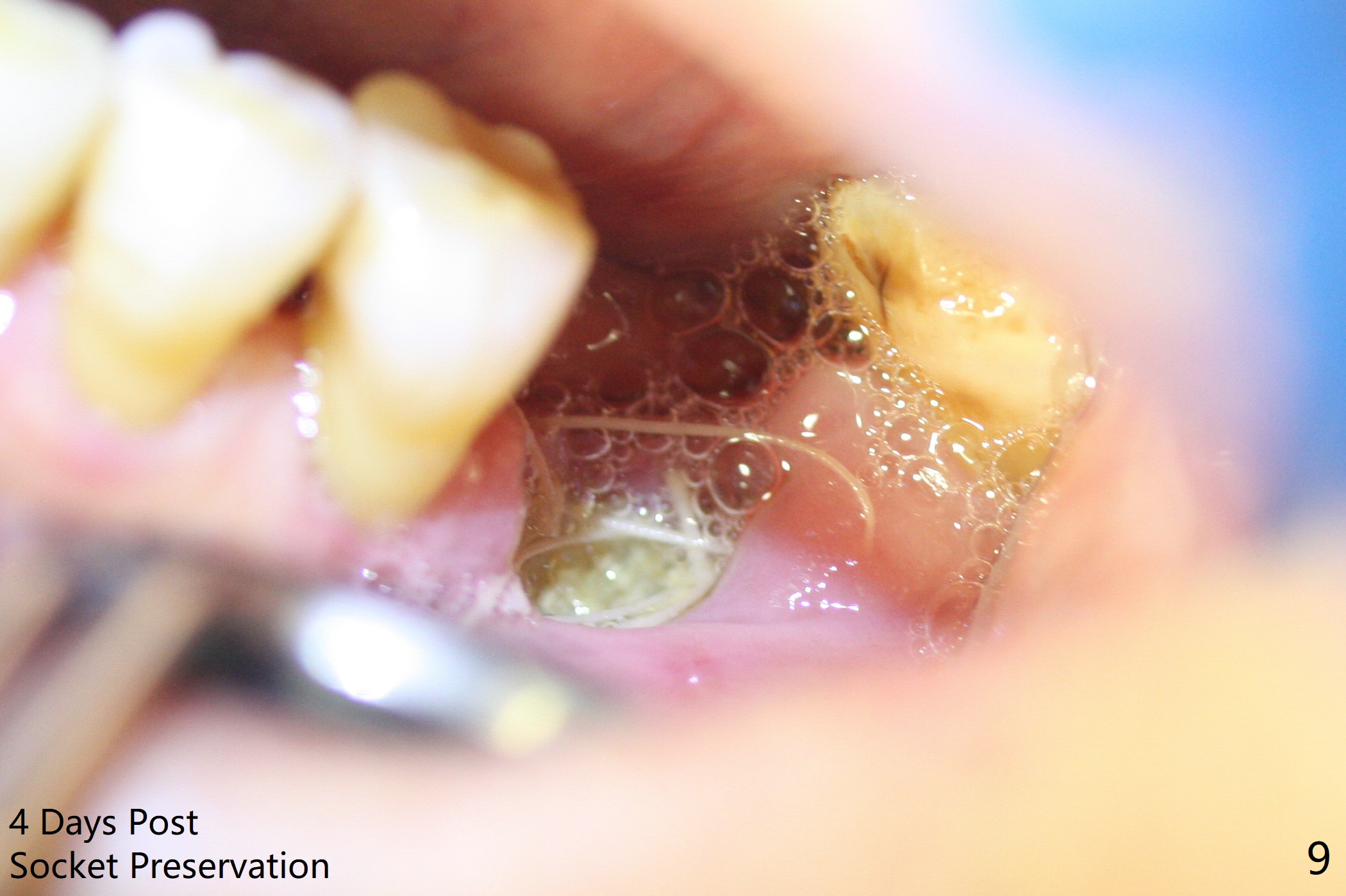
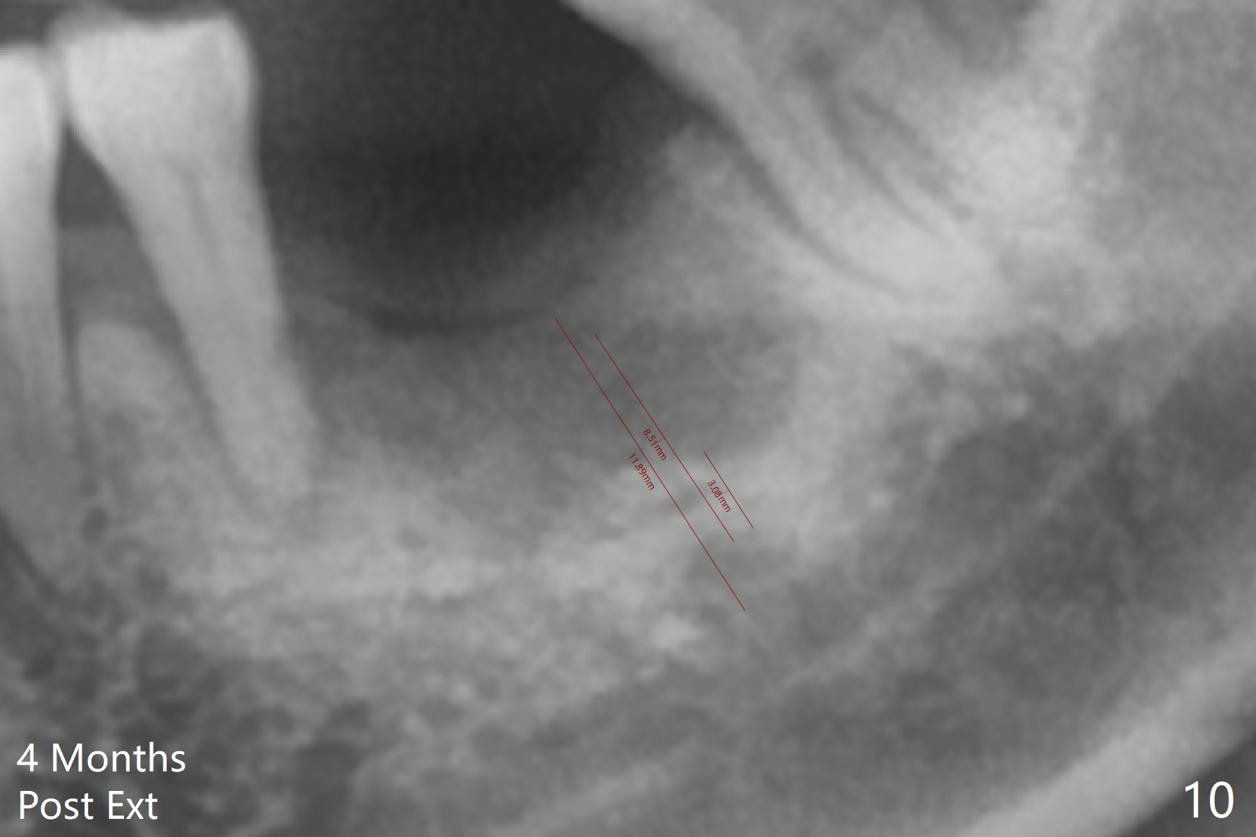
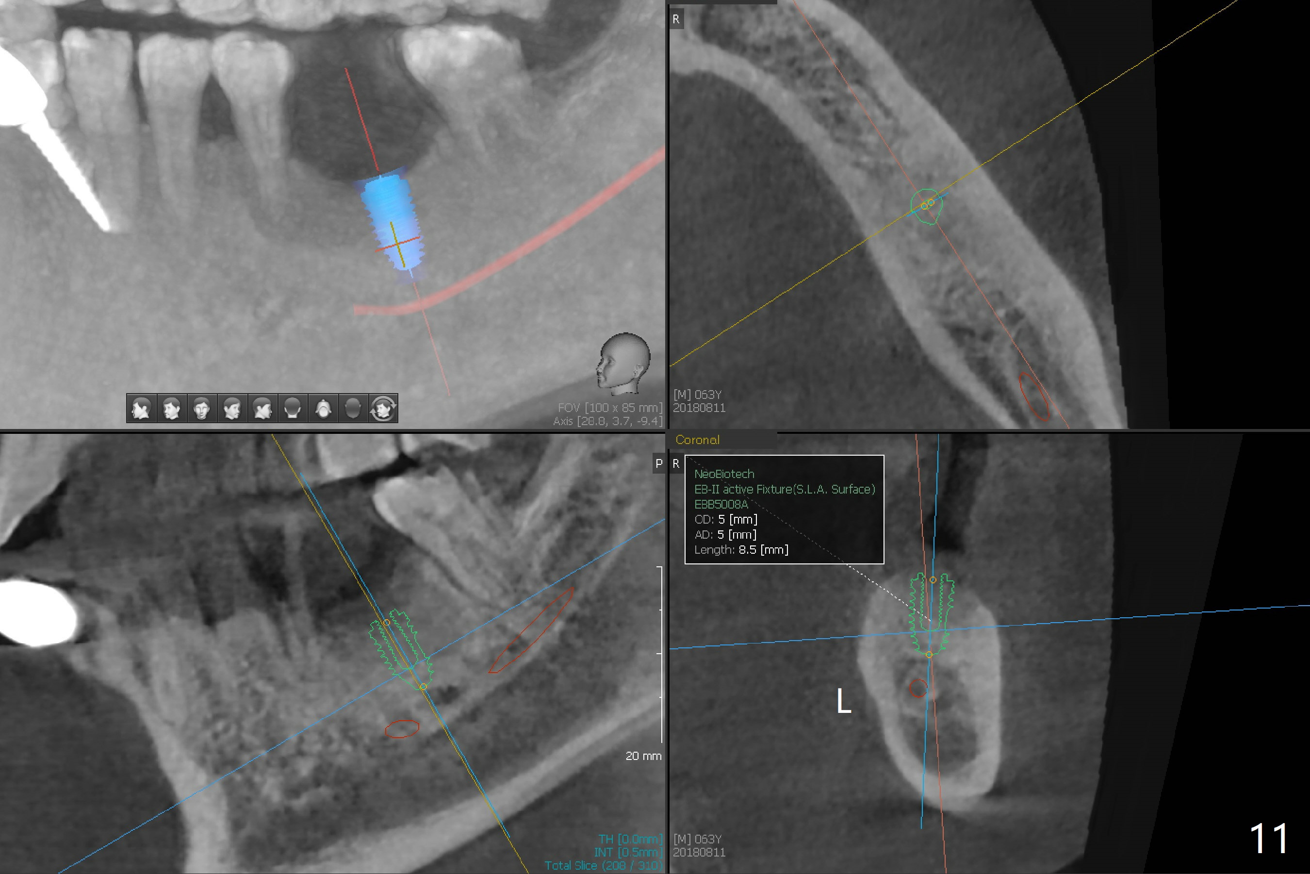
A 63-year-old man is a diabetic with history of good control. He masticates unilaterally with missing teeth #2,3 and 24 (Fig.1 (taken 5 years ago)). The latter has been restored with an implant. Last February the mesial root of the tooth #19 was found to have vertical fracture with bone loss until the base of the septum (Fig.2). The bone available for implantation is ~7-8 mm after root fracture (Fig.3,4). The initial osteotomy depth will be 7 mm (IS kit), followed by the calibrated parallel pin. The latter determines how many millimeters of an implant will be surrounded by the graft bone (Fig.4,5 (green: implant length; pink: cuff; blue: abutment length)). Since the buccal defect is severe with mesial root split (Fig.7 *), draw blood for PRF (2 large tubes). To save the remaining septum, place 2 of 1-piece implants on the either side of the septum (compare Fig.6,8). Take photos to compare buccal vs. lingual gingival recession. Tell the patient that the tooth #20 may be nonsalvageable. Since insurance preauthorization does not get approval for several times, the tooth #19 is extracted with socket preservation and periodontal dressing. The latter dislodges in 1 day. When the patient returns for follow up 4 days postop, the socket is exposed (Fig.9). An immediate implant should have been placed to keep the graft in place. PRF also helps. In fact preauthorization letter arrived 1 day earlier. It appears that guided surgery is indicated for limited bone height. The bone graft appears to gain ~ 5 mm bone in 4 months (Fig.10), which allows to place a 5x8.5 mm implant (Fig.11).
Return to
Lower
Molar Immediate Implant, Prevent
Molar Periimplantitis (Protocols,
Table),
Armaments,
No Antibiotic (canceled)
Xin Wei, DDS, PhD, MS 1st edition 12/24/2017, last revision 08/21/2018