|
|
|
||
|
|
|
|
|
Bone Loss Dictates Position of Implant
Placement M
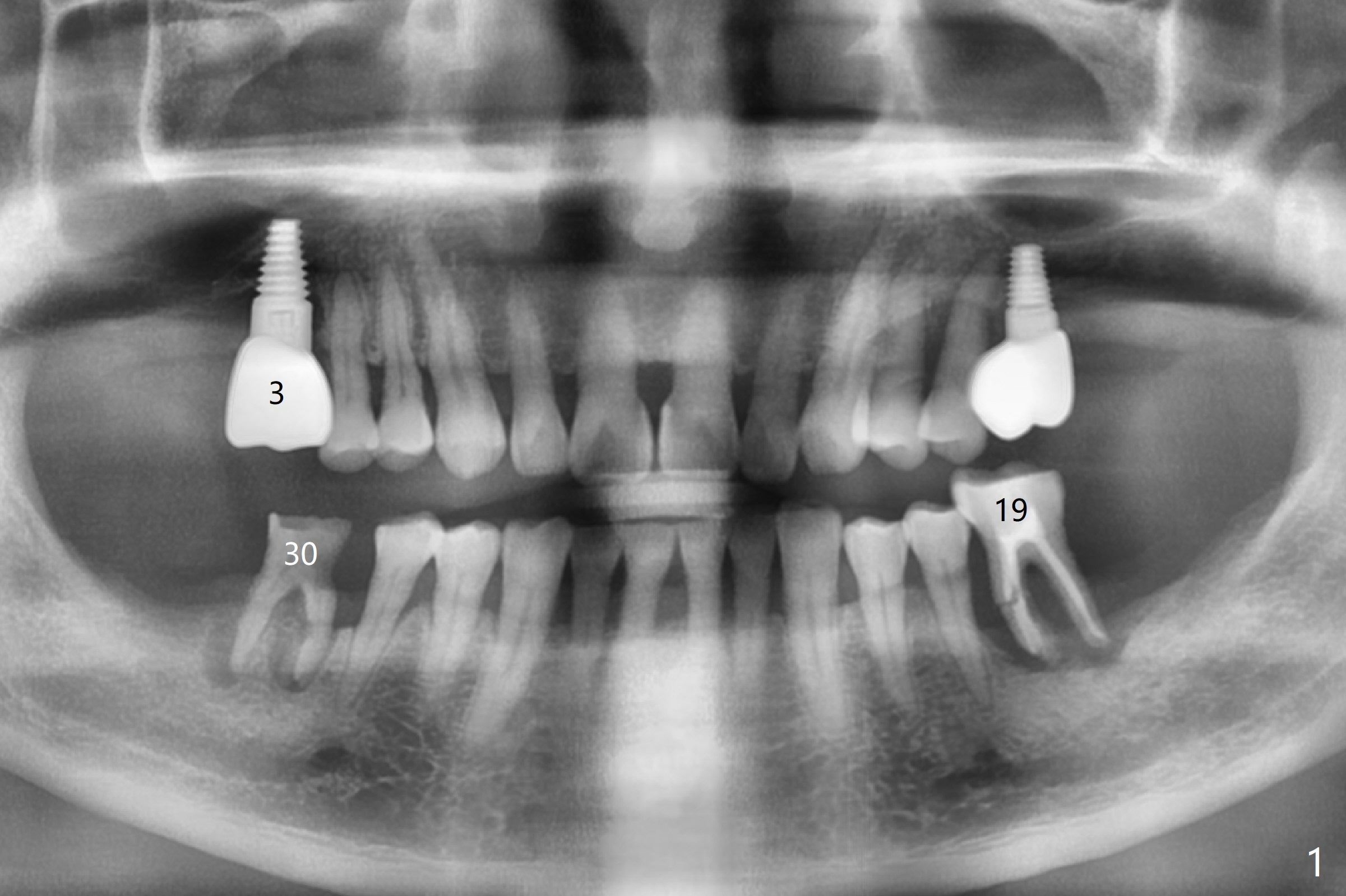
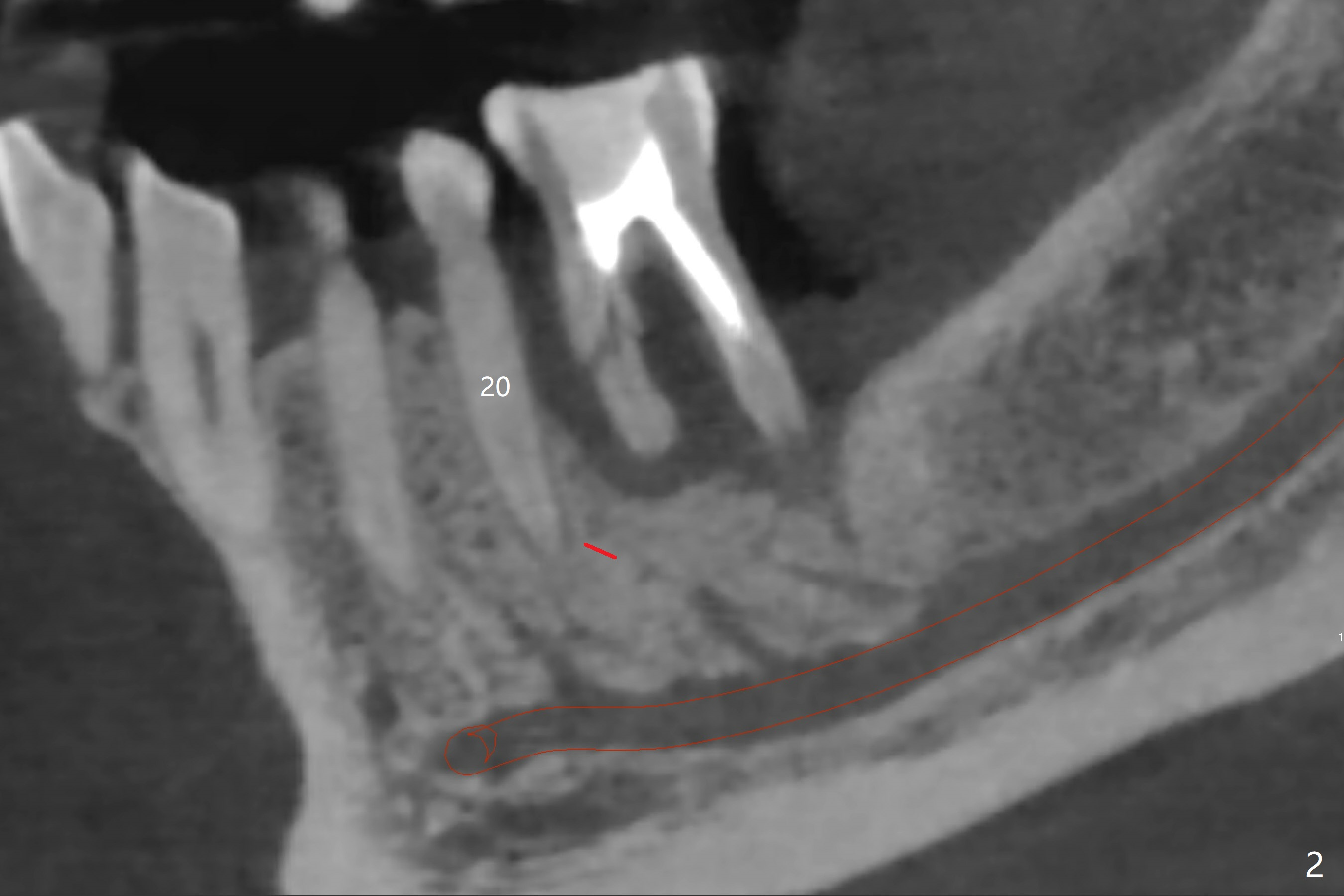
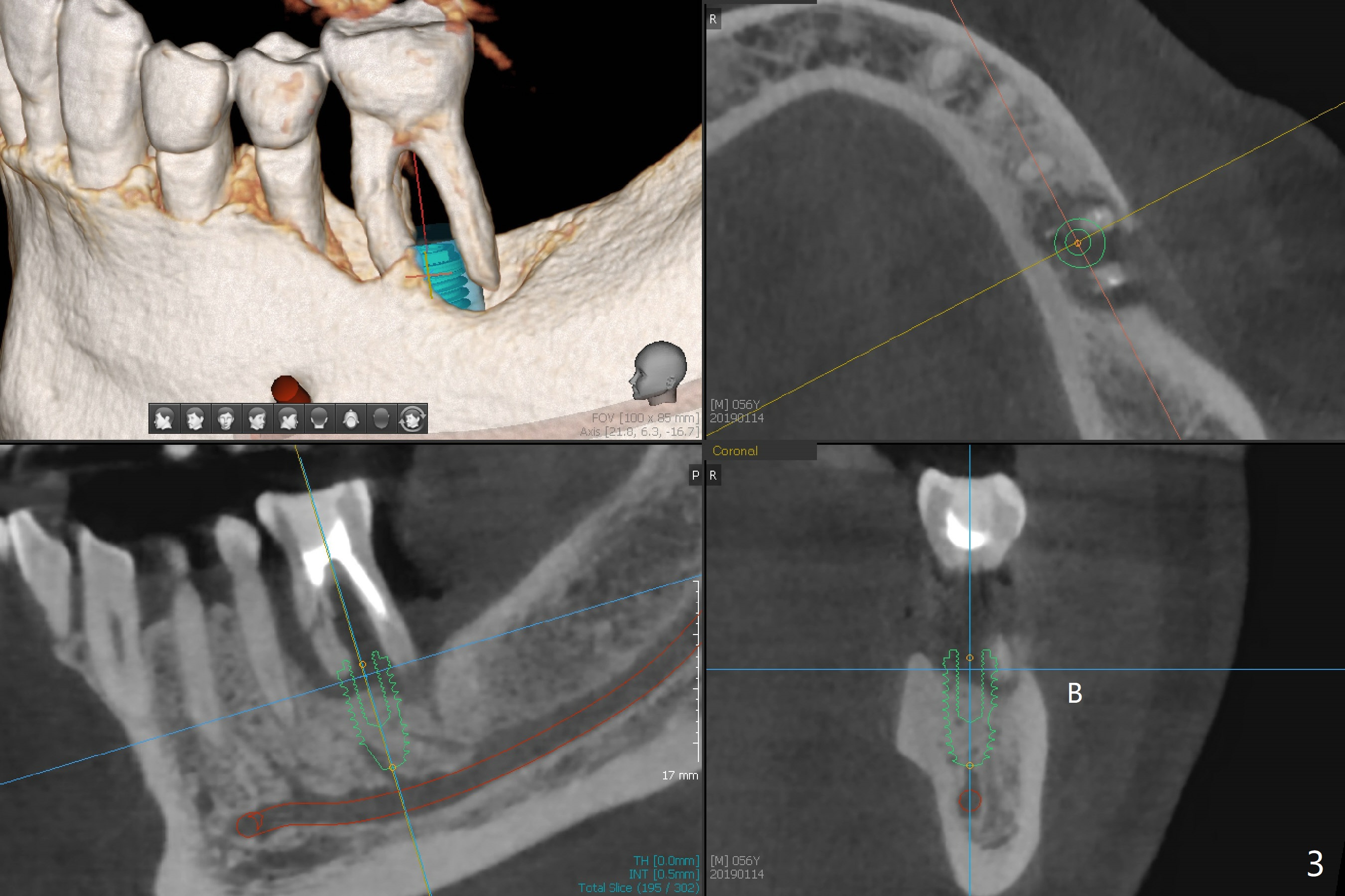
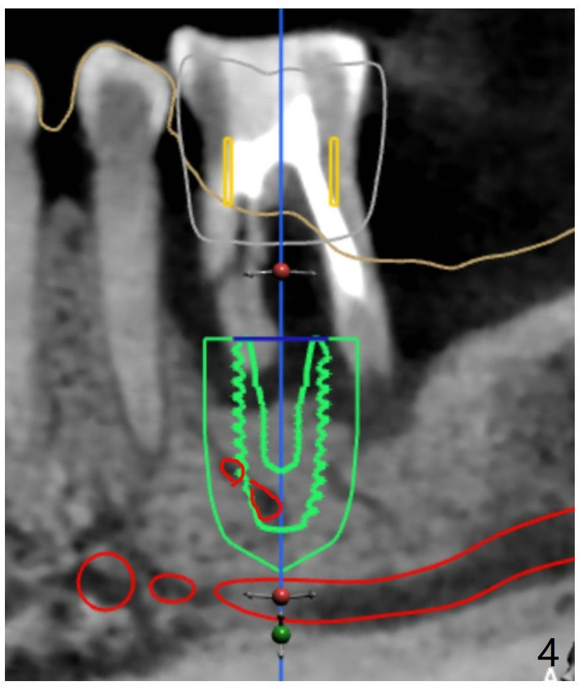
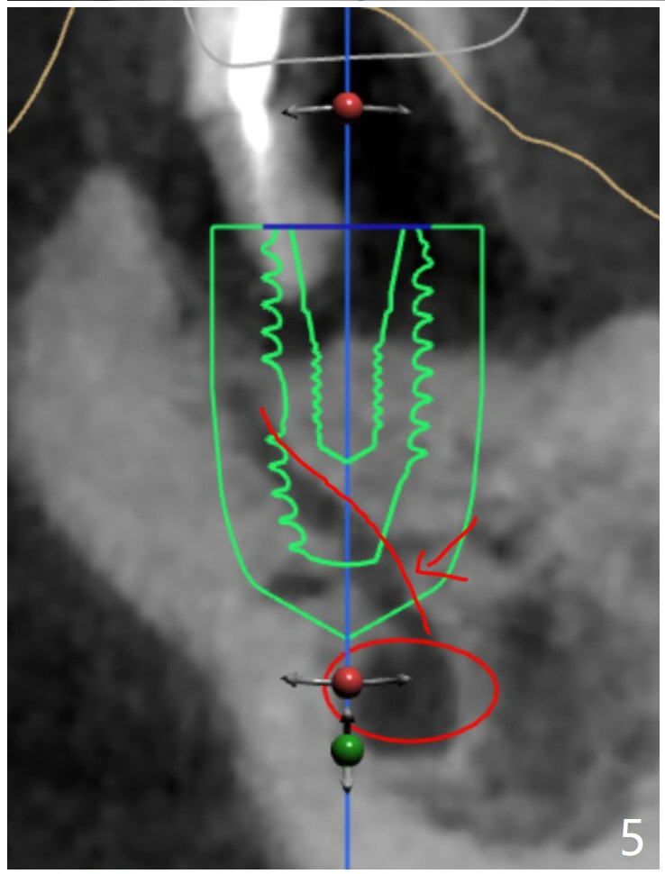
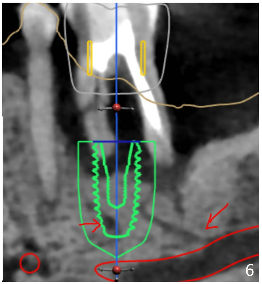
When the patient finally agrees to have the tooth #19 to be extracted for implant (Fig.1), there is also mesial bone loss associated with mesial root fracture (Fig.2). To avoid truncation of the blood vessels toward the apex of the tooth #20 (Fig.2 black shadow and red line; Fig.4-6 (lab design) red symbols), a 5x10 mm implant will be placed more distal than the earlier design when the septum was present. The implant is also to be placed slightly higher than the native bone to improve crown/implant ratio; i.e., equical to the buccal, lingual and distal crests (Fig.3-6). Allograft will be placed in the gaps. After distal placement with minor axial change, new treatment plan is born. Return to Lower Molar Immediate Implant, Prevent Molar Periimplantitis (Protocols, Table), Trajectory, Weichat Xin Wei, DDS, PhD, MS 1st edition 01/14/2019, last revision 12/28/2019