|
|
|
|
|
|
|
|
|
|
|
|
|
|
|
|
|
|
Improve Mini-implantation
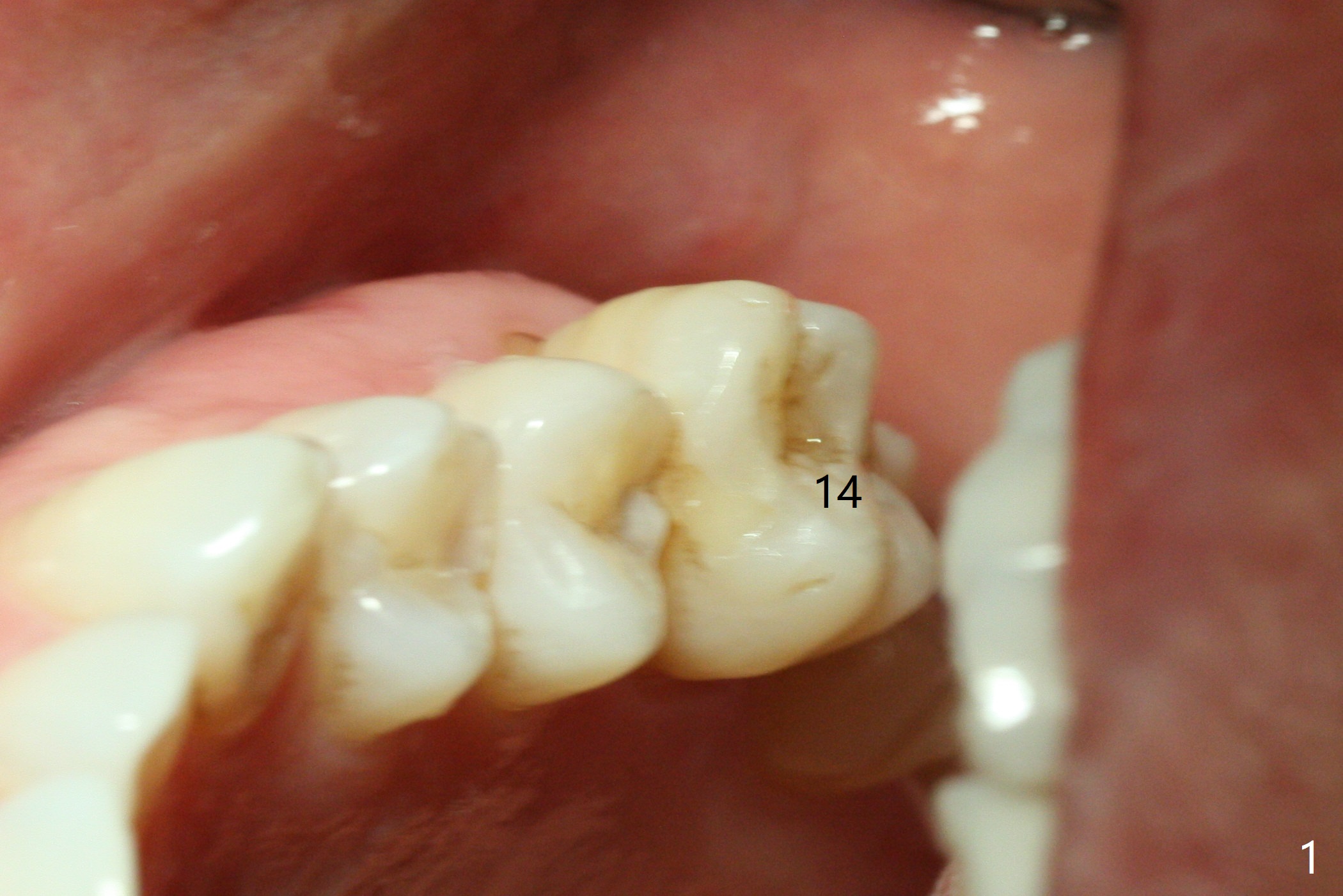
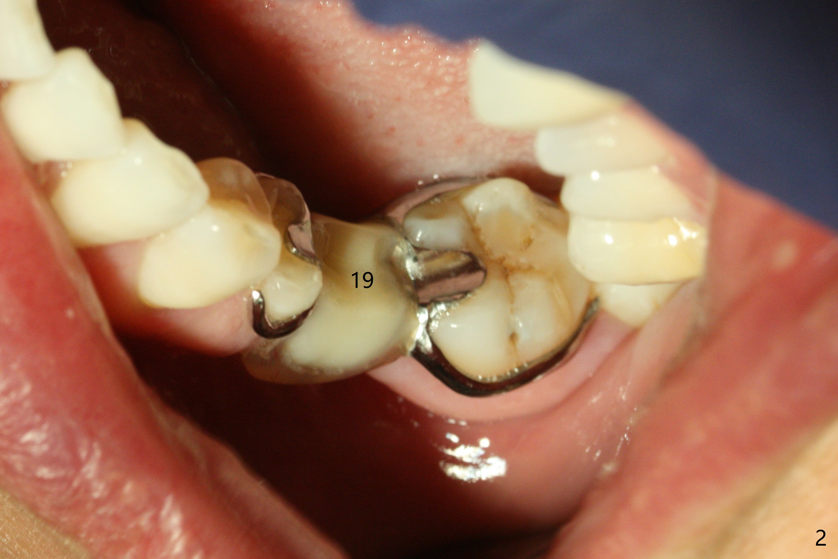
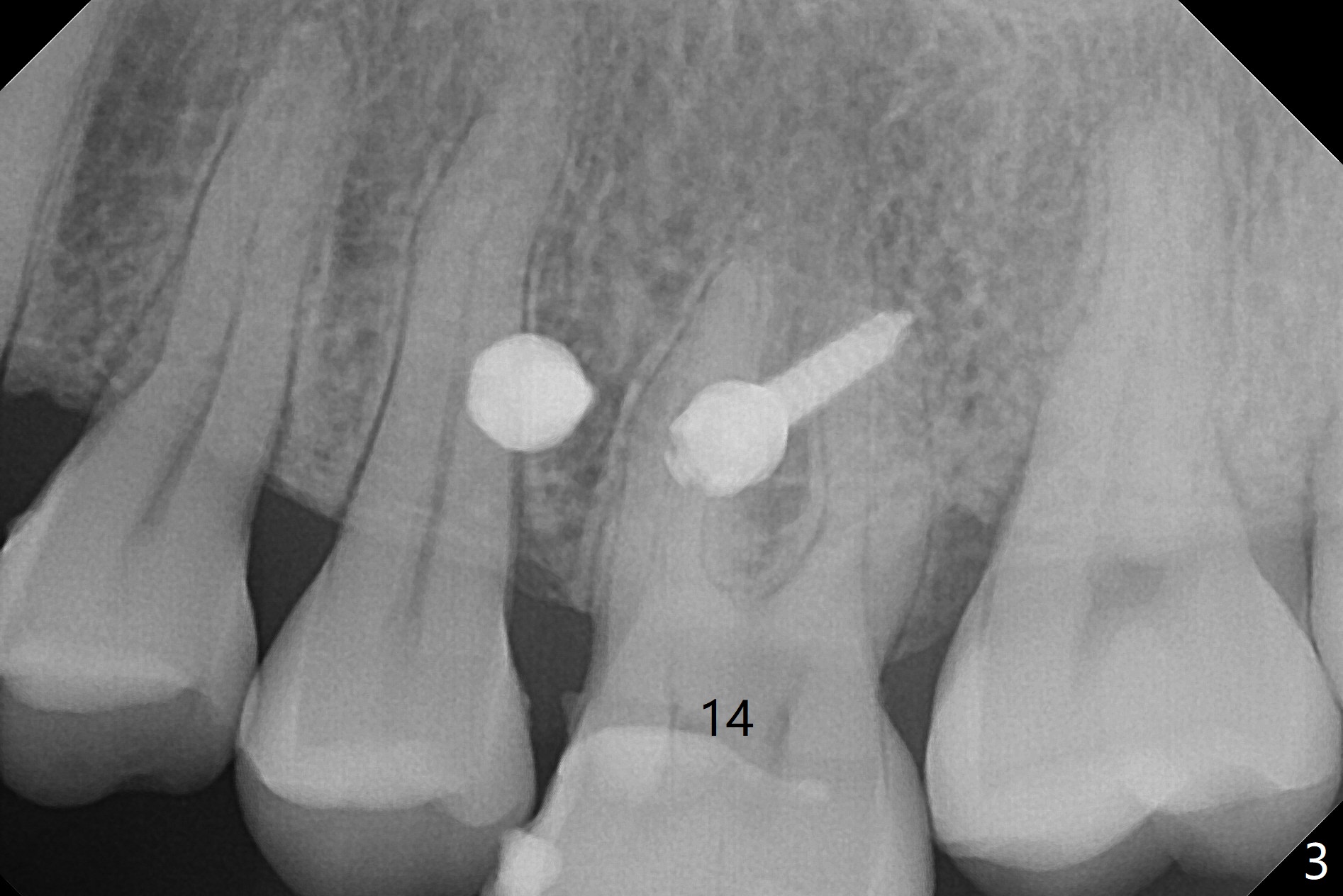
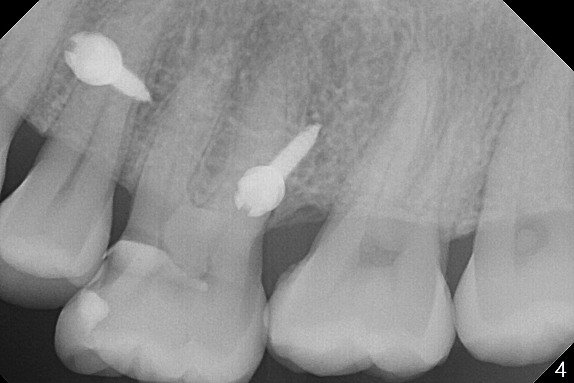
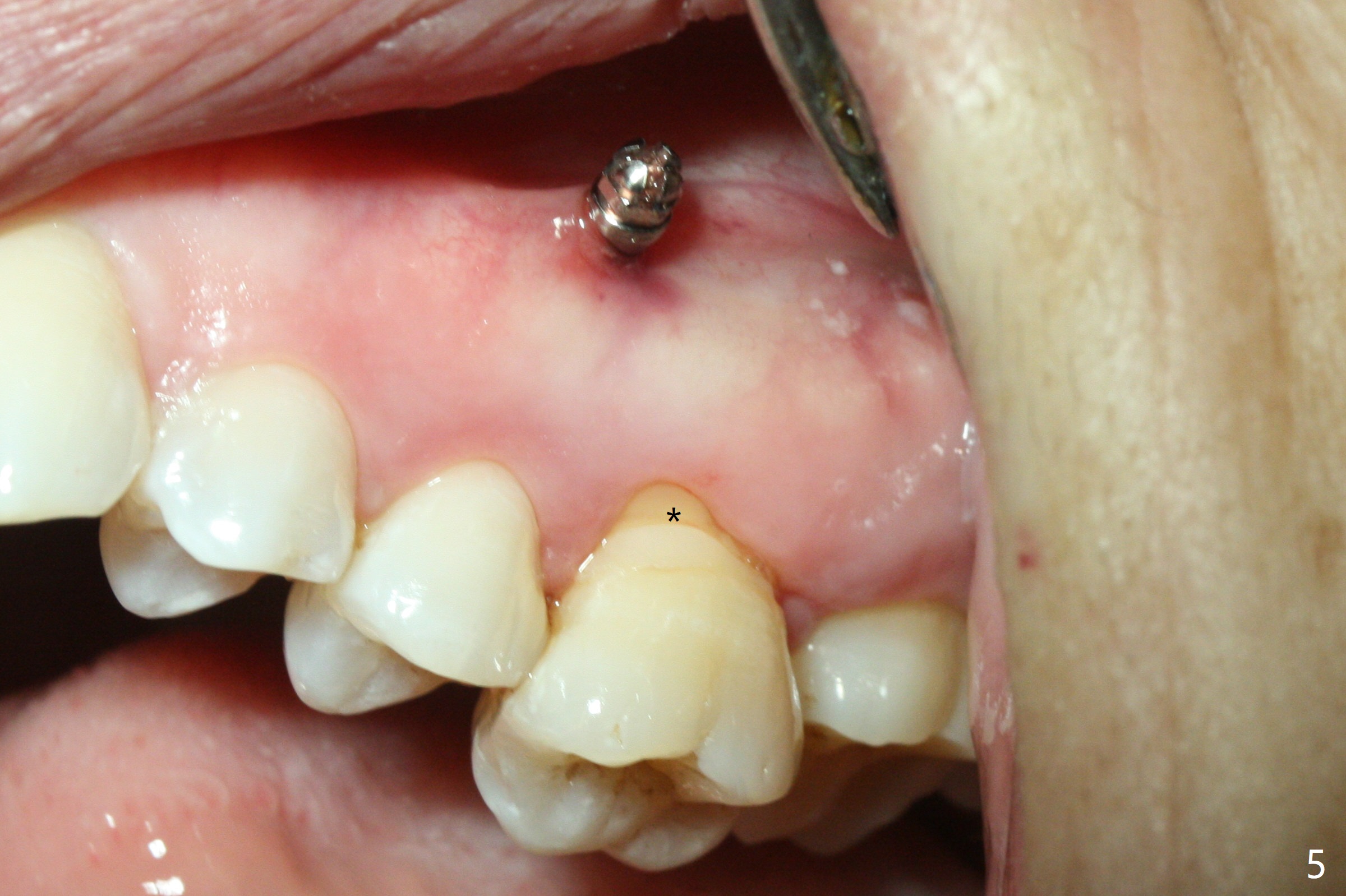
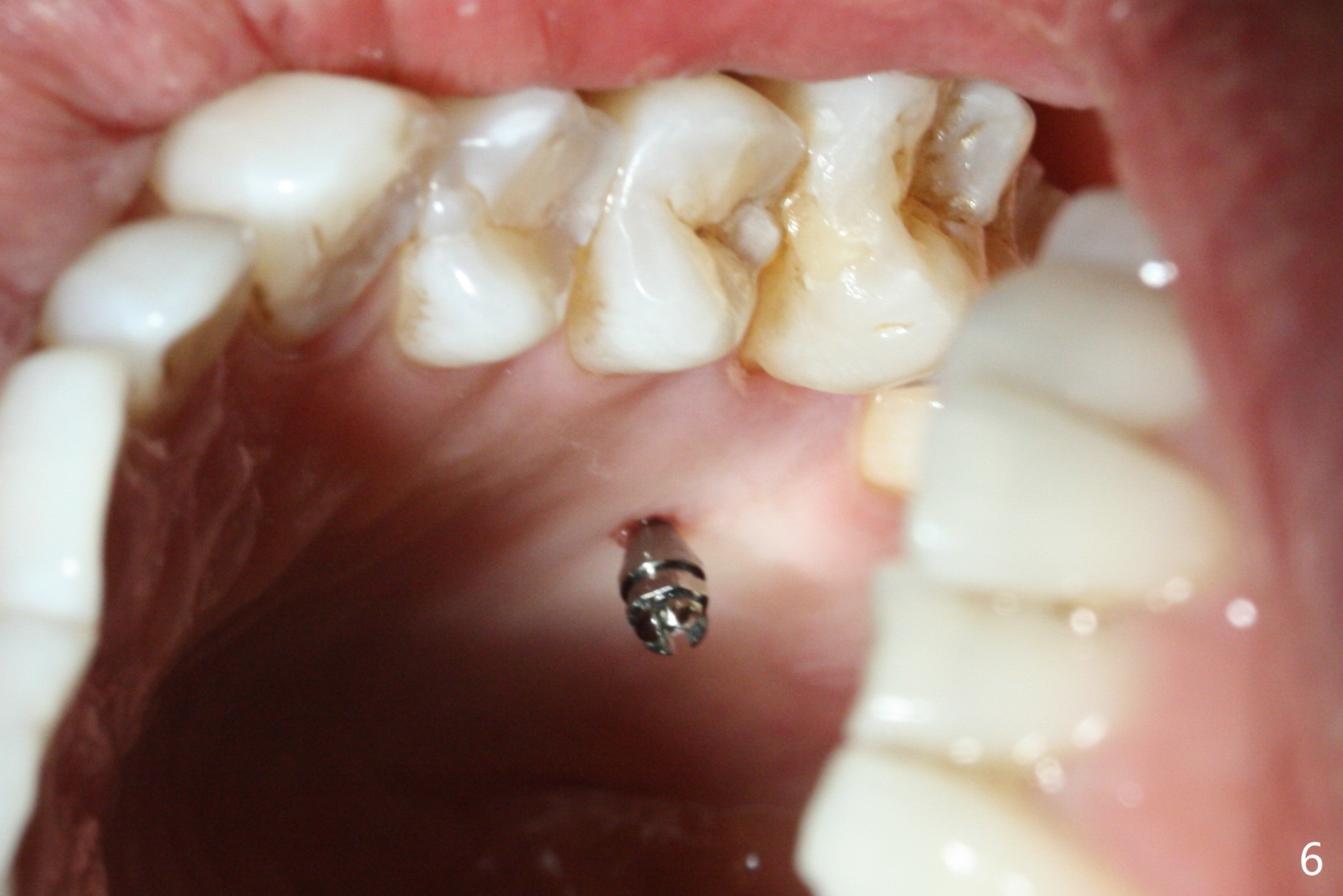
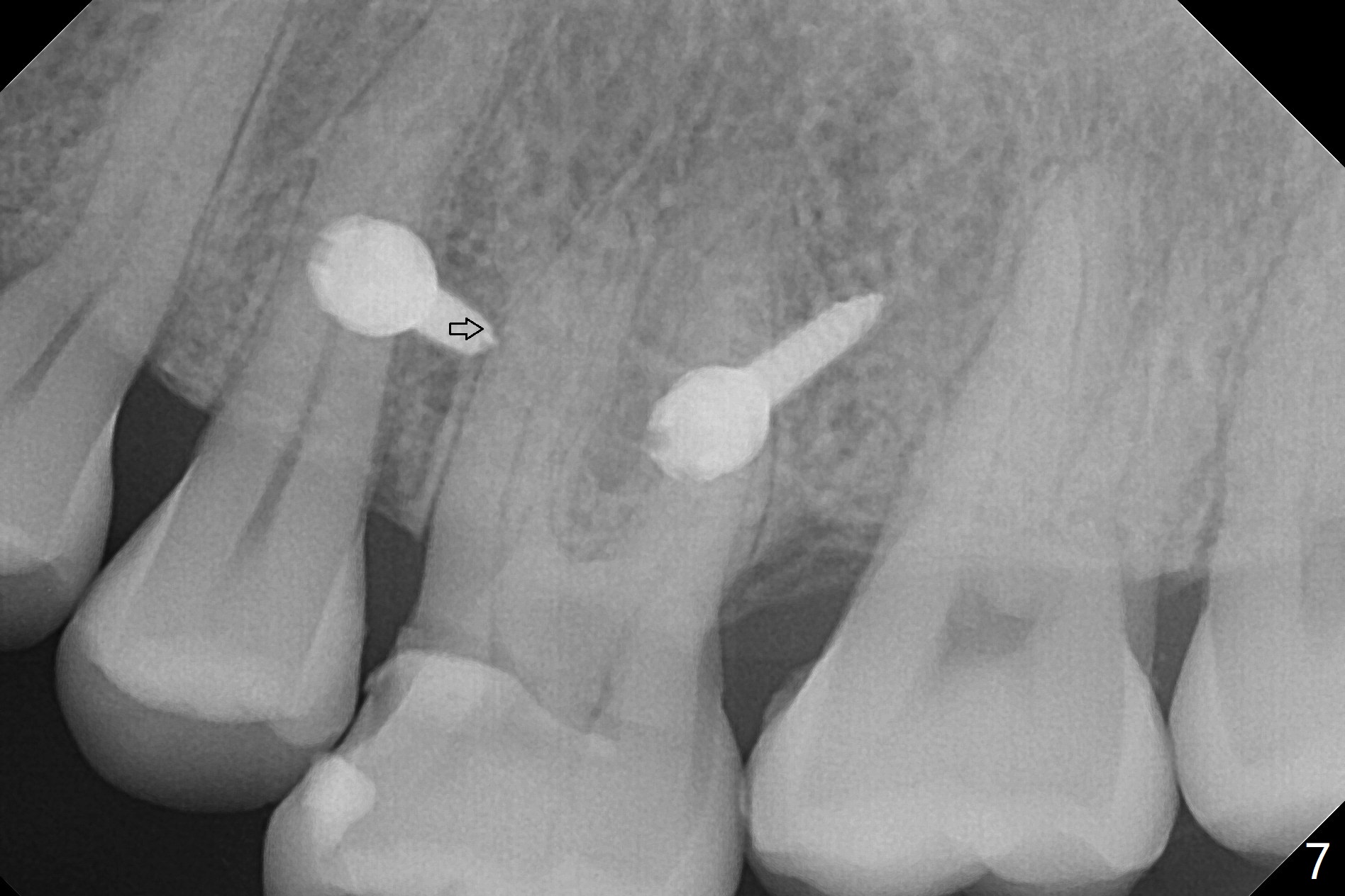
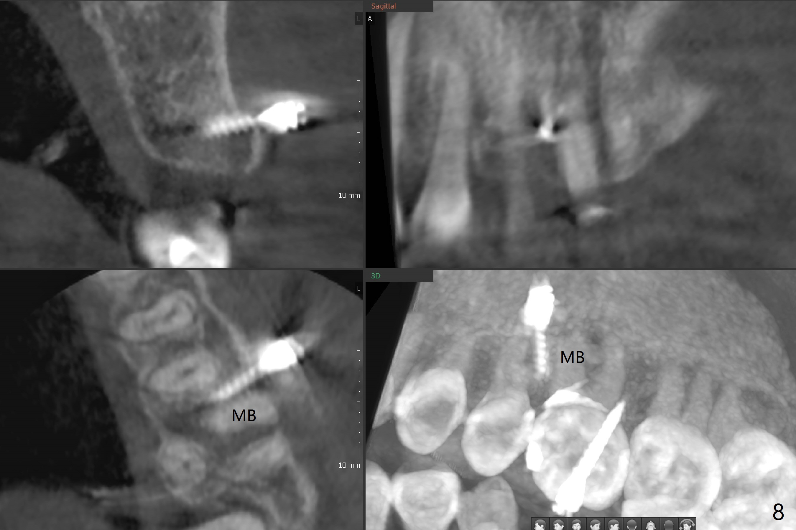
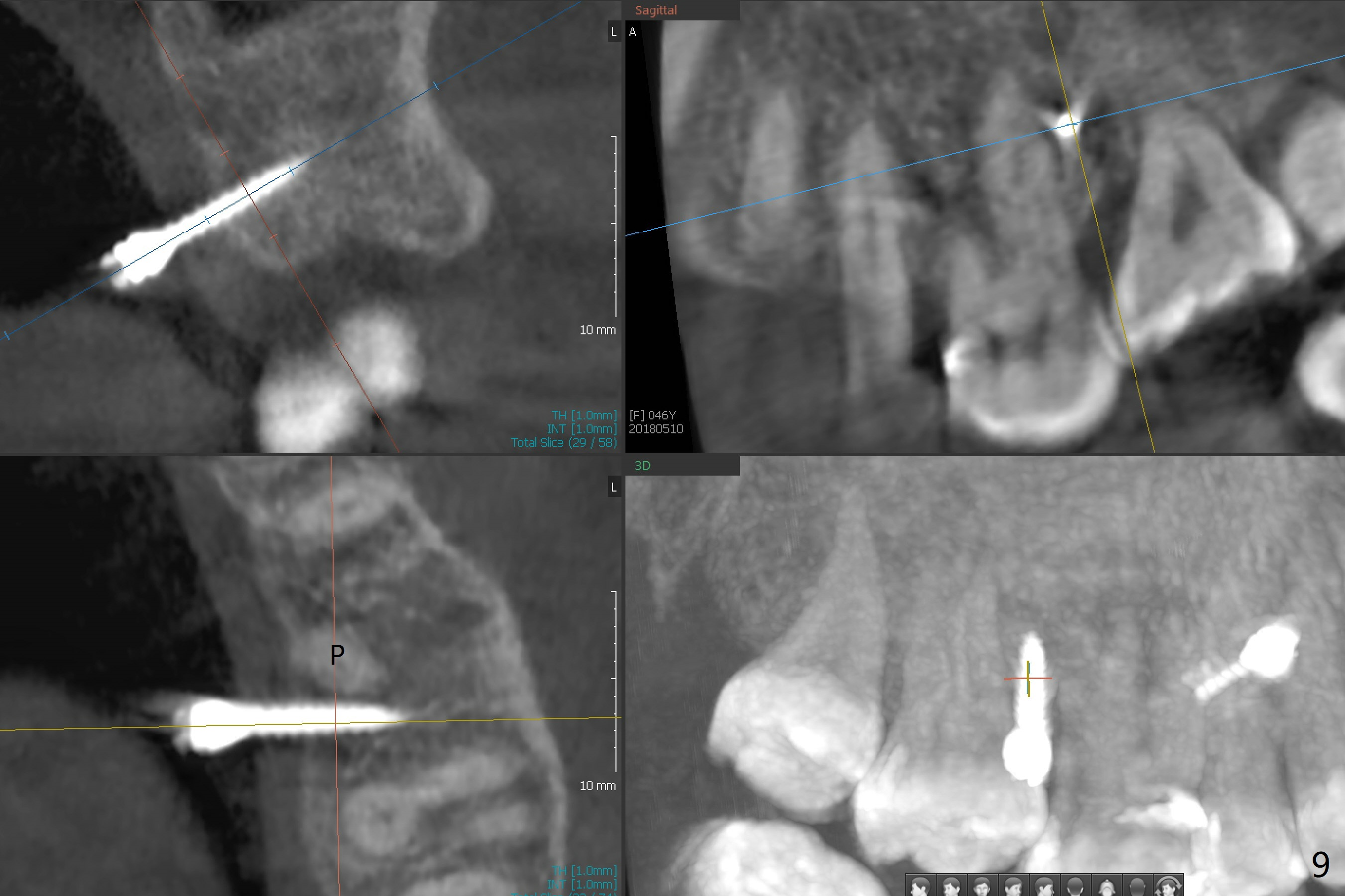
Fourteen days prior to implantation at #19 to replace a flipper (Fig.2), two minimplants are placed to intrude the supraerupted opposing tooth (#14, Fig.1). After use of minimal amount of local anesthetic (to keep proprioceptive in case root surface violation), the miniimplants are inserted ~ half of the length initially (Fig.3,4). The mesiobuccal (MB) one seems to be better positioned than the distopalatal (DP) one. When the implants are completely seated (Fig.5,6), three of PAs are taken, which suggests contact of the MB implant to the MB root of the tooth #14 (Fig.7 arrow). Immediately postop CT confirms approximation of MB and DP implants to the MB and P roots, respectively (Fig.8,9). The trajectory of these implants remain unchanged. Twelve days postop, the patient returns, uncomfortable with the palatal implant. After deep placement to bury the cuff (Fig.6) without local anesthesia (bone having no innervation), the patient feels better.
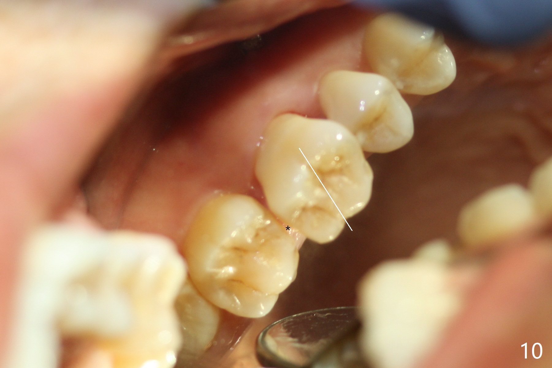
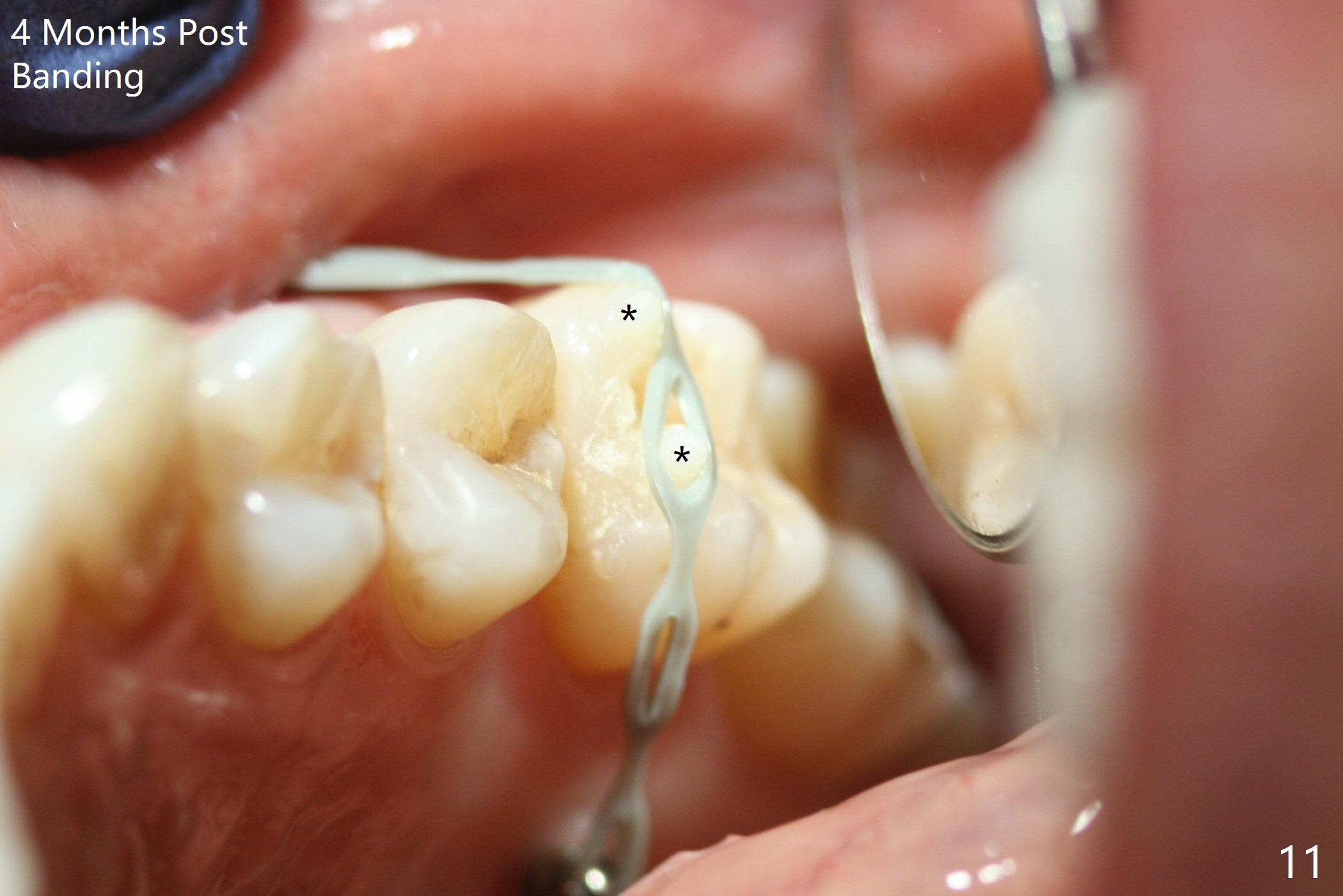
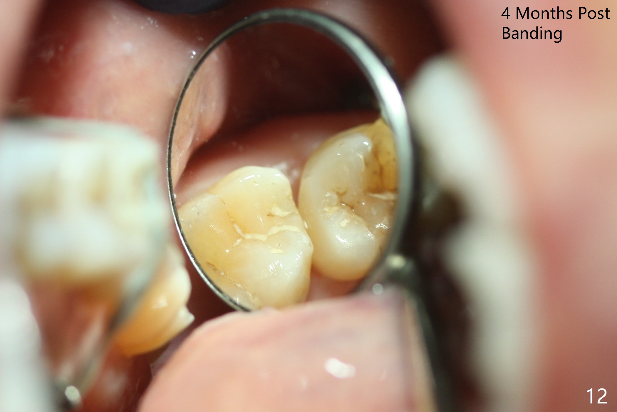
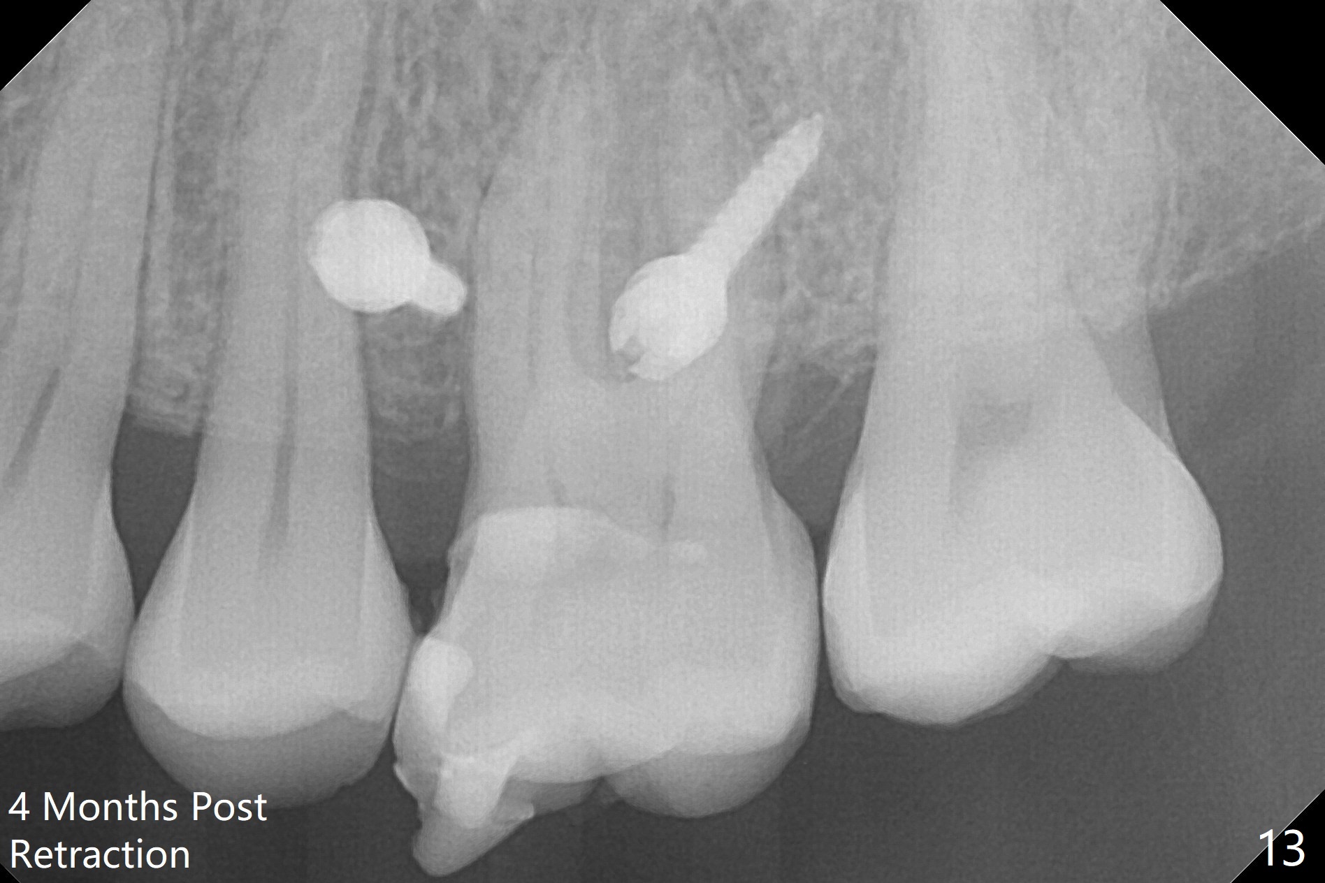
It looks as if the tooth #3 is blocked by the neighboring teeth. The proximal surfaces are reduced 1.5 months post orthodontic intrusion. Fourteen days later, the gap exists distal (Fig.10 *). It appears that the power chains placed on the natural groove of the tooth (white line) mesializes the tooth. Then the power chains are placed mesial to the mesiopalatal cusp by placing 2 of composite (Fig.11); one month later, the distal gap closes (Fig.12). The patient feels less pressure against the teeth anterior to the tooth #14. At the same time, the latter appears to have been intruded. The tooth appears to have been intruded radiographically 4 months post retraction (Fig.13).
Return to
Lower
Molar Immediate Implant, Armaments,
Ortho Cases,
Guide
Xin Wei, DDS, PhD, MS 1st edition 05/10/2018, last revision 09/19/2018