|
|
|
|
|
|
|
|
Bruxism, Long Bone and Severe Bone Loss
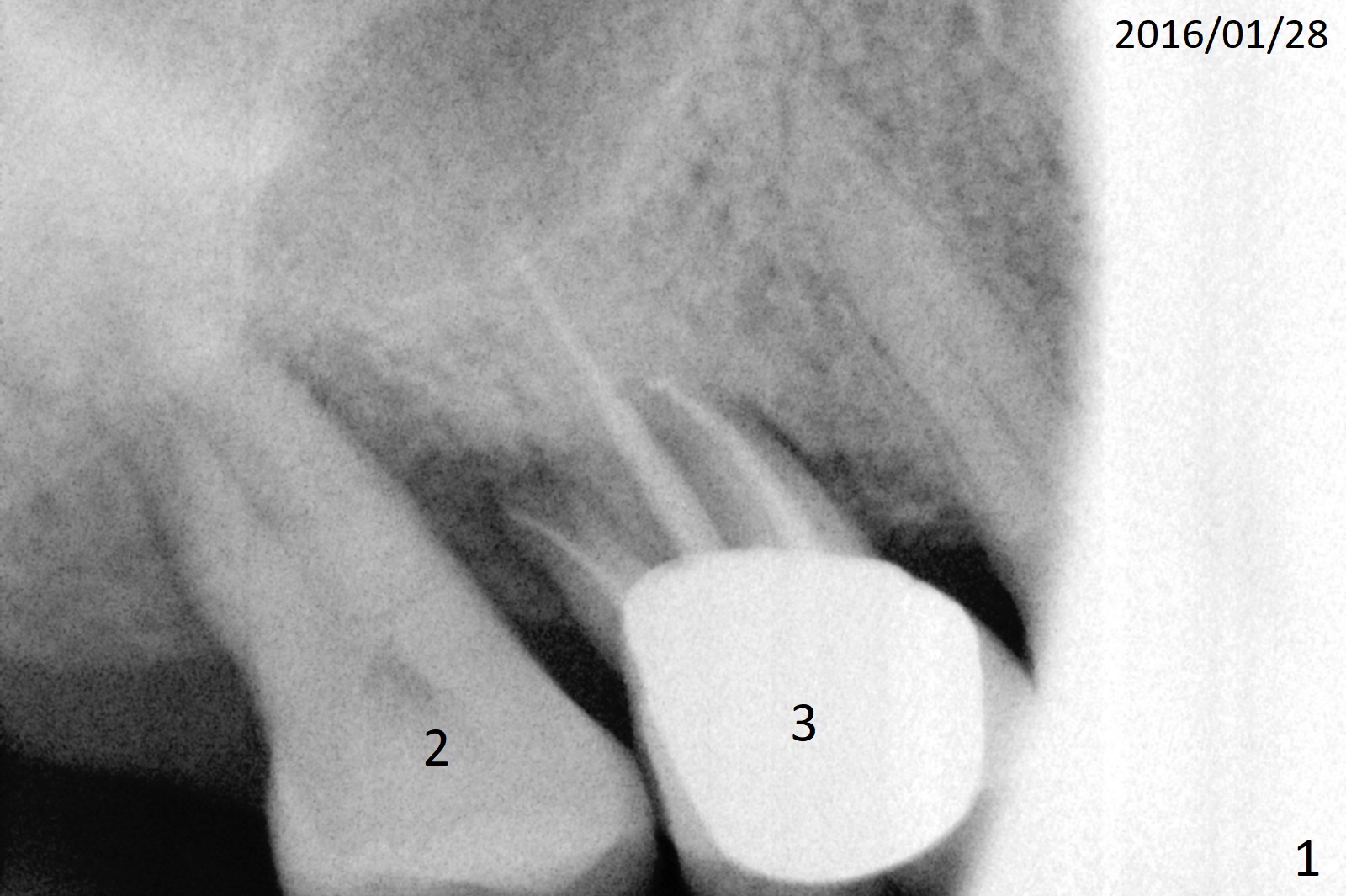
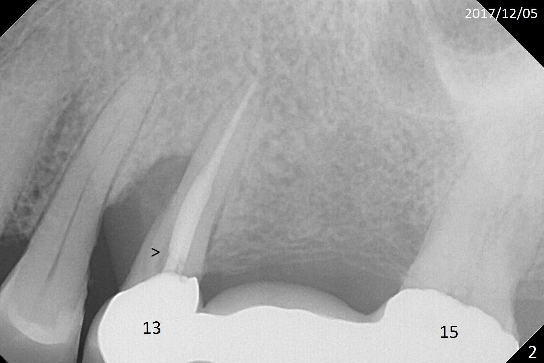
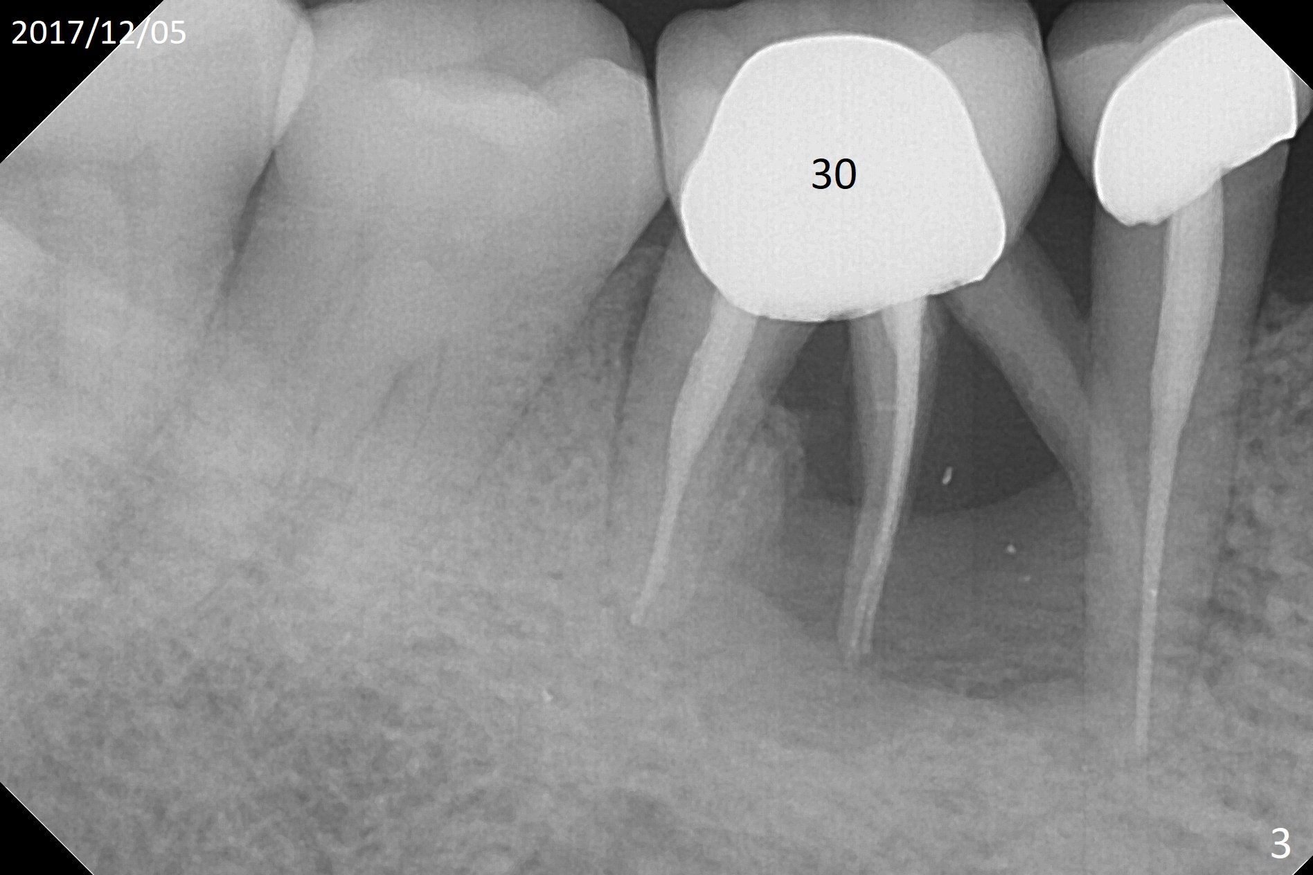
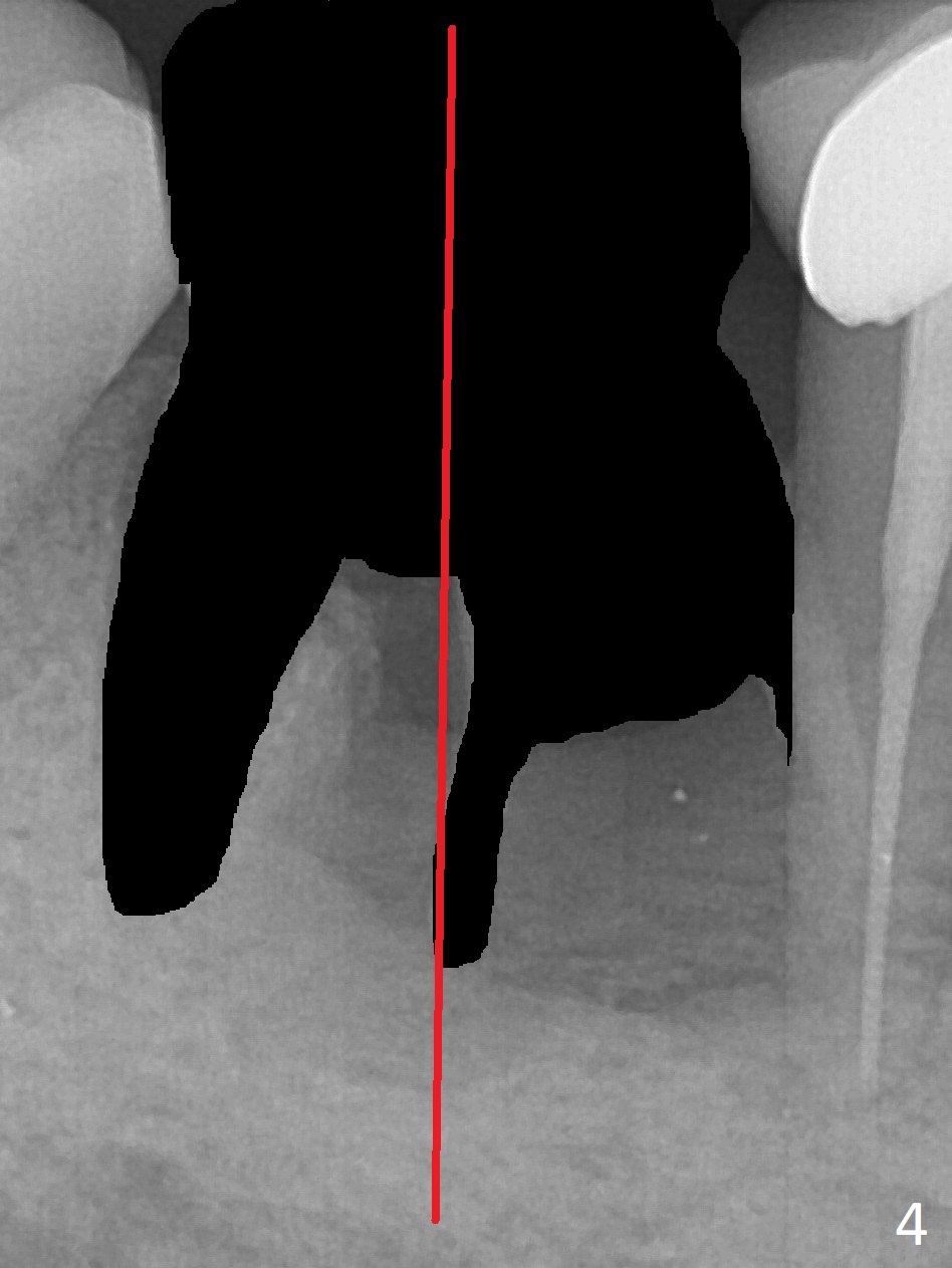
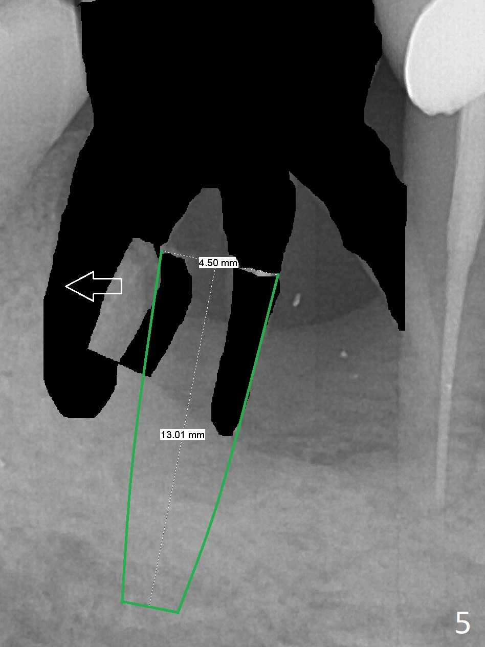
A 66-year-old woman is a bruxer/grinder. She loves chewing bone and eating nuts. The tooth #2 has exfoliated on its own, while the tooth #3 had periradicular bone loss (Fig.1). The upper left FPD is loose because of root fracture at #13 (Fig.2 >). Note the long alveolus. The mesial root of the tooth #30 has been fractured with severe bone resorption for the last 34 months (Fig.3). After extraction, initiate osteotomy in the middle of the socket irrelevant of the septum (Fig.4 red line). Take a PA or panoramic X-ray with a parallel pin immediately. When drills approaches the septum, use Magic Expanders to push the septum distal prior to resuming drills so that a 4.5x13 mm implant (Fig.5 green) is supported by the distally-displaced septum (arrow). The small implant is chosen so that there is room for bone graft, since the buccal and/or lingual plates are most likely defective mesially.
Return to
Lower
Molar Immediate Implant, Prevent
Molar Periimplantitis (Protocols,
Table), Armaments,
Metronidazole
Xin Wei, DDS, PhD, MS 1st edition 12/05/2017, last revision 12/05/2017