|
|
|
|
|
|
|
|
|
|
Place Original Implant Lingually
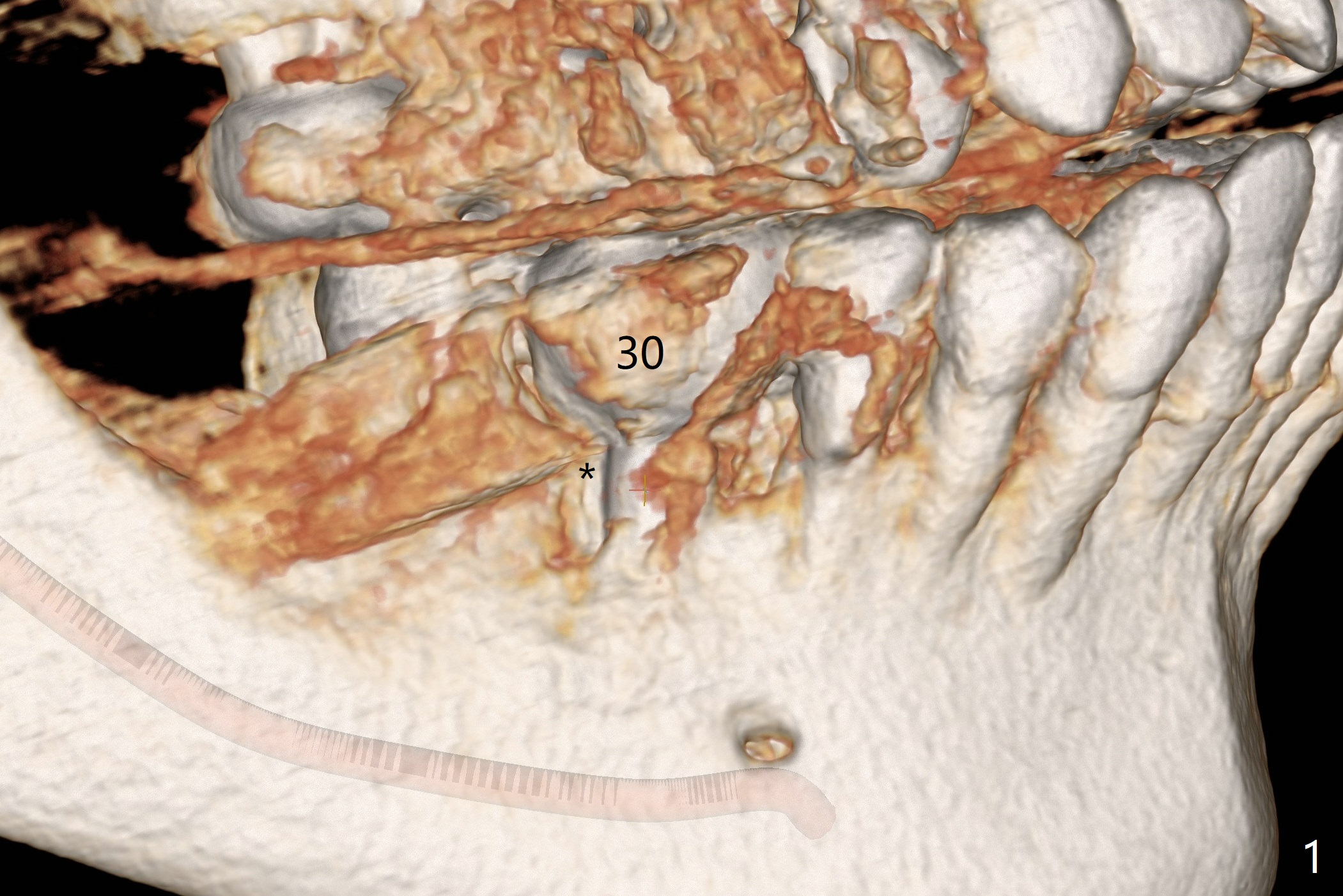
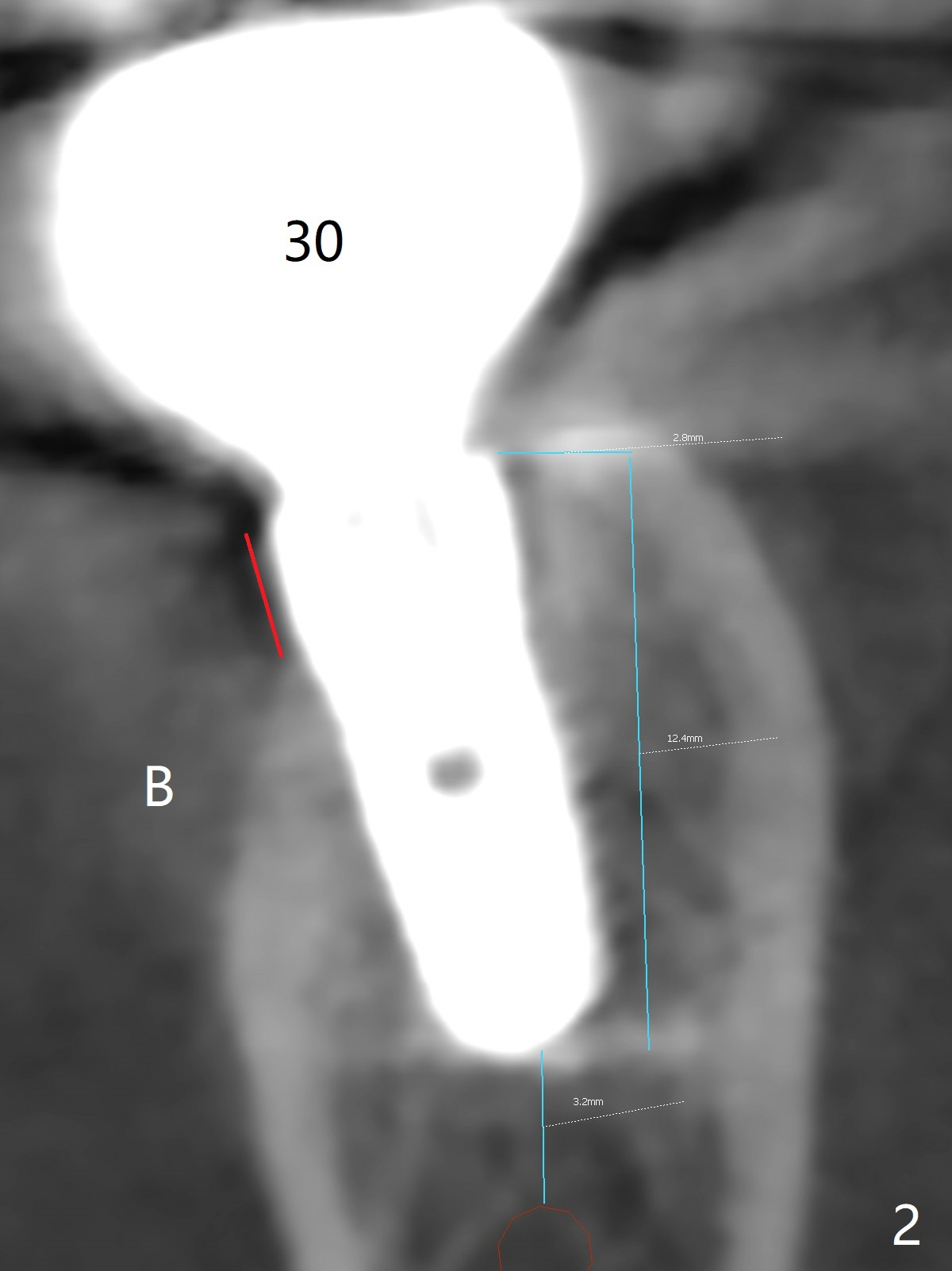
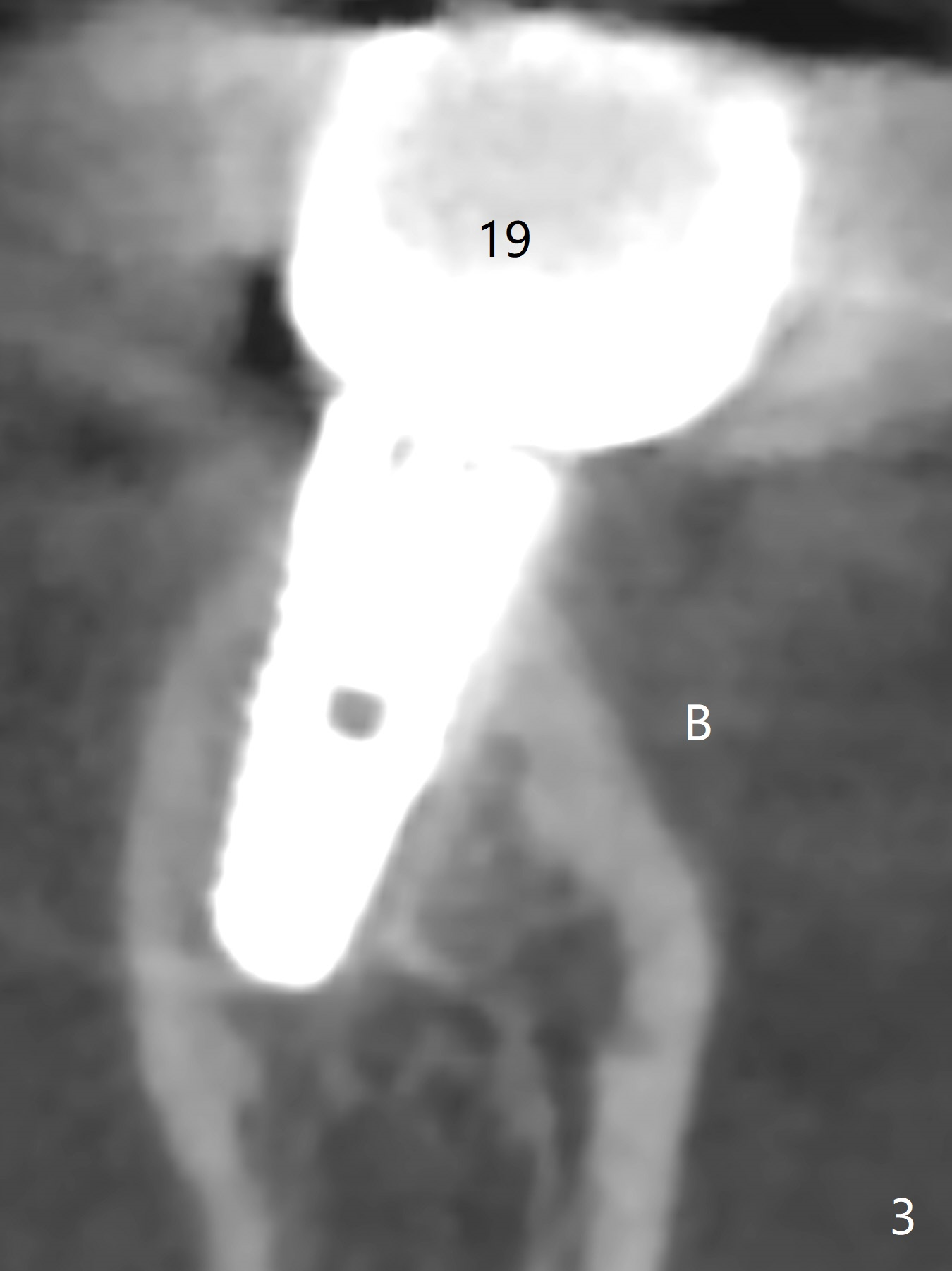
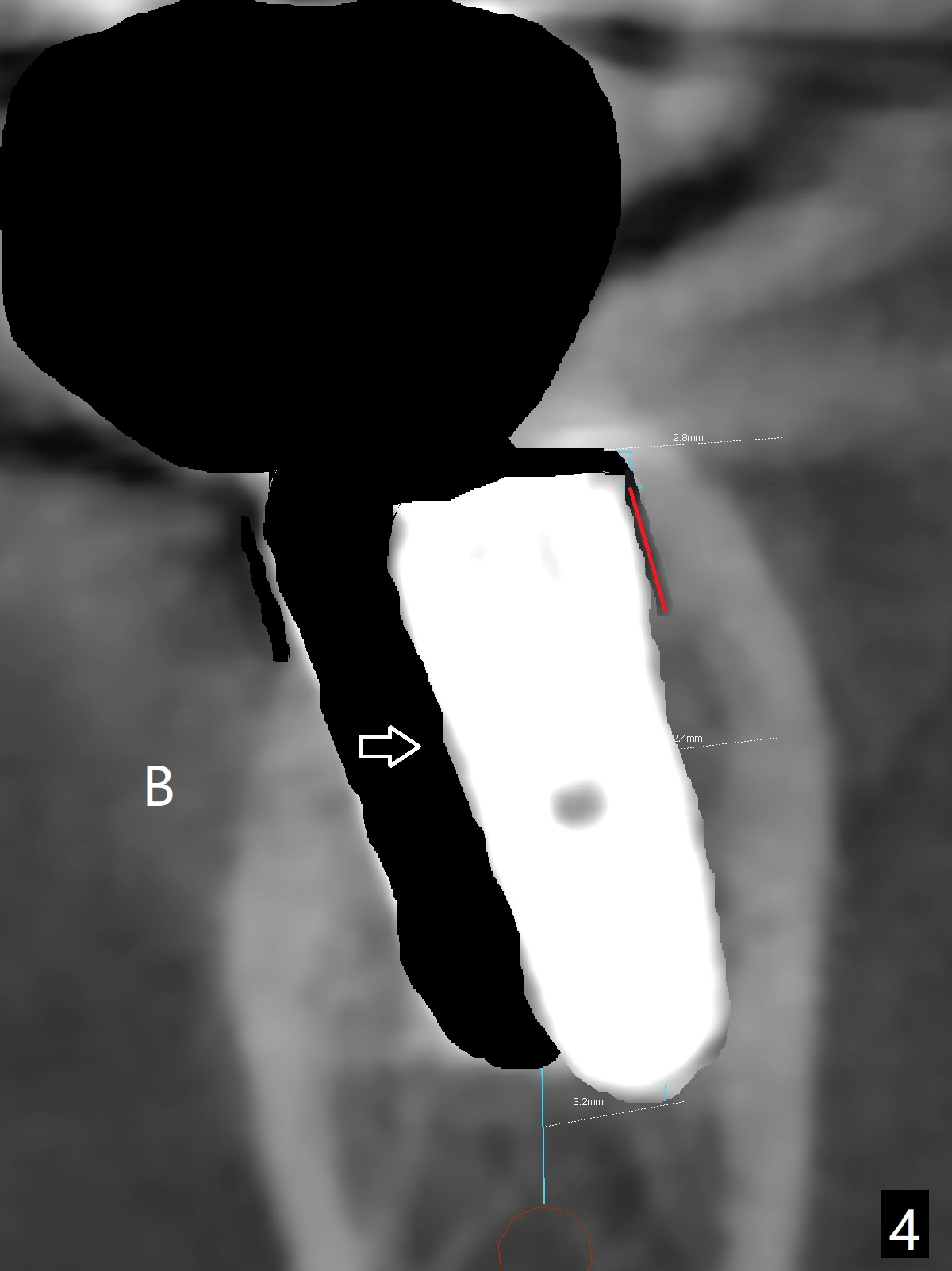
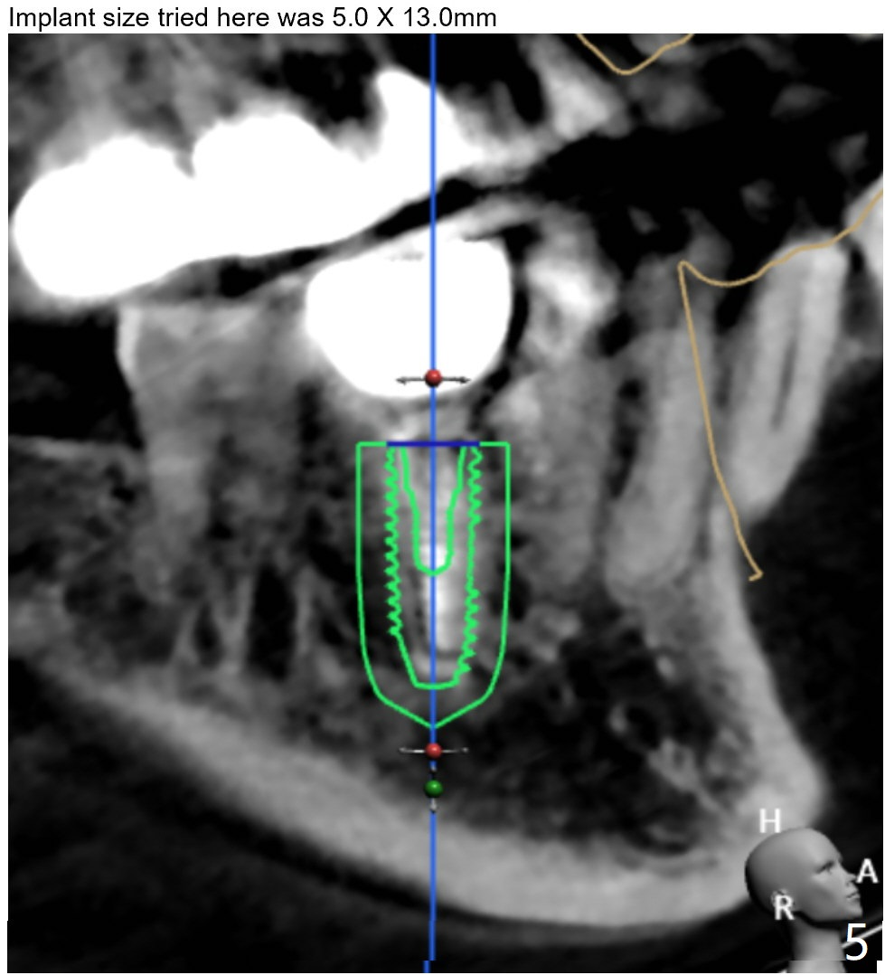
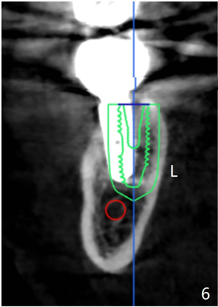
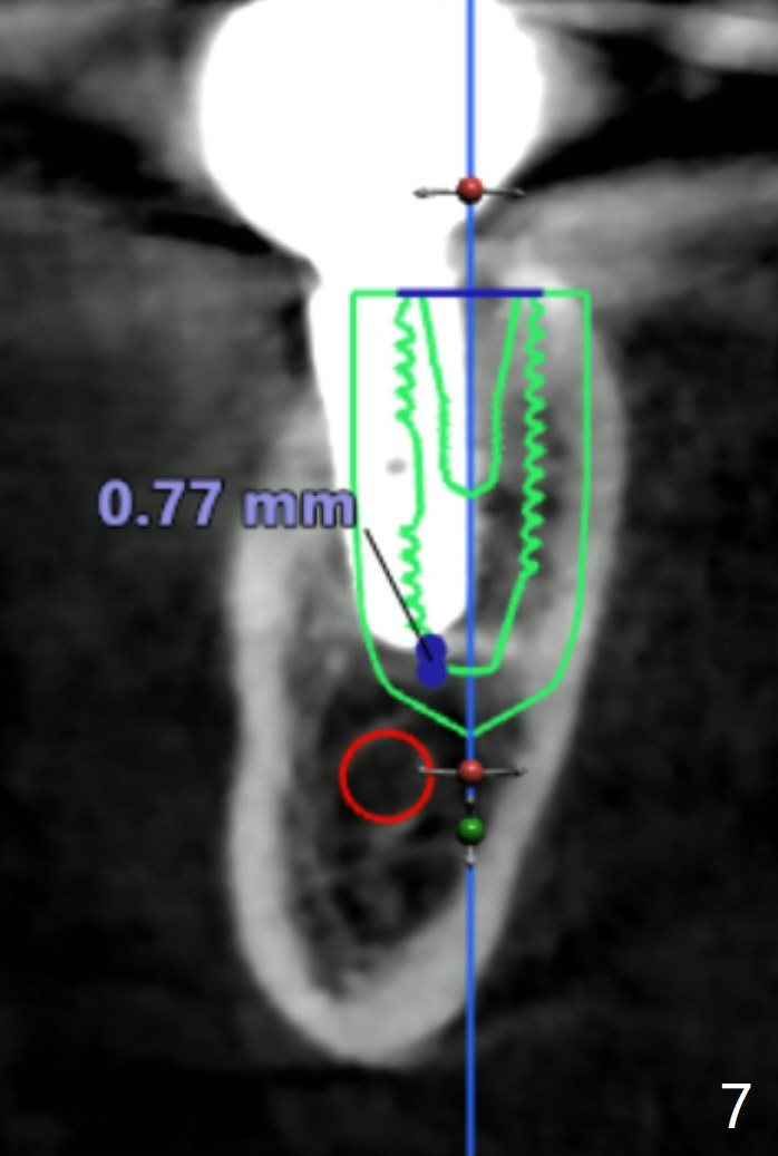
Twenty six months post cementation, periimplantitis develops buccally at #30 (Fig.1) due to buccal (B) placement (Fig.2) with buccal thread exposure (red line), as compared to the same sized implant at #19 (Fig.3). After removal of the crown and abutment and incision, use Titanium brush to clean the exposed threads. Following implant removal and removal of the lingual bone, place the same implant lingually (Fig.4 arrow) with the used-to-be-exposed surface facing lingual (Fig.4 red line). The buccal gap will be filled with autogenous bone harvested lingually and allograft. Use 2-3 pieces of PRF membranes to close the wound, i.e., to bury the implant. A 5x13 mm implant (Fig.5) placed lingually (Fig.6 L) appears to be unable to gain more than .77 mm native bone (Fig.7). Lab declines to make a guide.
Return to
Lower
Molar Immediate Implant, Prevent
Molar Periimplantitis (Protocols,
Table), Armaments,
Trajectory,
Clindamycin Metronidazole
No Antibiotic
14
Xin Wei, DDS, PhD, MS 1st edition 02/06/2018, last revision 04/12/2020