|
|
|
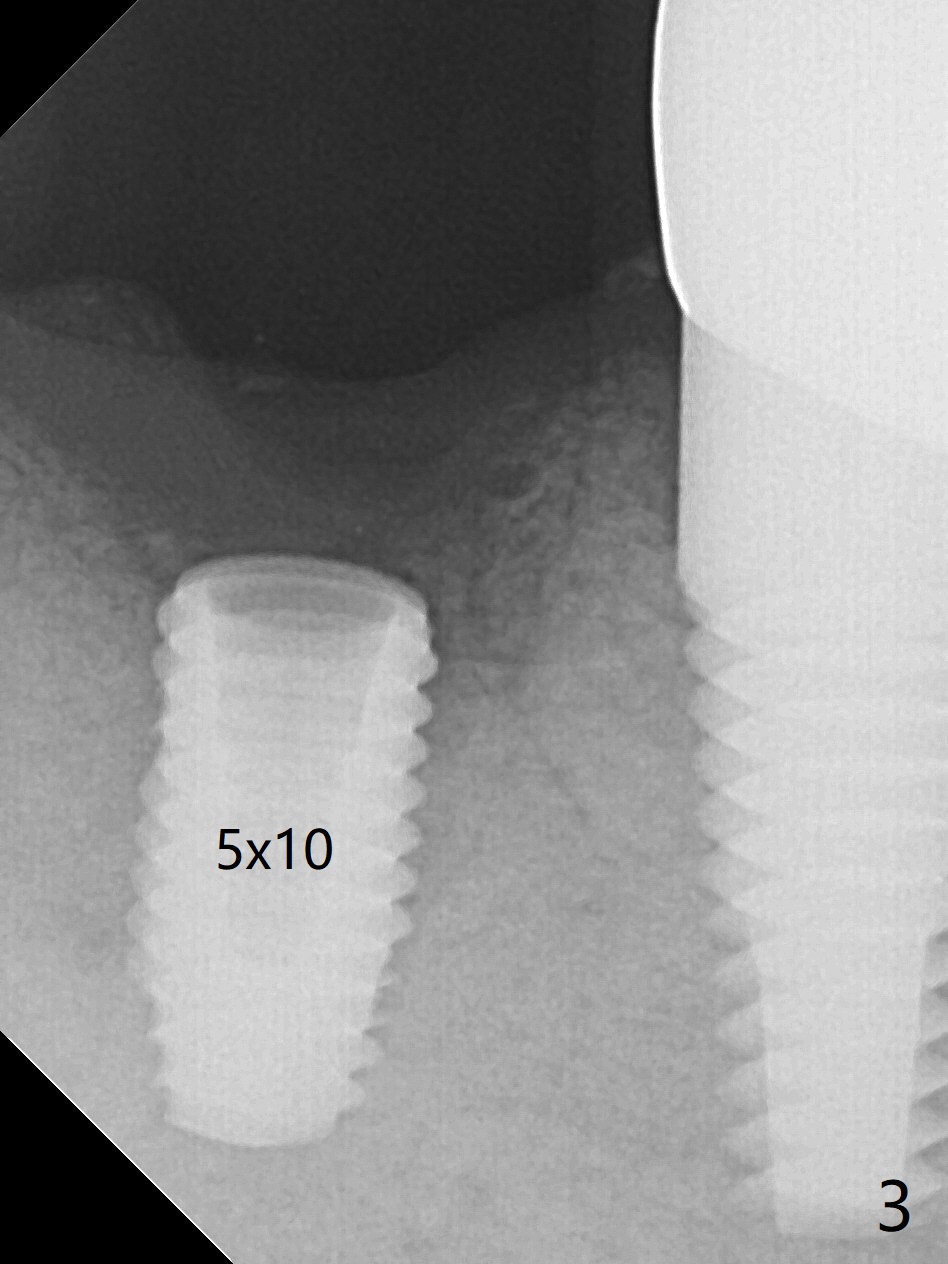
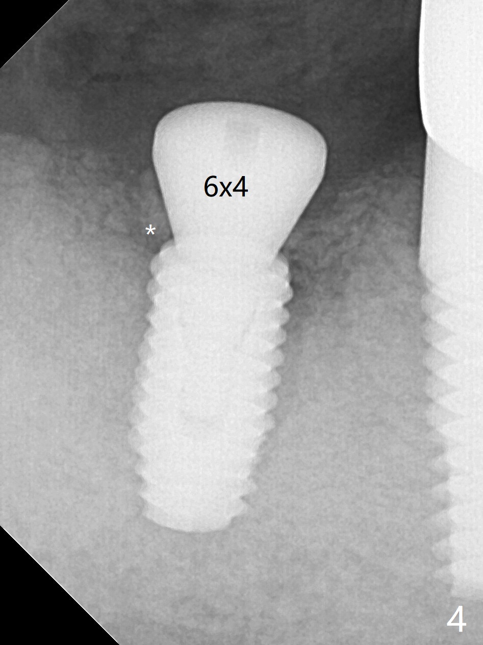
Guided surgery resumes as planned to remove the lingual and apical bone. The buccal bone looks porous from the osteotomy opening. Mixture of autogenous and allograft is packed against the bone plate from the osteotomy before implant placement, but the 5x10 mm implant is unable to be seated completely. After removal of the bone graft from the osteotomy site and use of 4x11.5 mm drill for ~ 1 mm, the implant is seated with ~ 60 Ncm (Fig.3). With buccal incision, bone graft is placed over the distobuccal exposed implant thread (Fig.4 *) following placement of a 6x4 mm healing abutment.
Post-Implant Bone Graft Last Next Xin Wei, DDS, PhD, MS 1st edition 08/01/2019, last revision 08/01/2019