|
|
|
|
|
|
|
|
|
|
|
Socket Preser-vation with rhPDGF-BB I
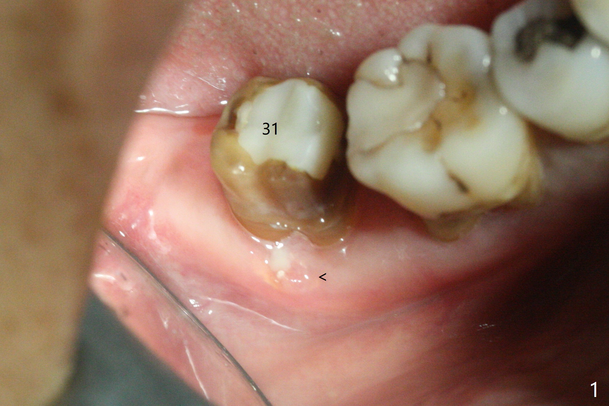
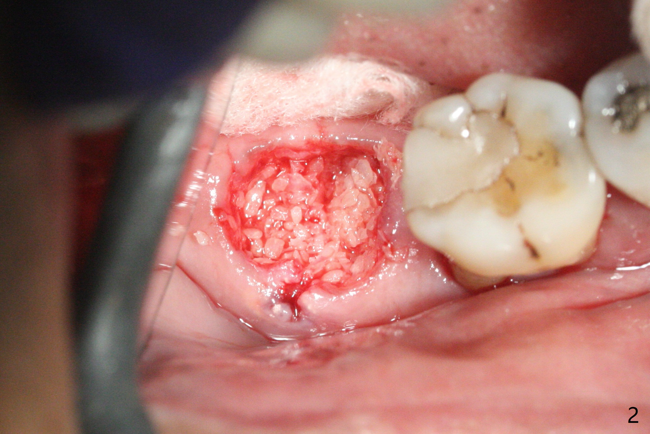
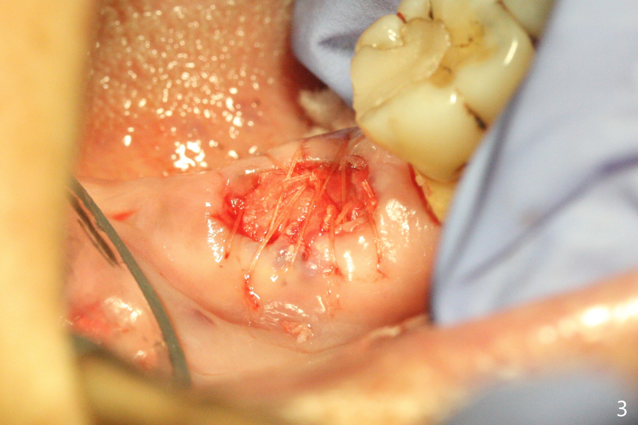
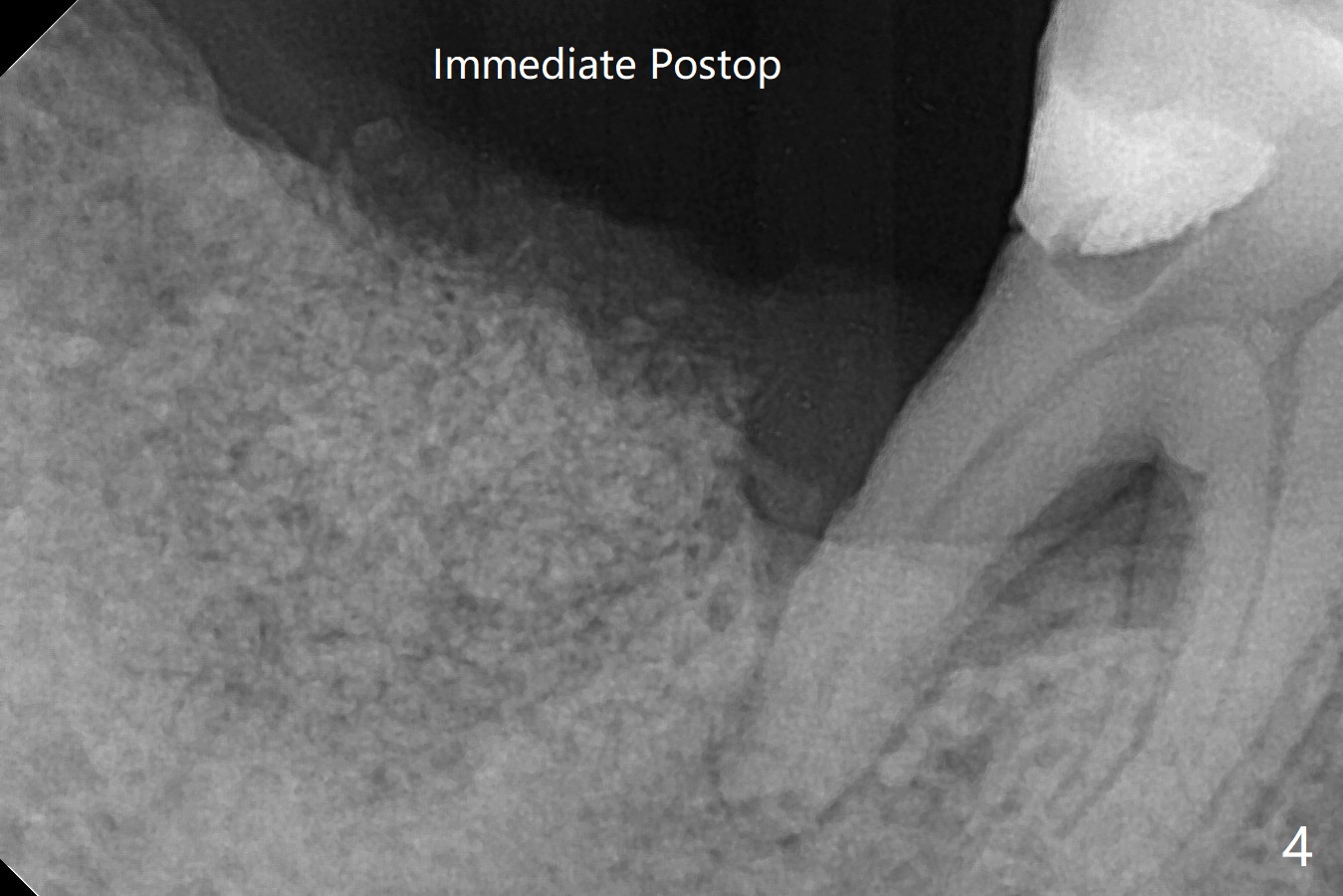
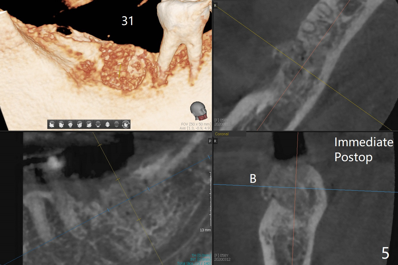
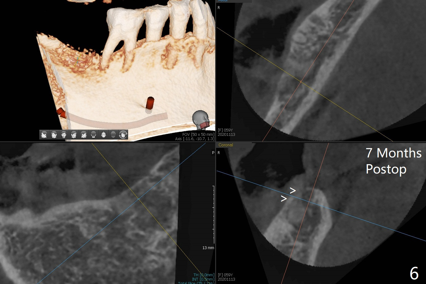
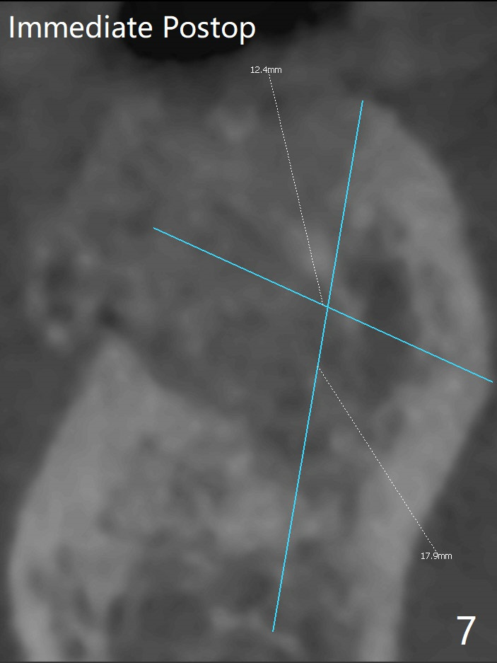
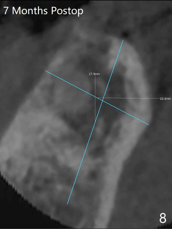
After extraction of the tooth #31 with mesial root fracture and a buccal fistula (Fig.1 <) and debride-ment, cortical: cancellous (50: 50) mineralized allograft (.5-1 mm) saturated with ~ .3 ml of .3 mg/ml of rhPDGF-BB (one component of GEM21S) is placed in the socket. Amazingly bone graft granules seem to stick to each other (semi sticky bone (Fig.2), as compared to PRF). The socket opening is covered with a piece of Osteogen plug and 12x12 mm Amnion-Chorion Allograft, followed by 4-0 PGA suture (Fig.3). The bone graft is packed as apical (Fig.4) and buccal (Fig.5 B) as possible. The patient will return to soft tissue healing check in a week. CT will be taken to determine whether the buccal plate is repaired 4 months postop. Watch Video. In fact COVIT 19 delays her return. The buccal plate seems to have reformed 7 months postop (Fig.6 >). The width and height of the ridge remains basically the same (compare Fig.7 and 8).
Return to
No Deviation
Plug
GEM21S
Cases
Xin Wei, DDS, PhD, MS 1st edition
03/12/2020, last revision
11/13/2020