|
|
|
|
|
|
|
|
|
|
Mini Implant Placement with Several Steps of Adjustment
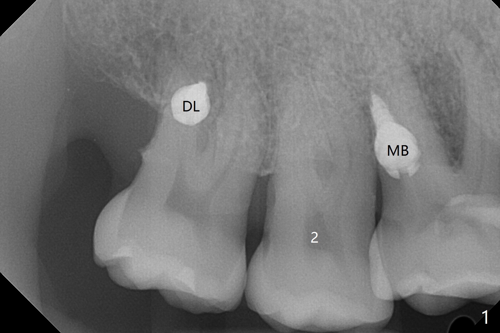
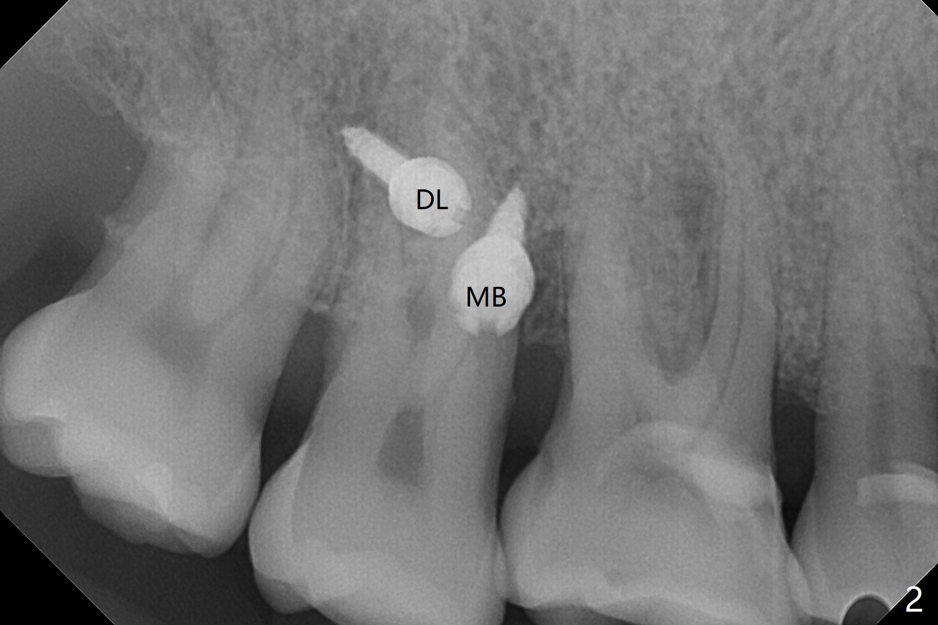
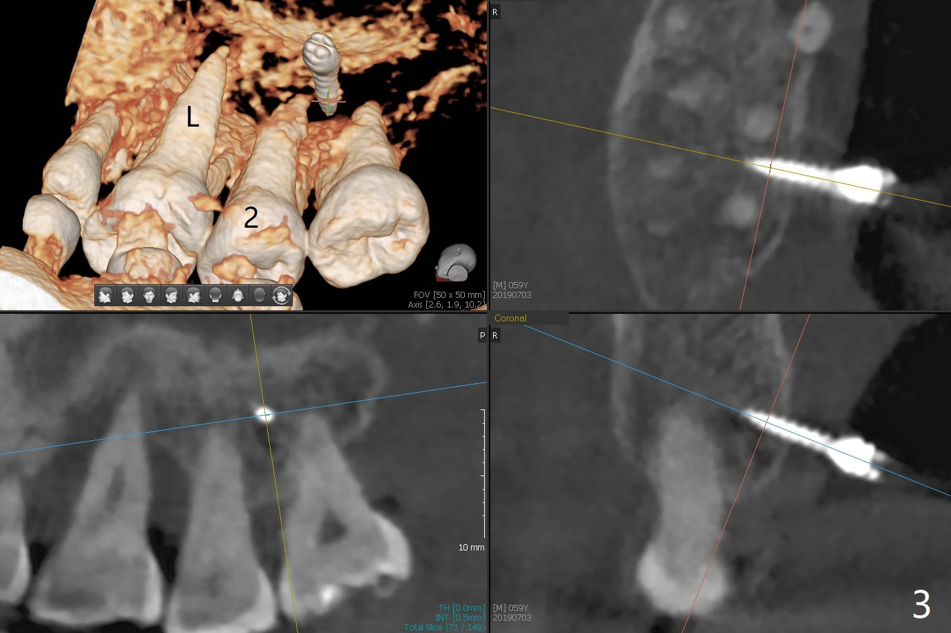
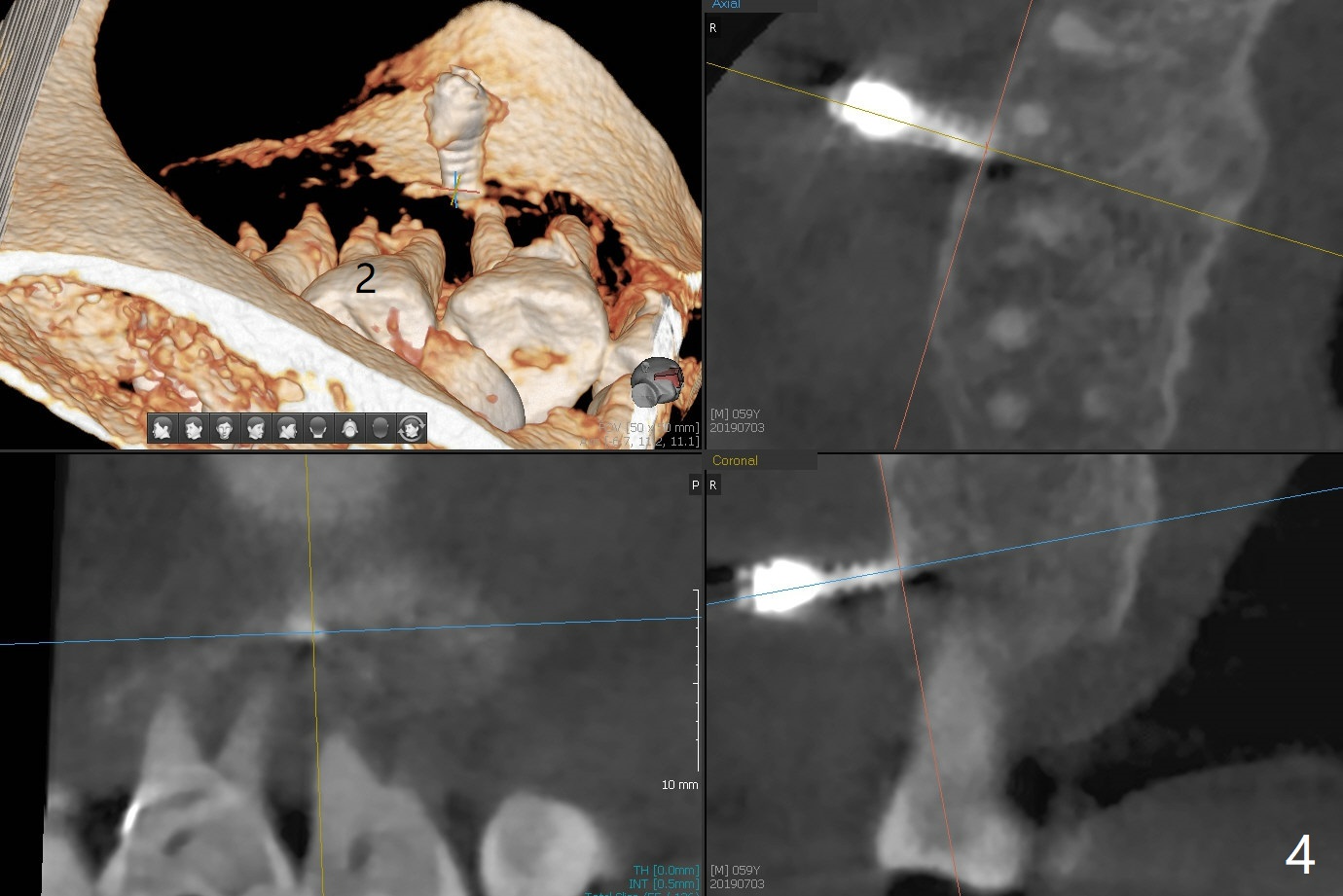
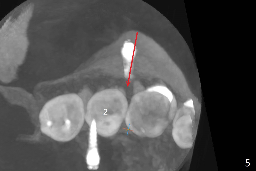
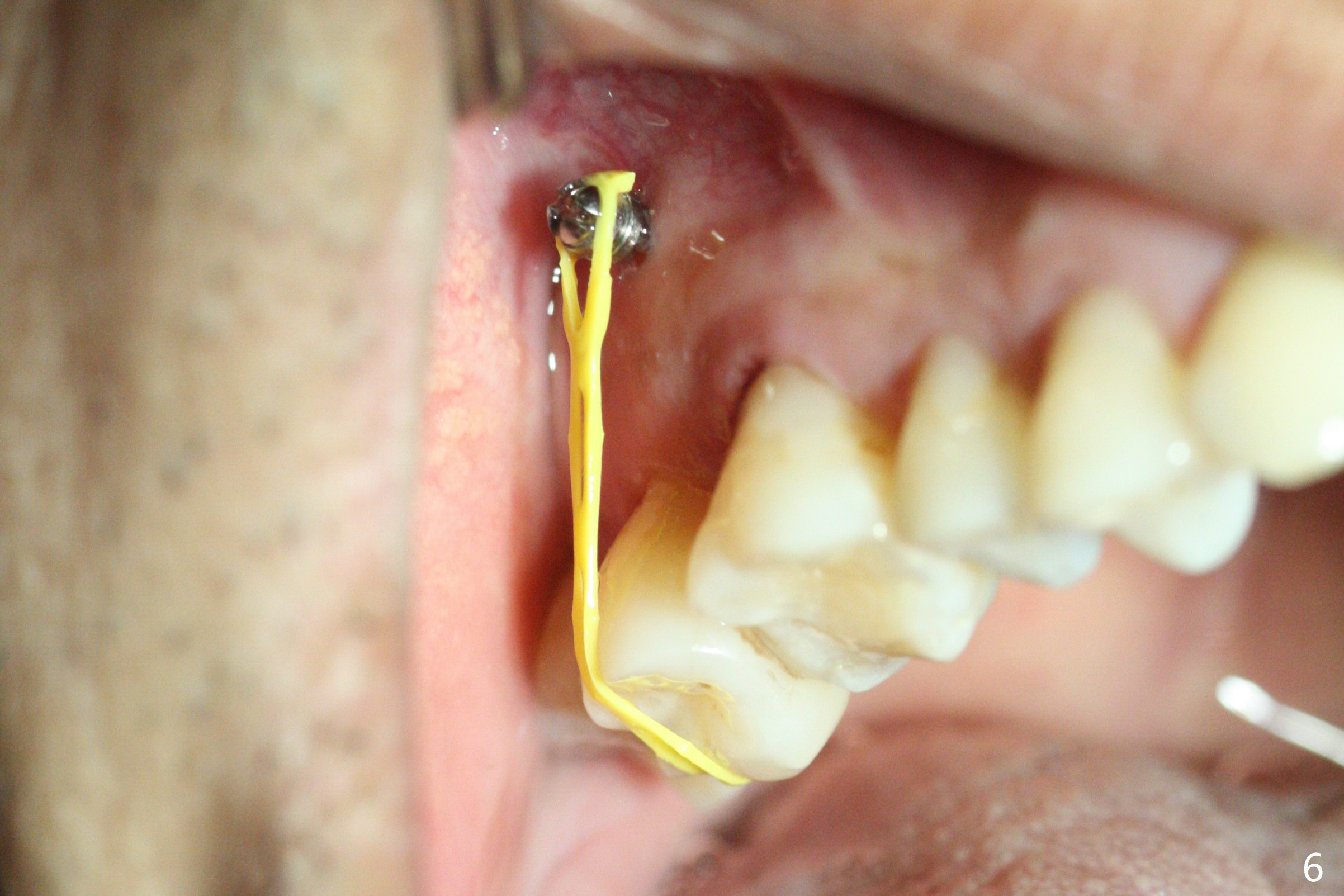
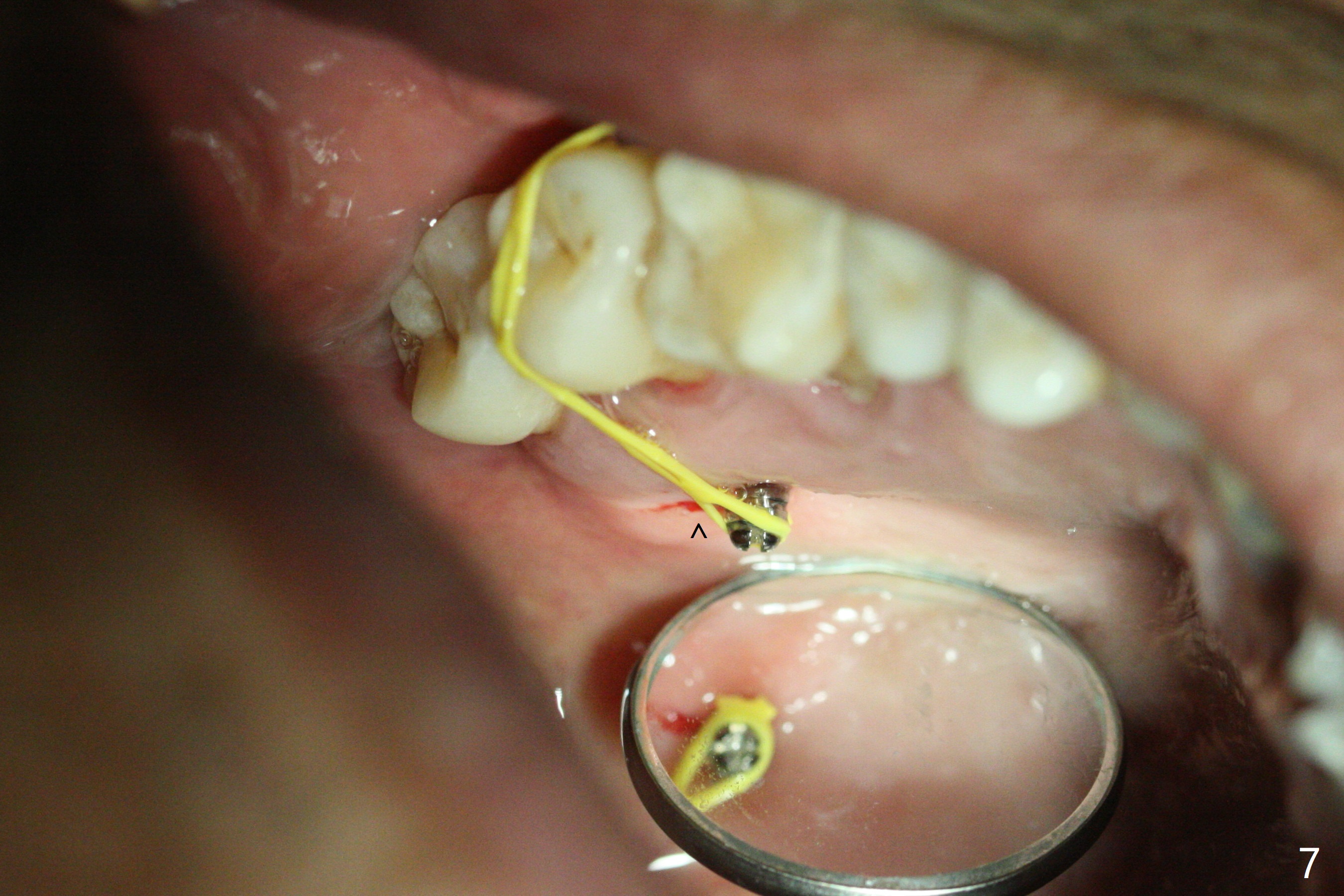
When 6 and 8 mm Tomas pins are initially placed mesiobuccal (MB) and distolingual (DL) to the supraerupted tooth #2, PA is taken (Fig.1), which shows that the DL one seems too distal. The mini implant is removed and placed more mesial (Fig.2, 7 (^: original entry)). When the patient experiences a little pain as the DL implant is being placed deeper with minimal local anesthetic, CT is taken. In fact the latter is between the apices of the teeth #1 and 2 (Fig.3 (L: lingual view of 3D image)). The implant is later placed deeper with minor angular change as well as more anesthetic. There is an advantage to place the miniimplants apically; there is more space. But the MB implant is a little bit mesial (Fig.4). After withdrawal, the trajectory of the implant is changed somewhat as indicated by a red arrow in Fig.5). Note the apical placement of the minimplants (close to mucogingival junction, Fig.6,7).
Return to
Lower
Molar Immediate Implant,
Trajectory,
Ortho Cases
Xin Wei, DDS, PhD, MS 1st edition
07/03/2019, last revision
07/03/2019