.jpg)

|
|
|
|
|
|
|
|
|
|
|
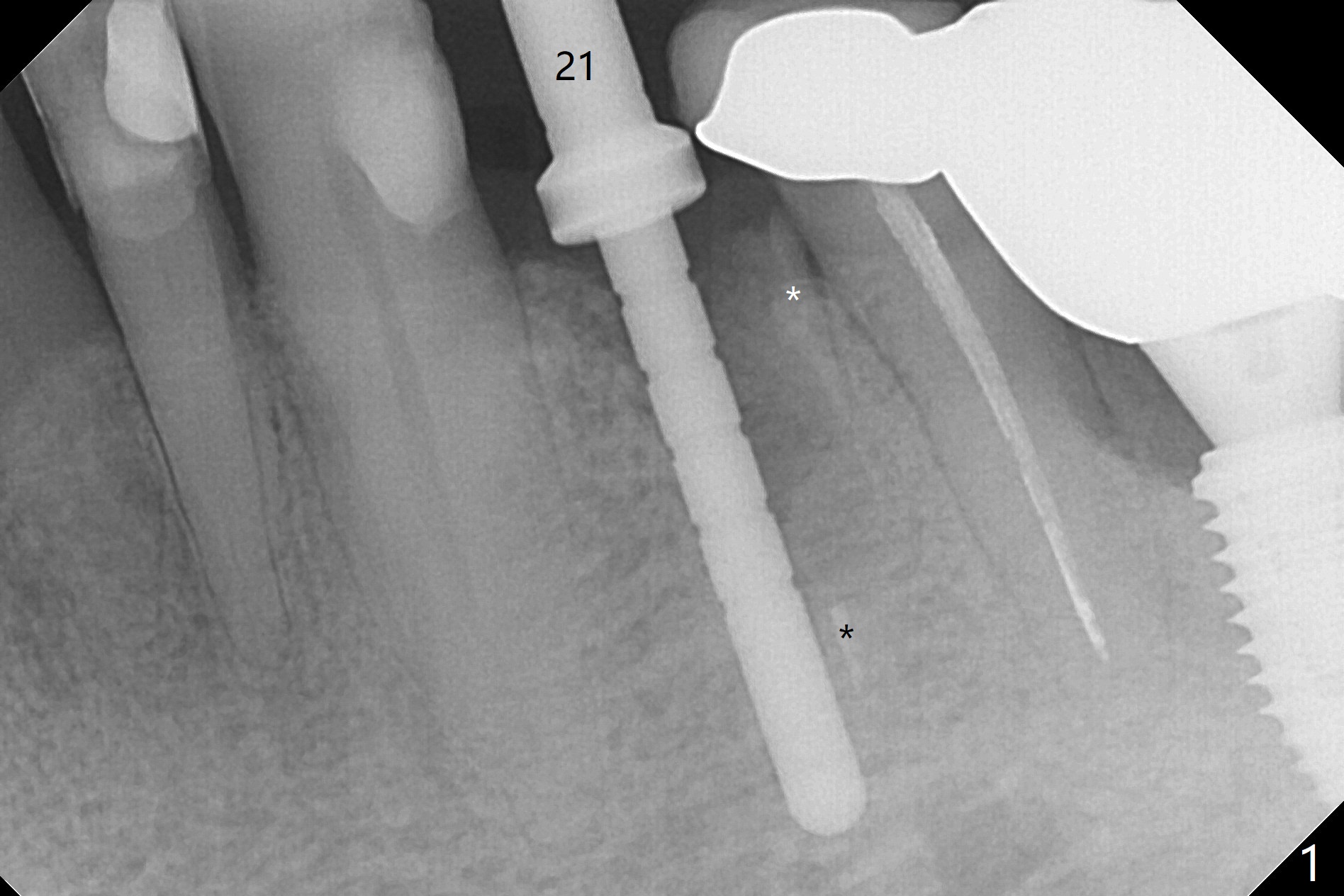
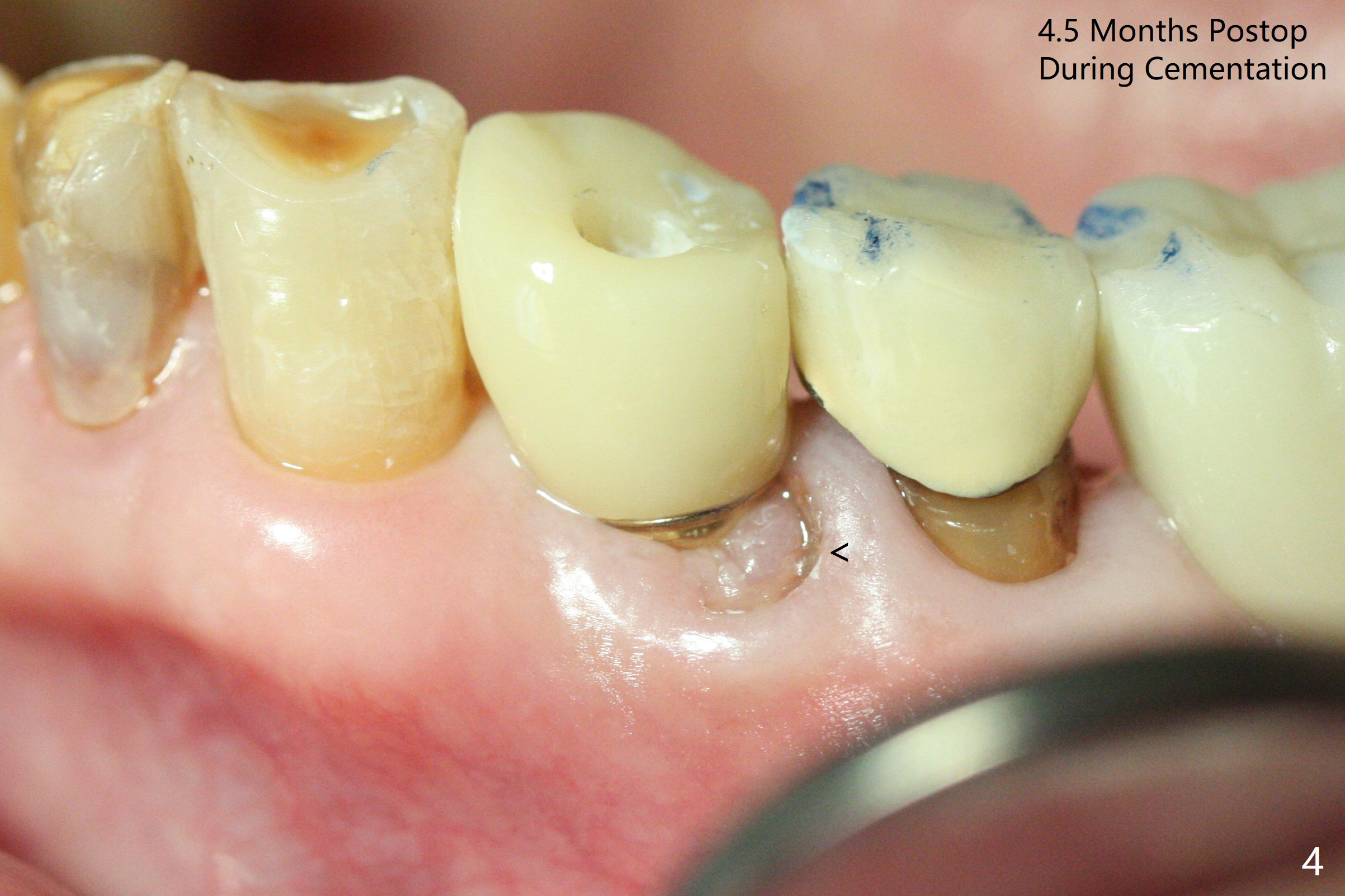
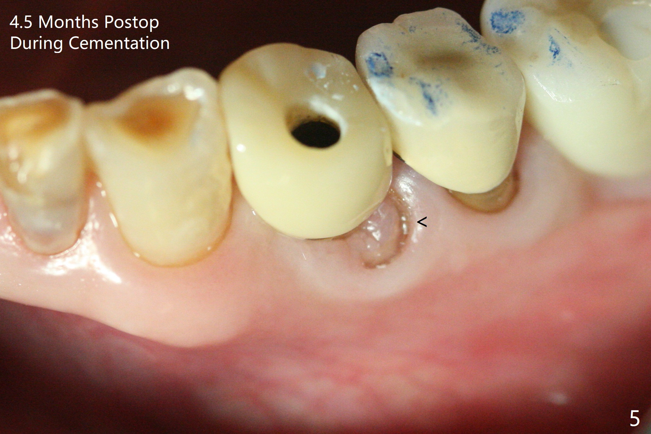
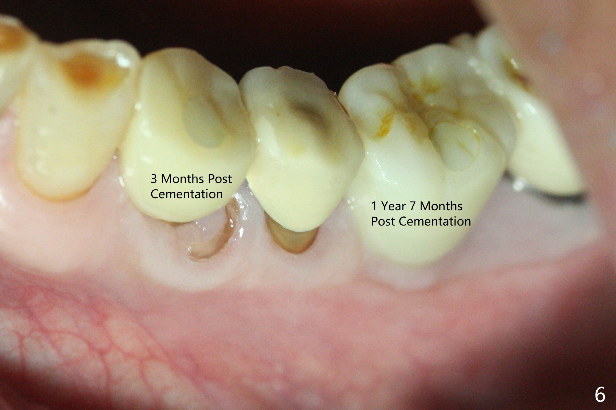
Socket Shield Because of Difficult Extraction
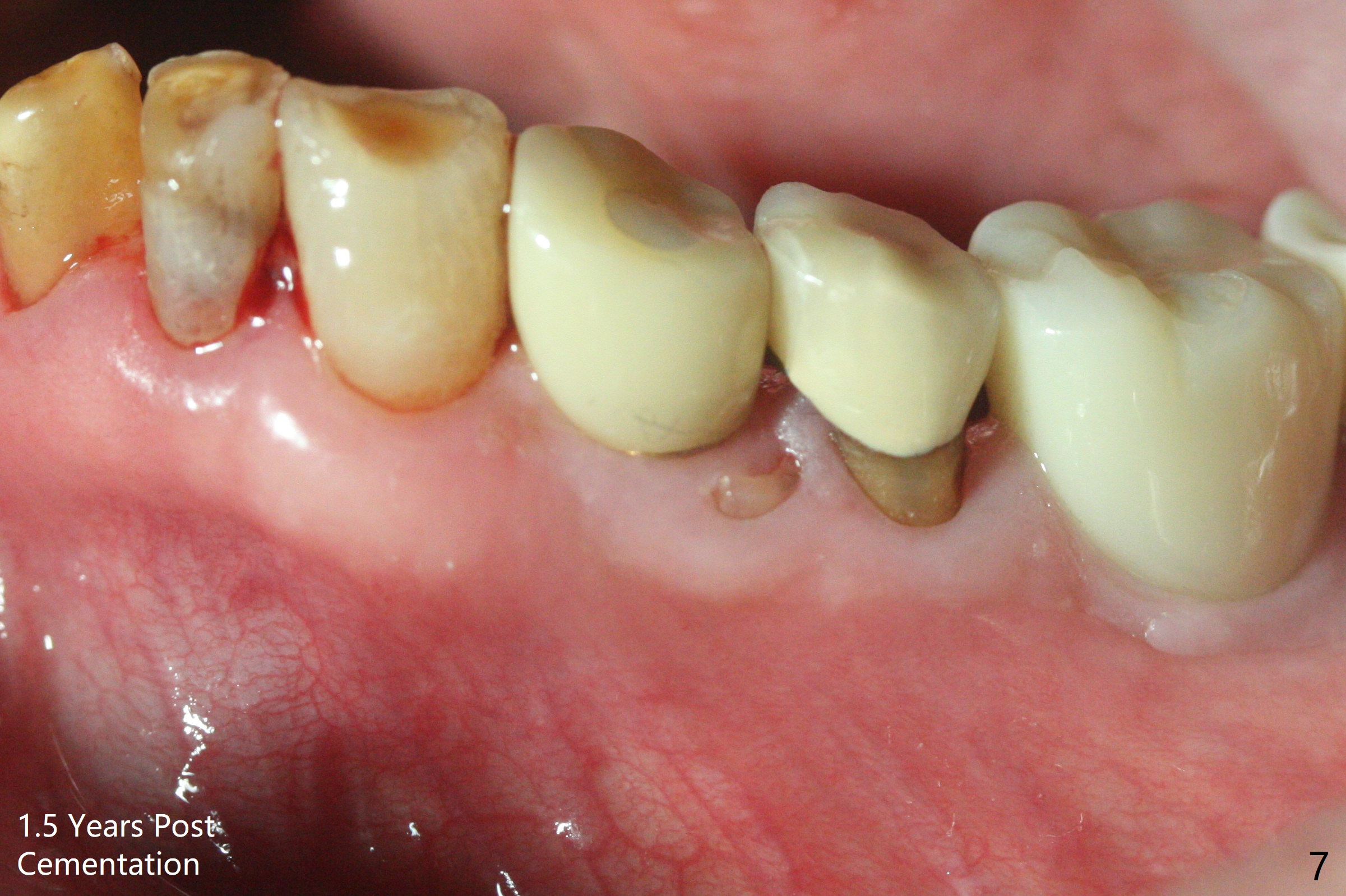
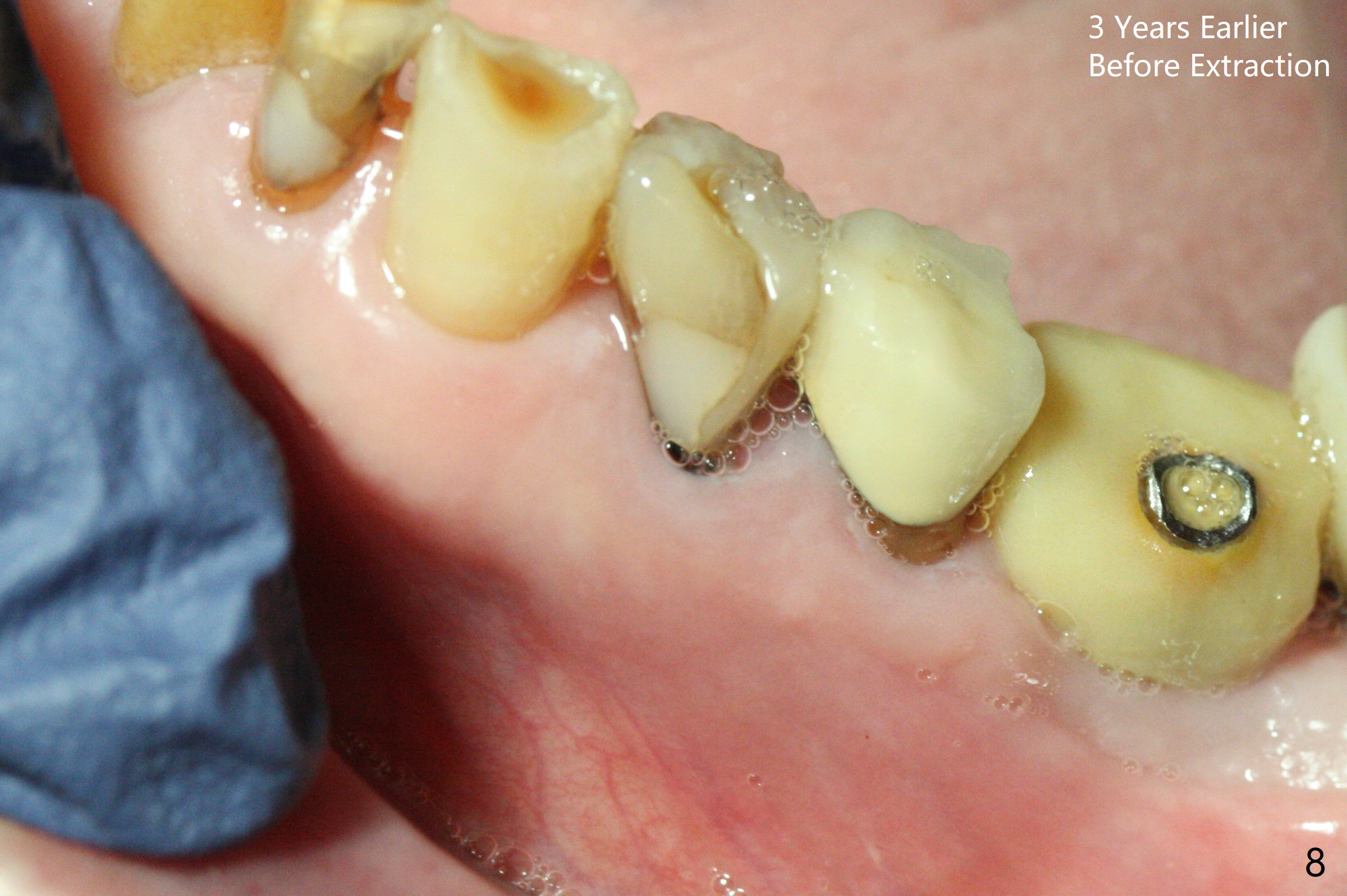
Extraction of the fractured tooth at #21 is as difficult as #19 (requiring incision). Because of the long root and hemorrhage, the apex of the tooth (Fig.1 black *) and a thin layer of the buccal shield (white *) remain when osteotomy is initiated. Due to hard bone, a 3.8x11.5 mm implant achieves insertion torque of ~35 Ncm (Fig.2); with placement of a 4.5x4(3) mm abutment, an immediate provisional is fabricated. There is limited remaining space for bone graft. The patient will return for final restoration 4 months postop; the buccal plate will be expected to have not collapsed! The implant seems to be osteointegrated, while the root piece (socket shield *) is exposed 4 months postop (Fig.3). The socket shield seems to be harmless, difficult to trim without local anesthesia and associated with no buccal plate concavity (Fig.4,5 <). With socket shield at #21, the crown looks normal, whereas the one at #19 without socket shield looks long, suggesting vertical bone loss early postop (before restoration, Fig.6).没有牙根处,角化龈少(图七,与术前(图八)对比)。
Return to
Lower Premolar
Immediate Implant,
Trajectory,
10,
31,15 Acrylic Dressing
Xin Wei, DDS, PhD, MS 1st edition
02/26/2019, last revision
02/21/2021