|
|
|
|
|
|
|
|
|
|
|
|
|
|
|
Trajectory Check
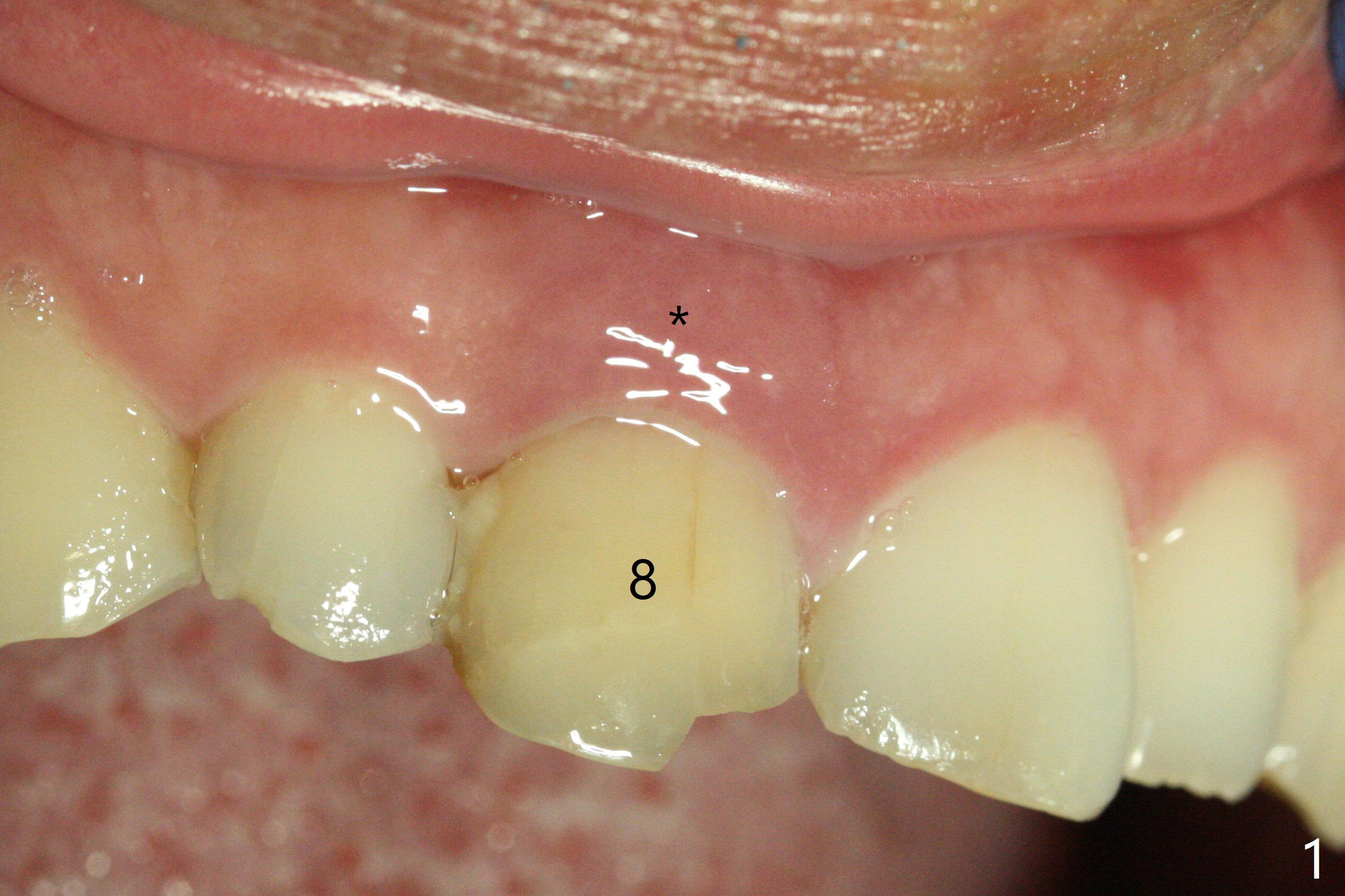
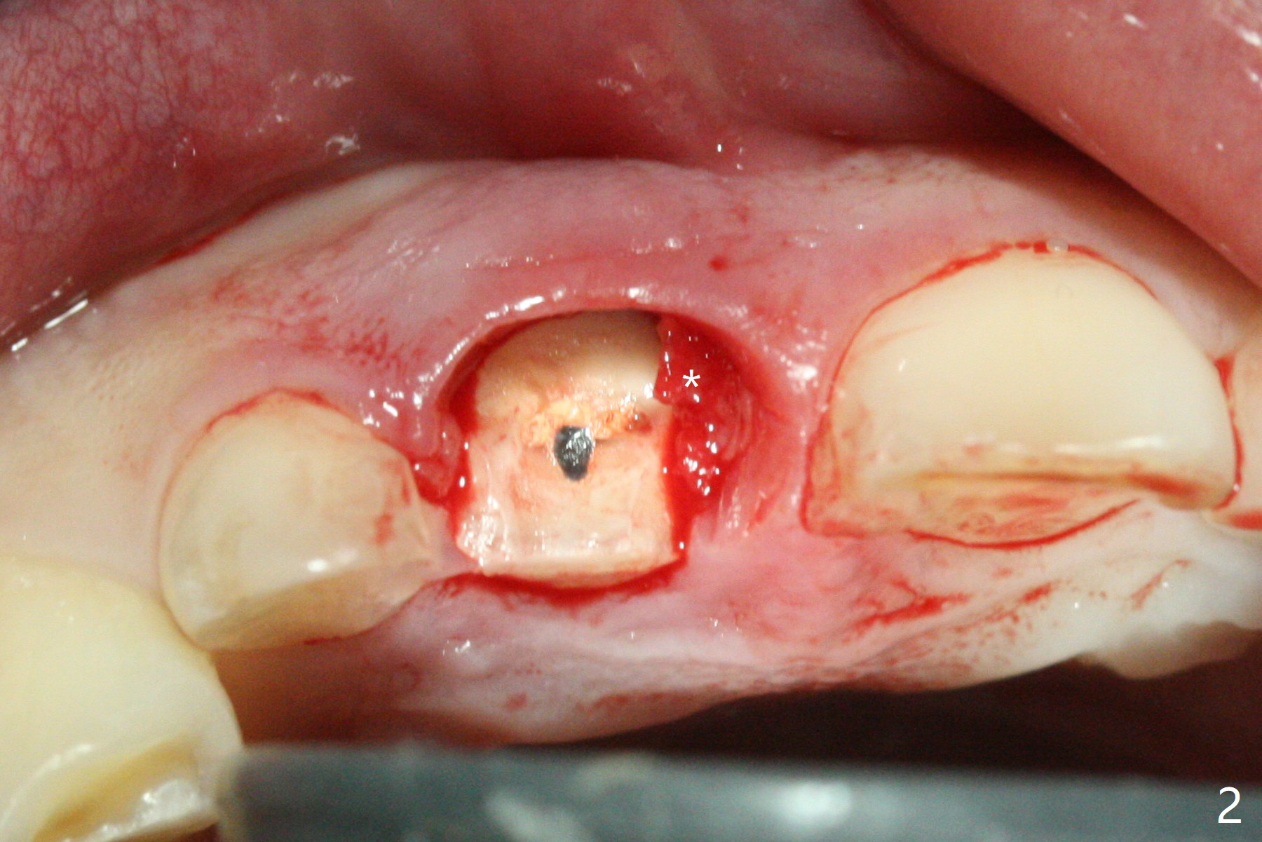
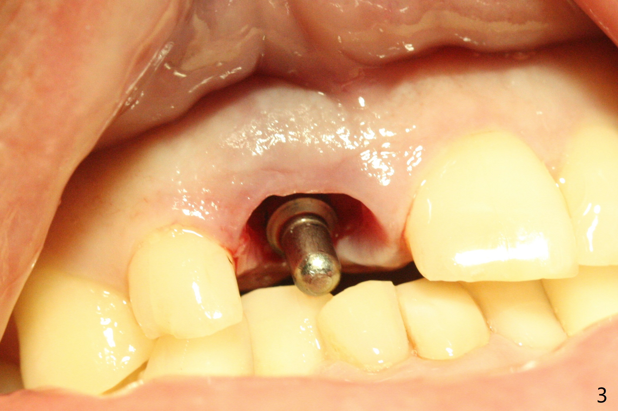
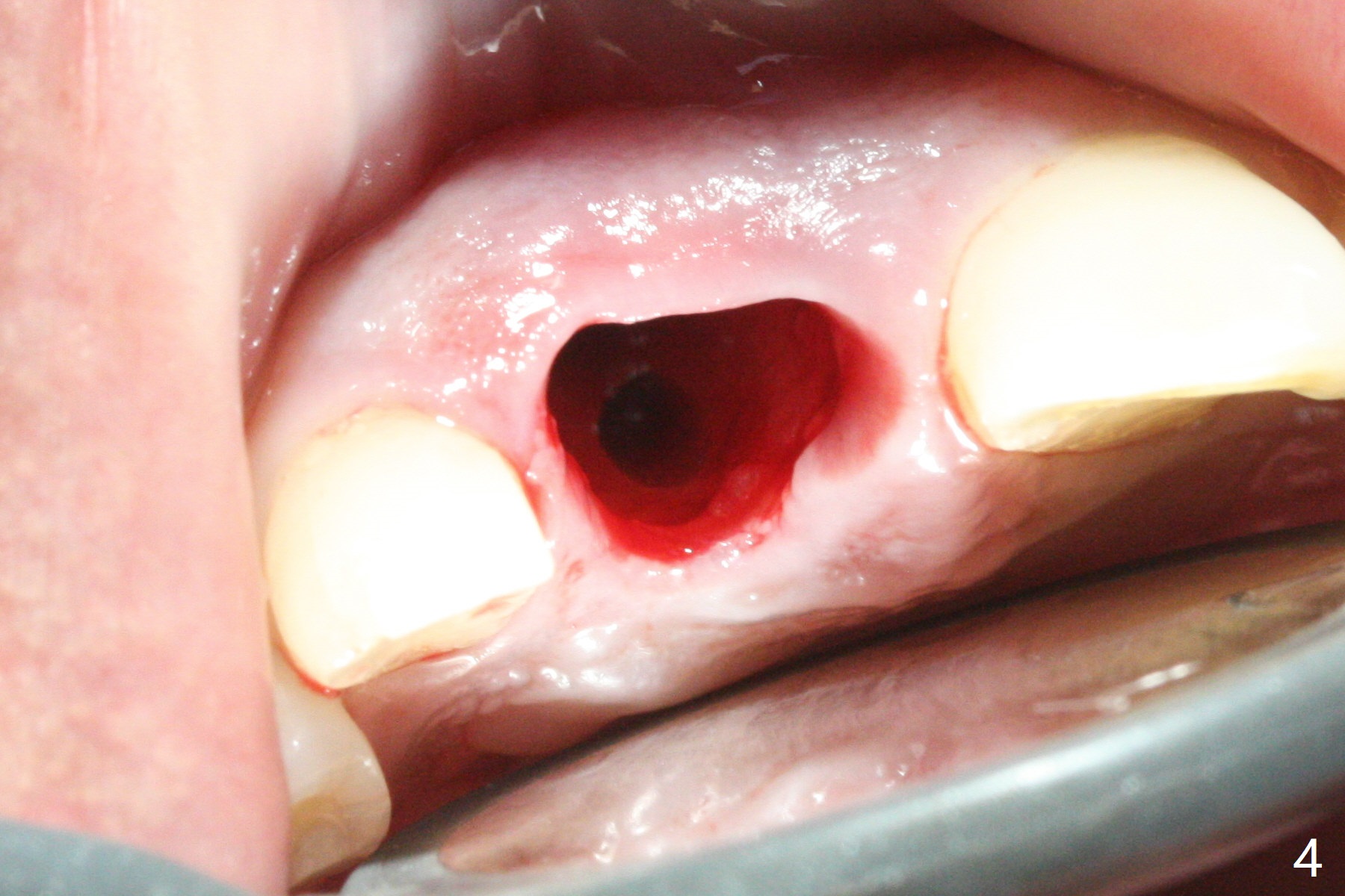
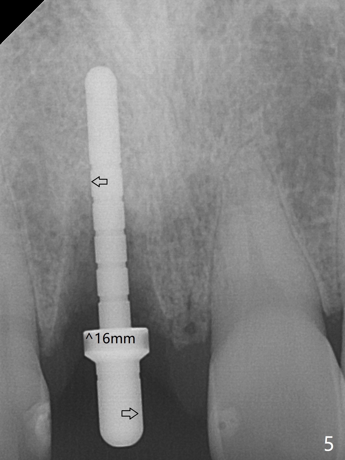
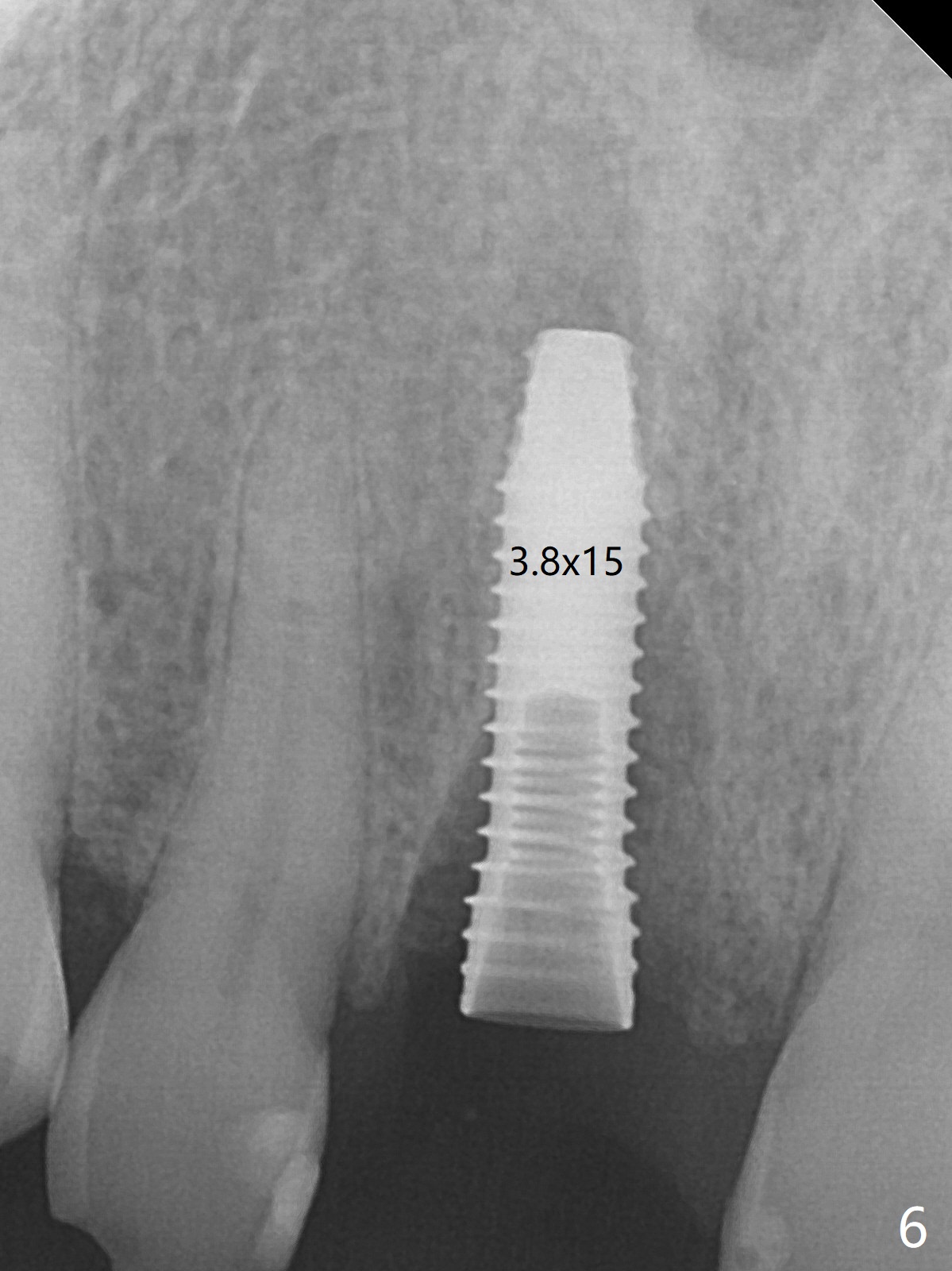
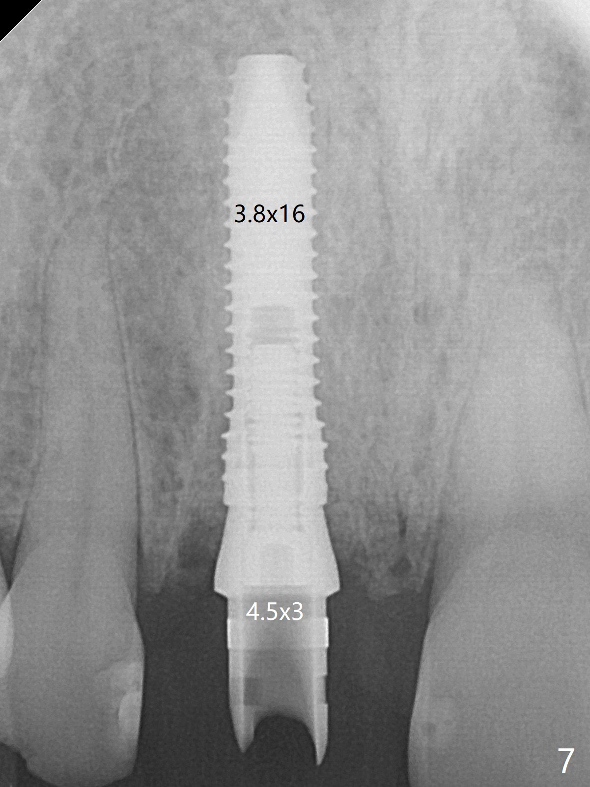
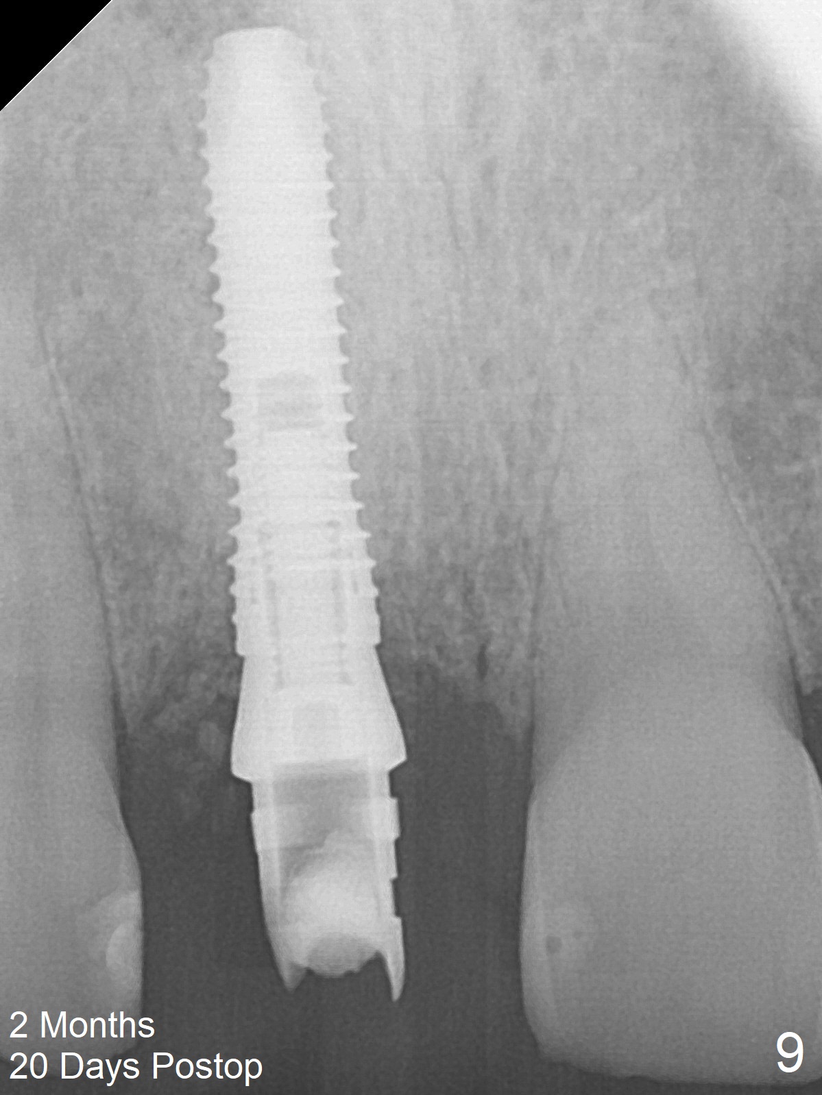
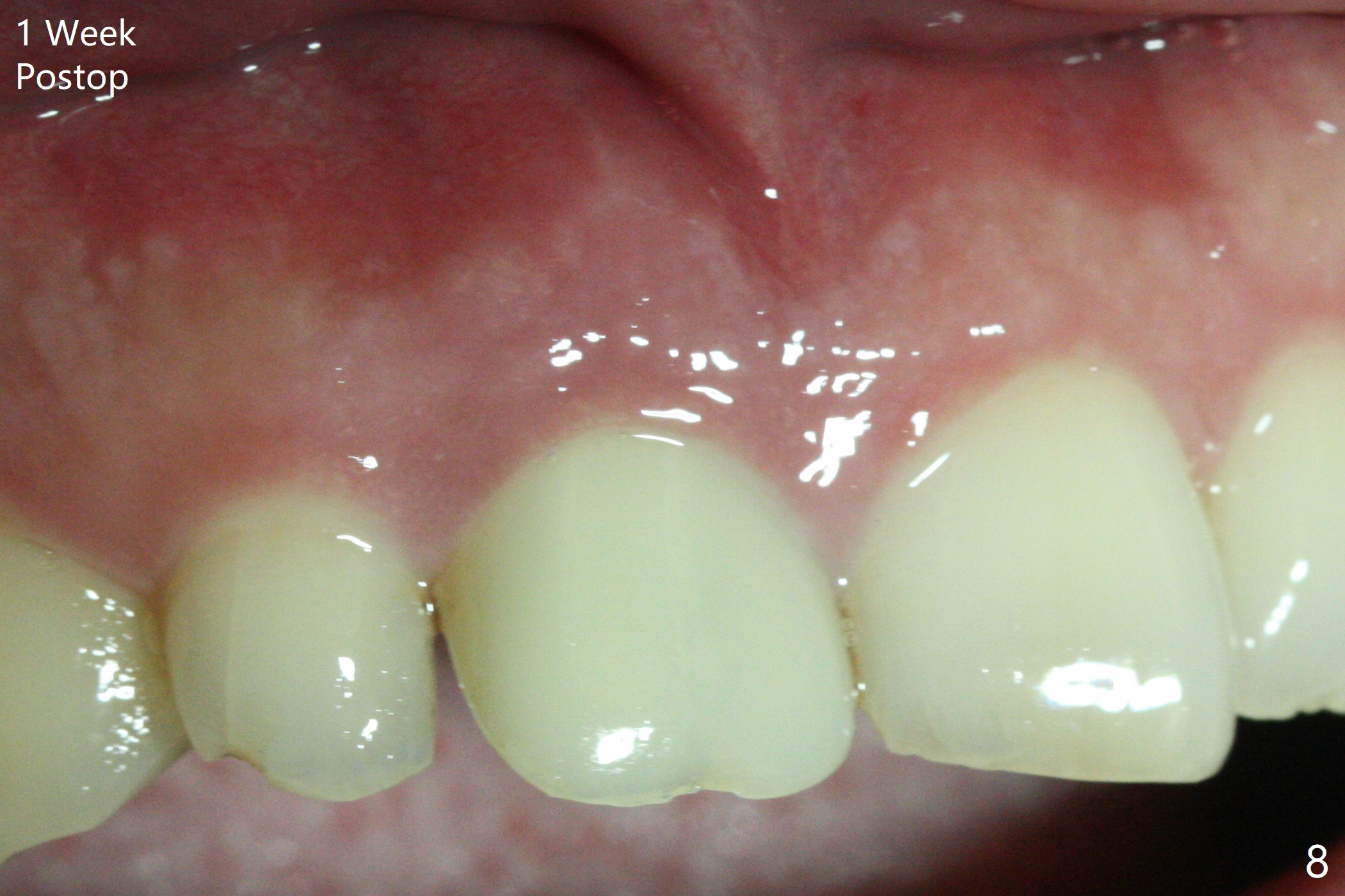
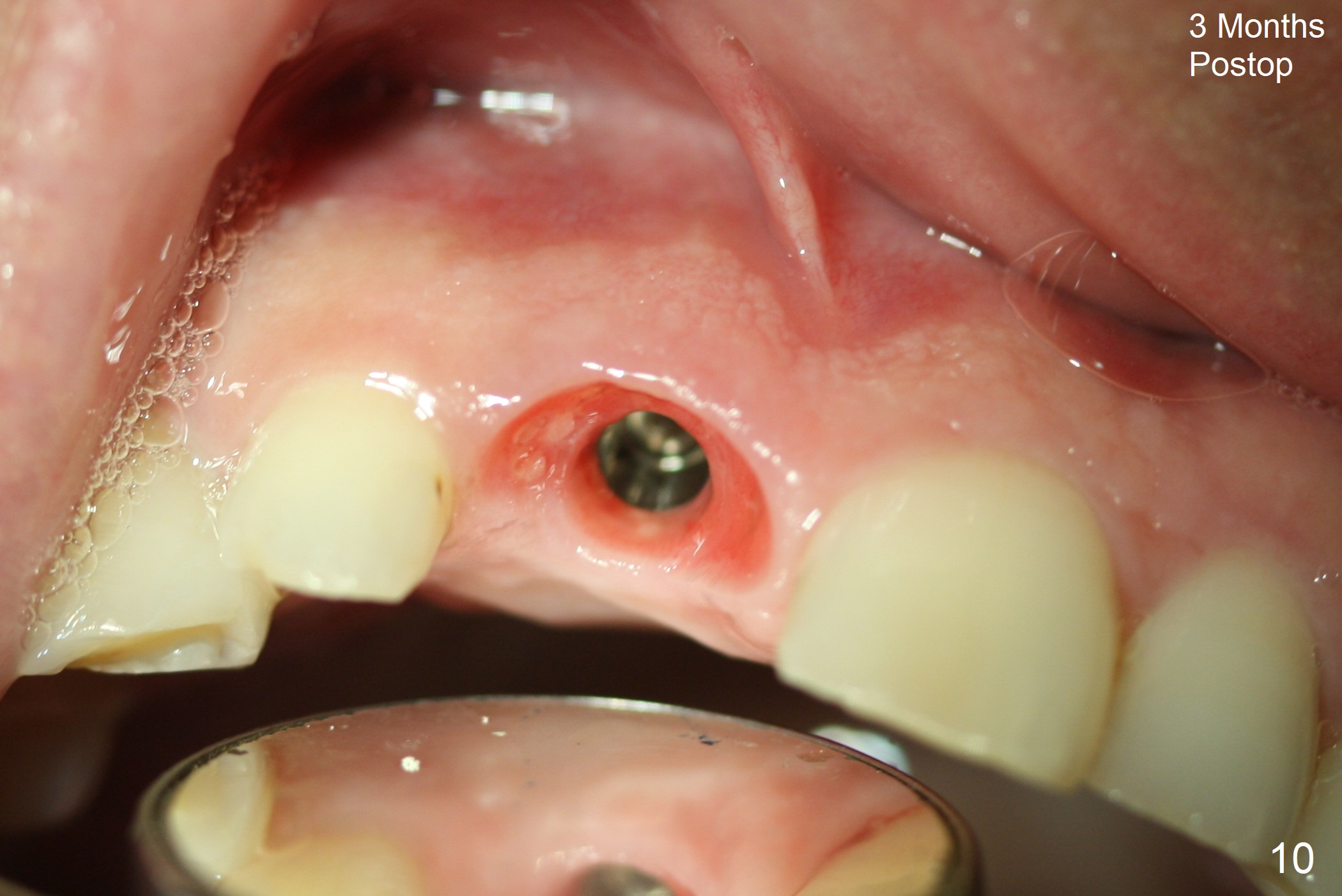
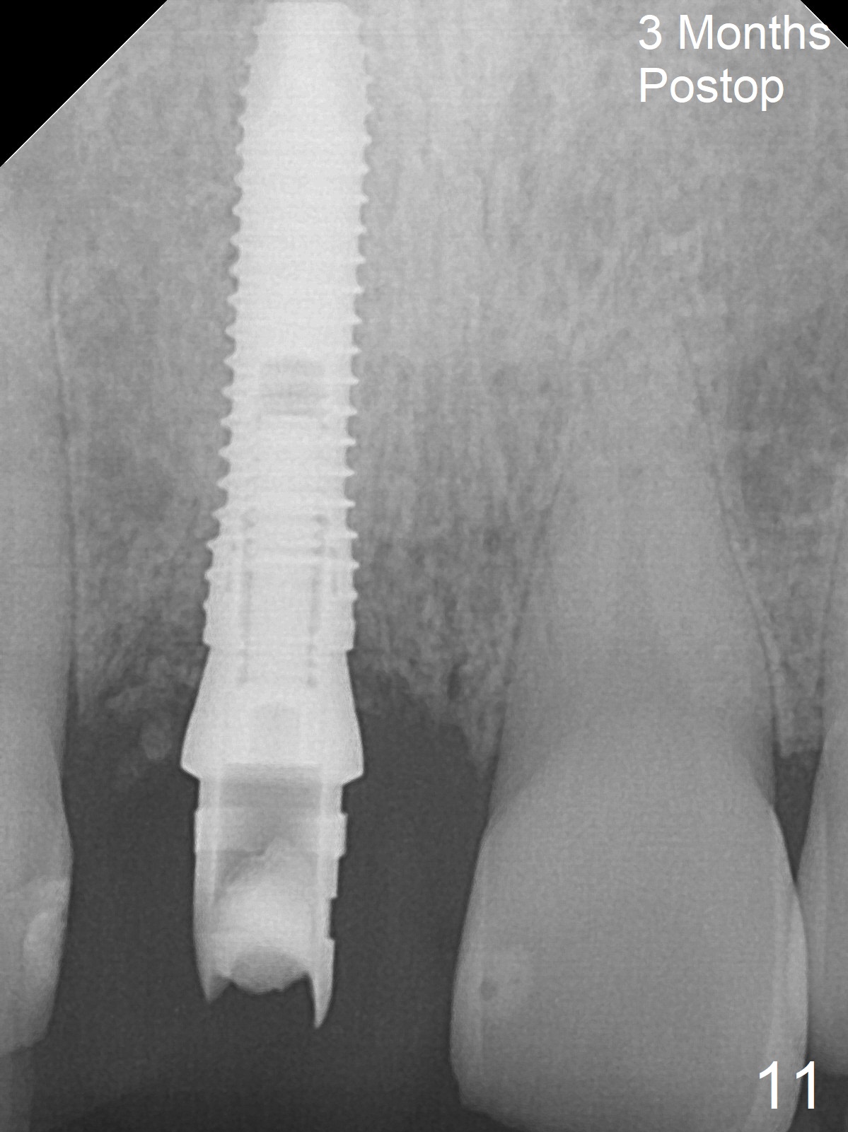
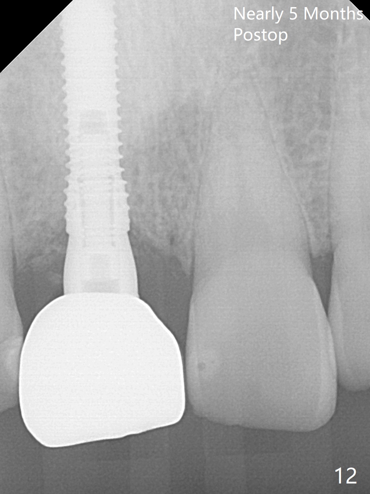
The buccal gingiva at #8 is erythematous (Fig.1: *), which is associated with the tooth fracture line (Fig.2 with granulation tissue: *). The buccal plate remains intact. Osteotomy is initiated in the palatal wall of the socket (Fig.3,4). The initial osteotomy depth is 16 mm (Fig.5); the trajectory is going to be adjusted as shown by arrows. The trajectory improves when a 3.8x15 mm dummy implant is placed (Fig.6). The definitive implant (3.8x16 mm) appears to be placed at an appropriate level (Fig.7). A 4.5x3 mm temporary abutment is inserted for an immediate provisional. As routine, Vera Graft is placed in the buccal gap. The buccal gingival erythema reduces without tenderness 1 week postop (Fig.8). Although the provisional is unstable, there is no bone loss 2 months 20 days postop (Fig.9). Because of the loose provisional (partial detachment from the underlying temporary abutment), impression is taken earlier (3 months postop, Fig.10,11). Due to the pointed abutment tip, the crown is redone 3 times. By the time of cementation (nearly 5 months postop), the socket appears to have healed (Fig.12).
Return to
Upper
Incisor Immediate Implant,
Armaments
Xin Wei, DDS, PhD, MS 1st edition 03/01/2018, last revision 07/25/2018