,%20VeraGraft.jpg)
|
|
|
|
||
|
|
|
|
|
|
|
|
|
|
||
|
|
|
|
|
|
|
|
|
|
||
Tap with Soft Tissue Depth Mark
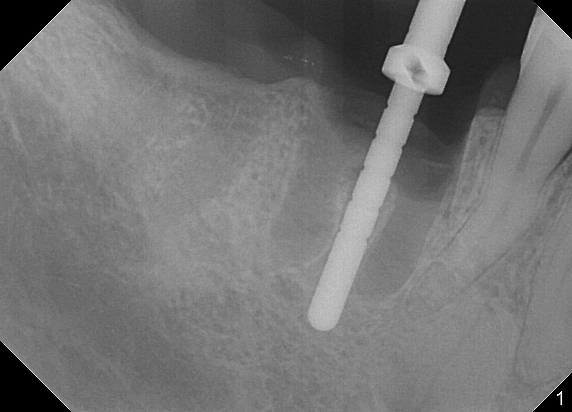
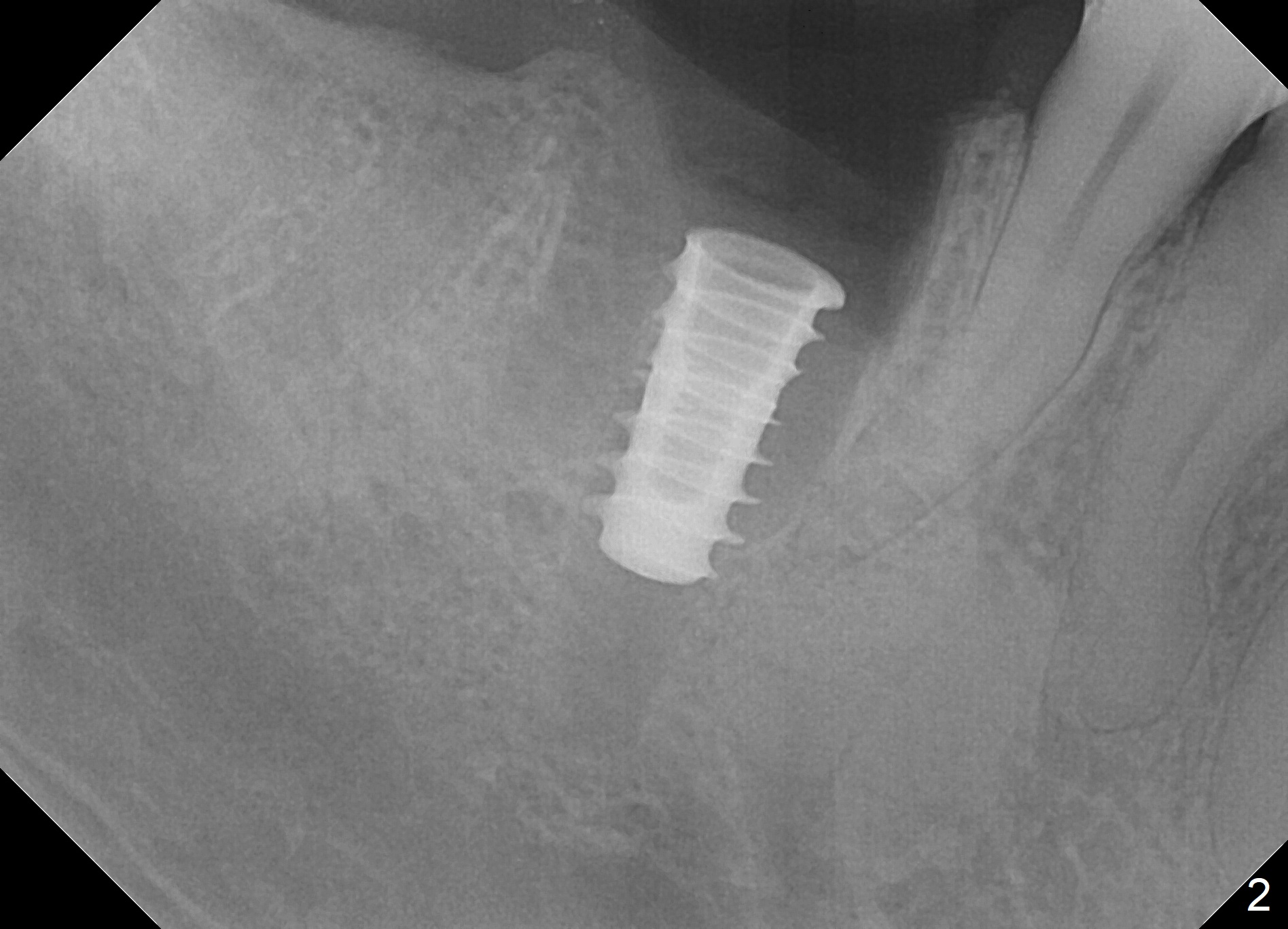
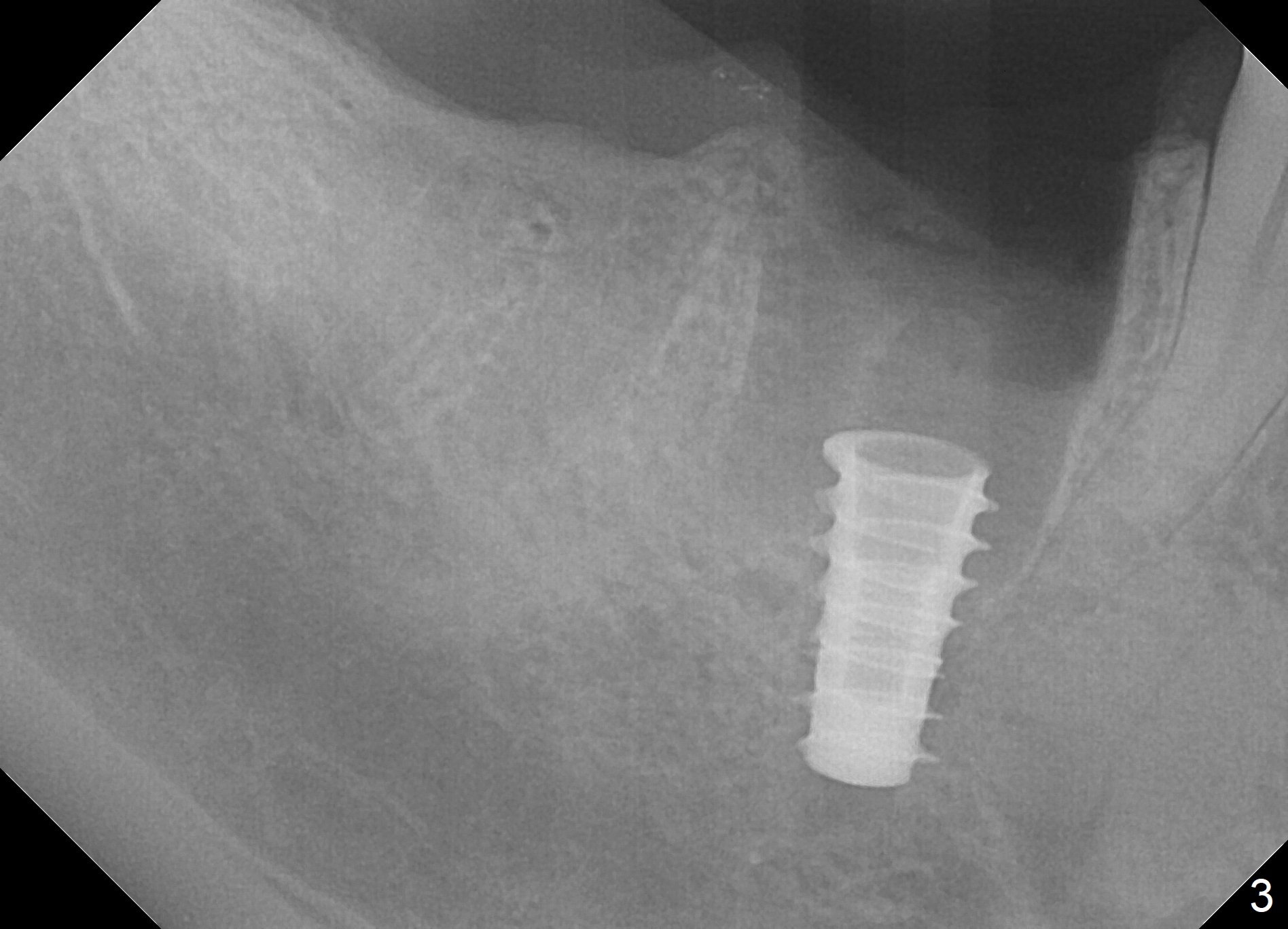
Extraction is difficult due to hard bone and brittle, curved roots of the tooth #30; initial depth in the septum is 14 mm (gingival level, Fig.1). Then the depth increases to 17 mm; with sequential osteotomy using 2.8 mm to 4.8 mm Magic Drills (MD), a 5x9 mm dummy implant is placed incompletely due to hard bone (Fig.2). After use of 5.3 mm MD for ~ 15 mm, the dummy implant is placed to the depth (Fig.3). The definitive IBS implant (5x13 mm) is placed with 50 Ncm; a 6.5x5.7(4) mm abutment is placed with allograft filling the gap (* and arrow).
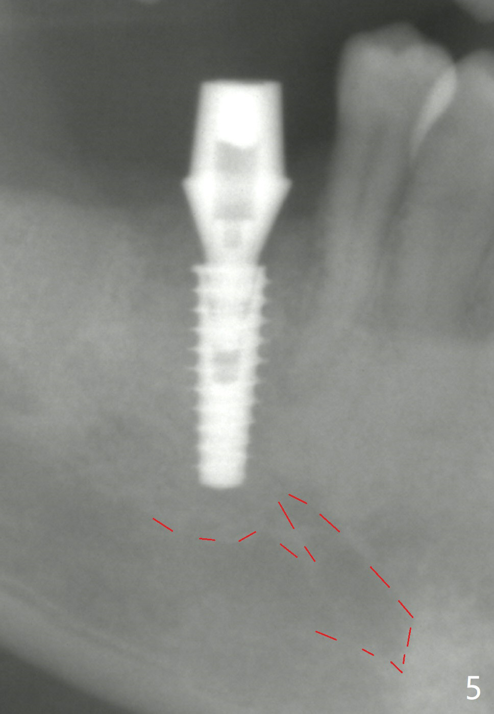
The final implant is placed a little too deep in this case. The problem can be avoided by using Tap with soft tissue depth mark and is solved by using a longer implant (5x13 mm) or having an abutment with longer cuff (5 or 6 mm). Postop there is tooth sensitivity. When the latter disappears, the patient feels the right jaw different. Panoramic X-ray taken 2 months postop shows that the implant has clearance from the Inferior Alveolar Canal (Fig.5 red dashed line).
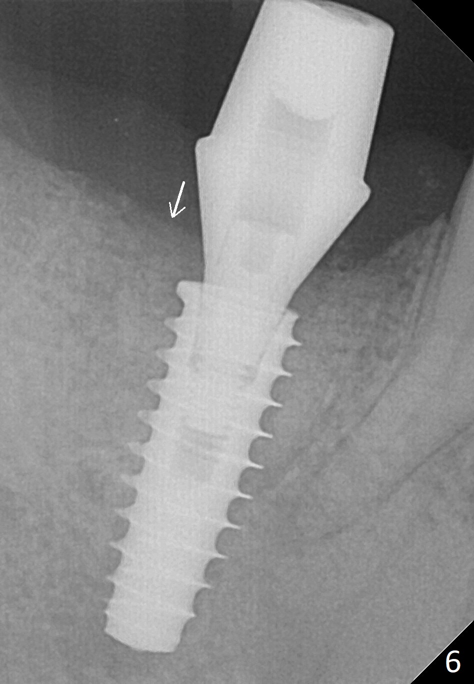
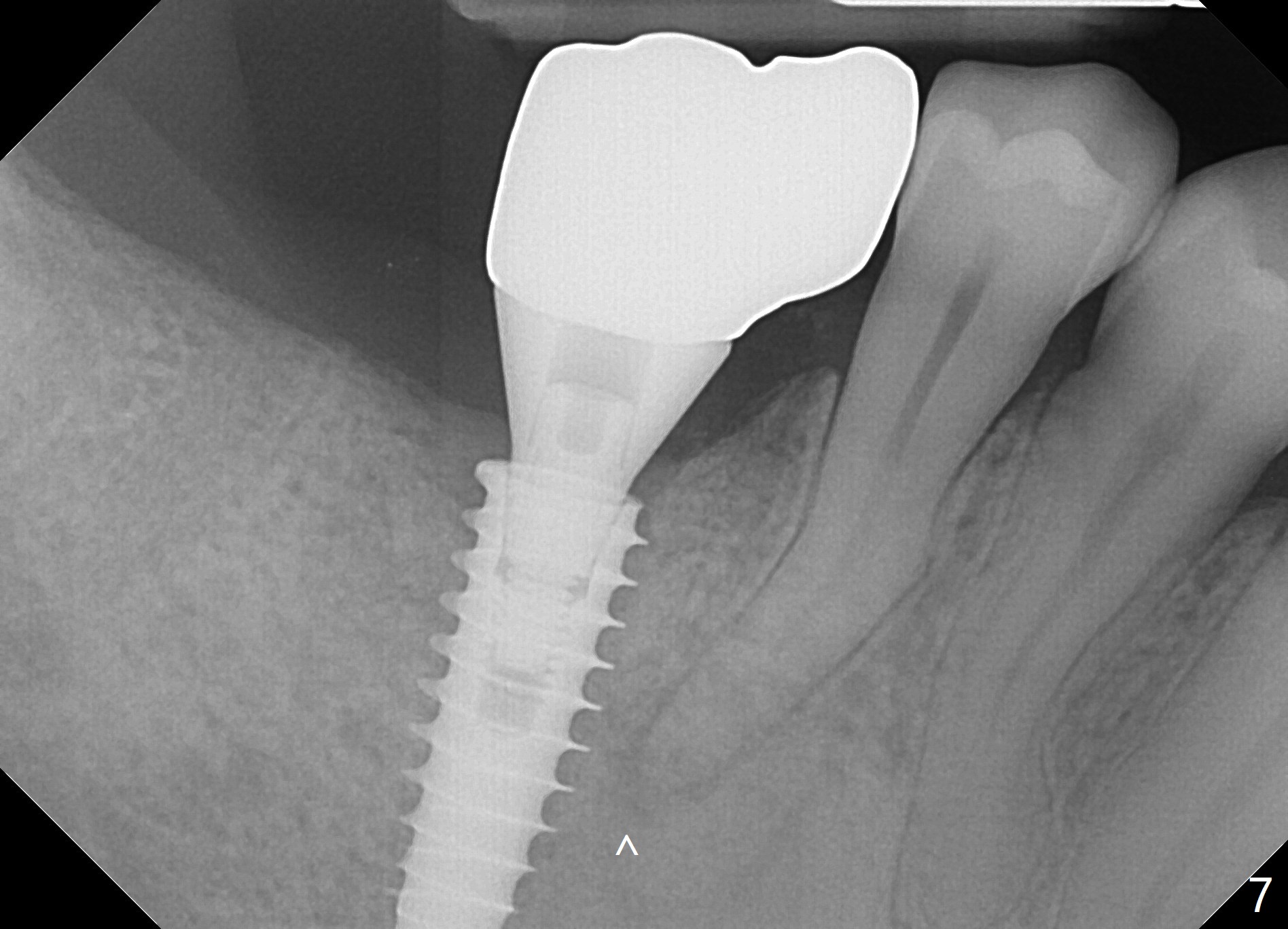
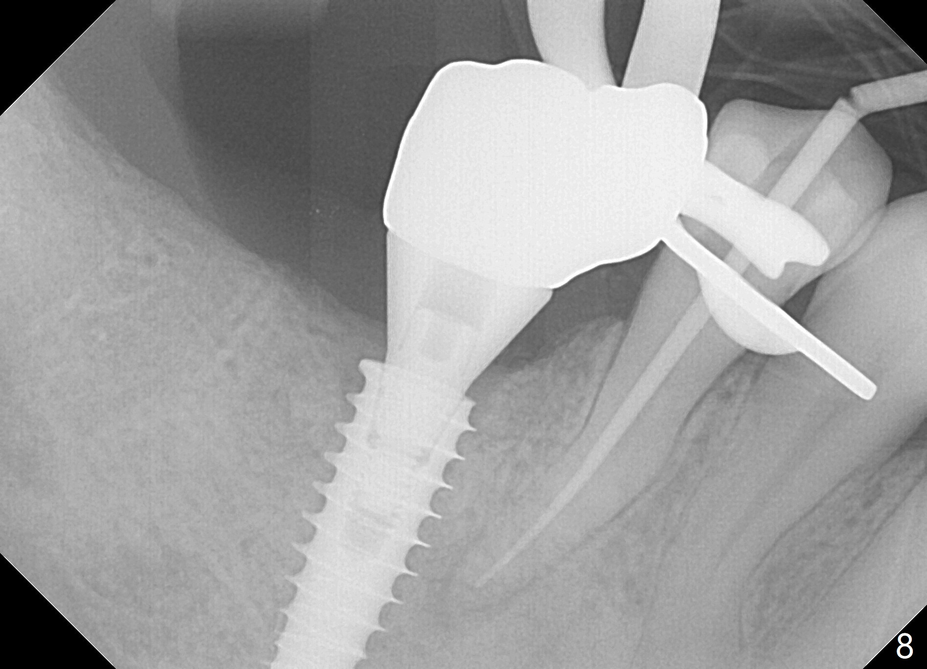
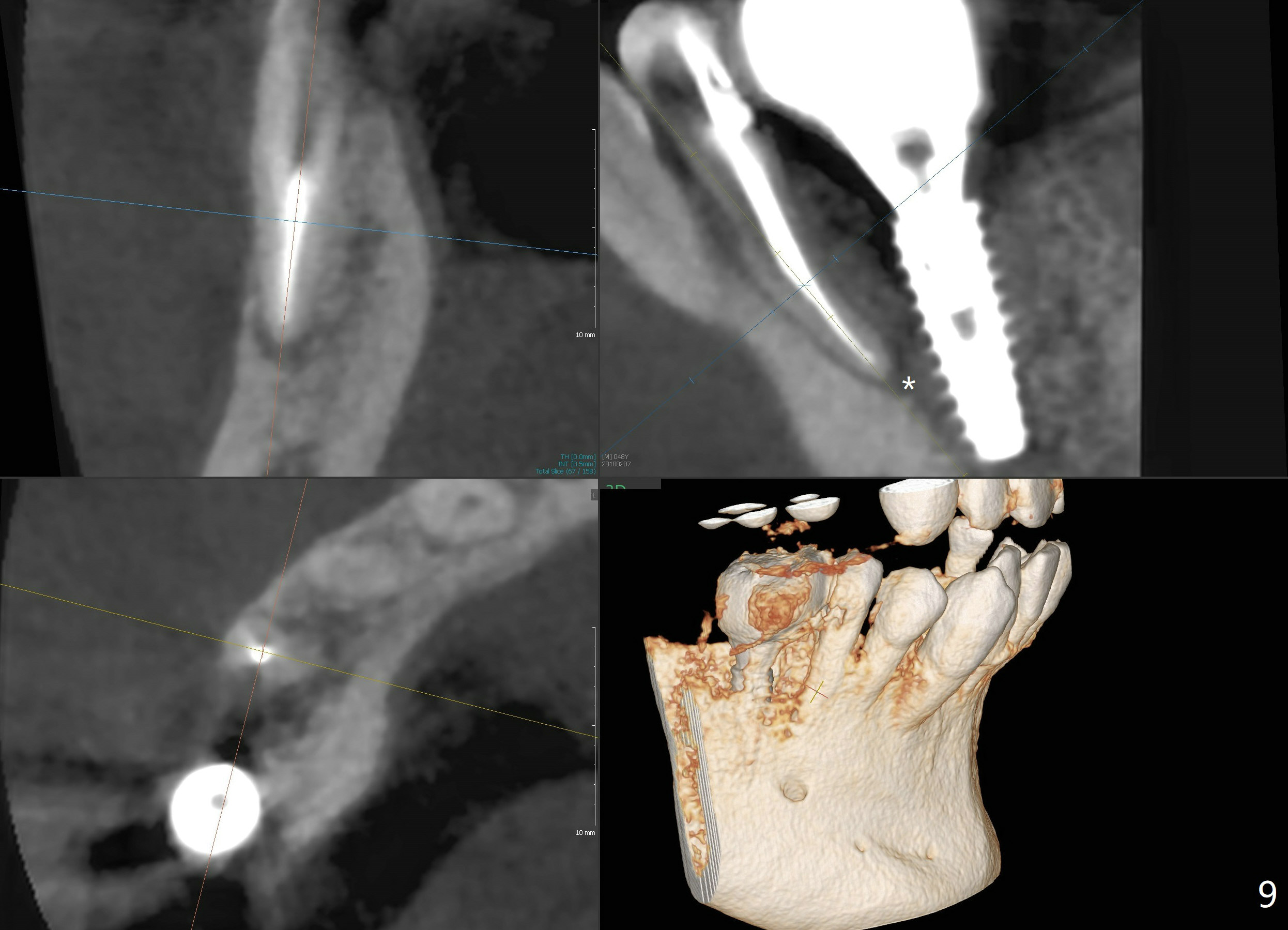
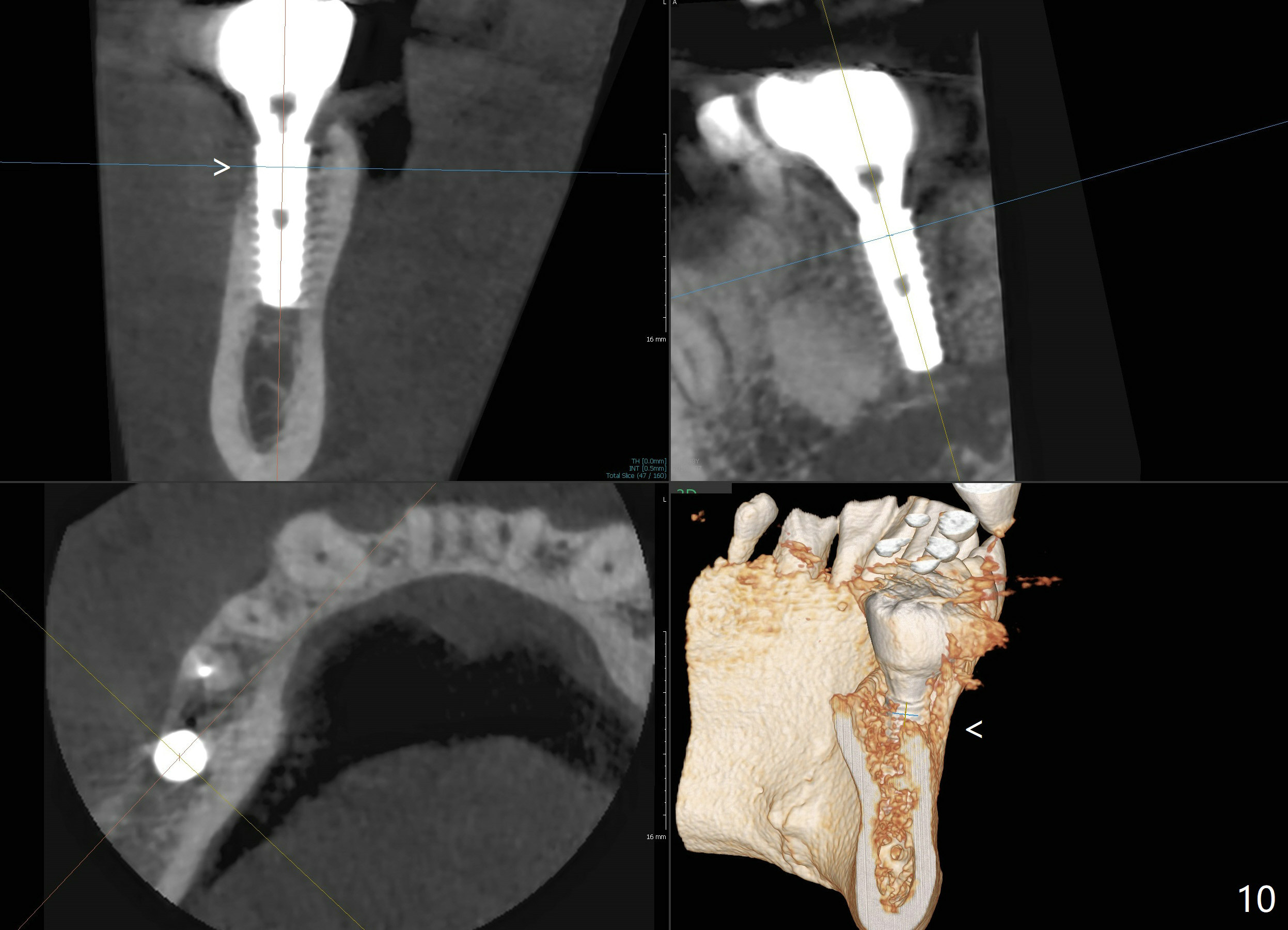
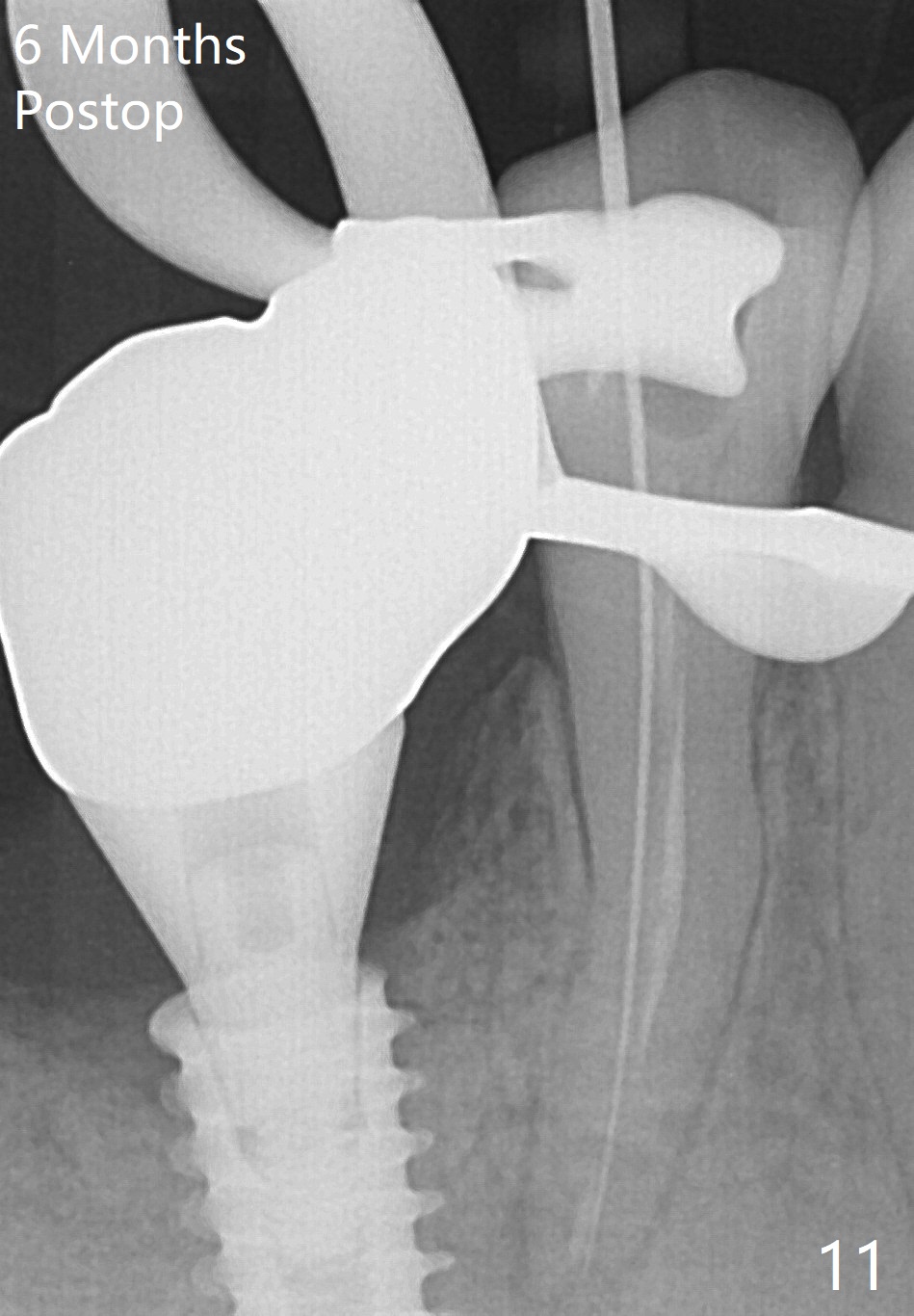
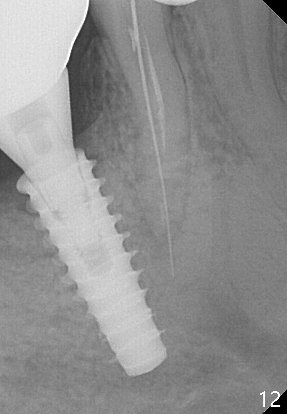
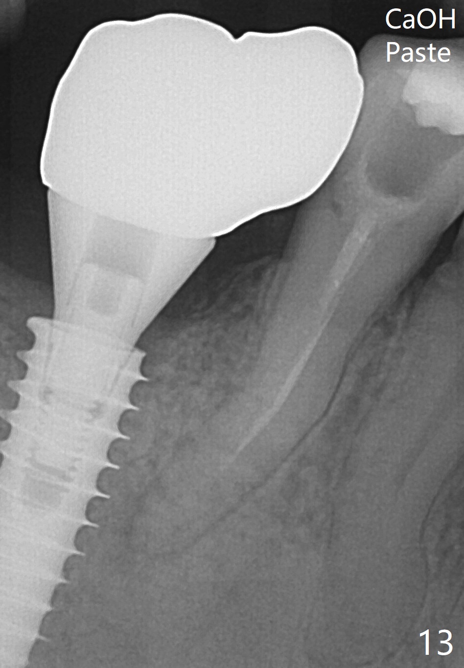
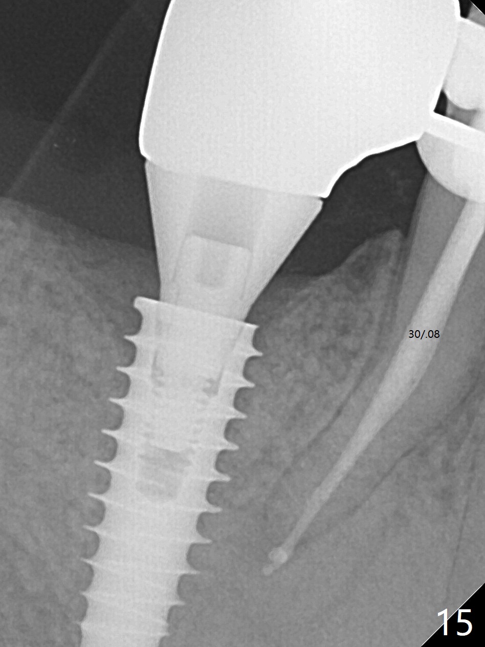
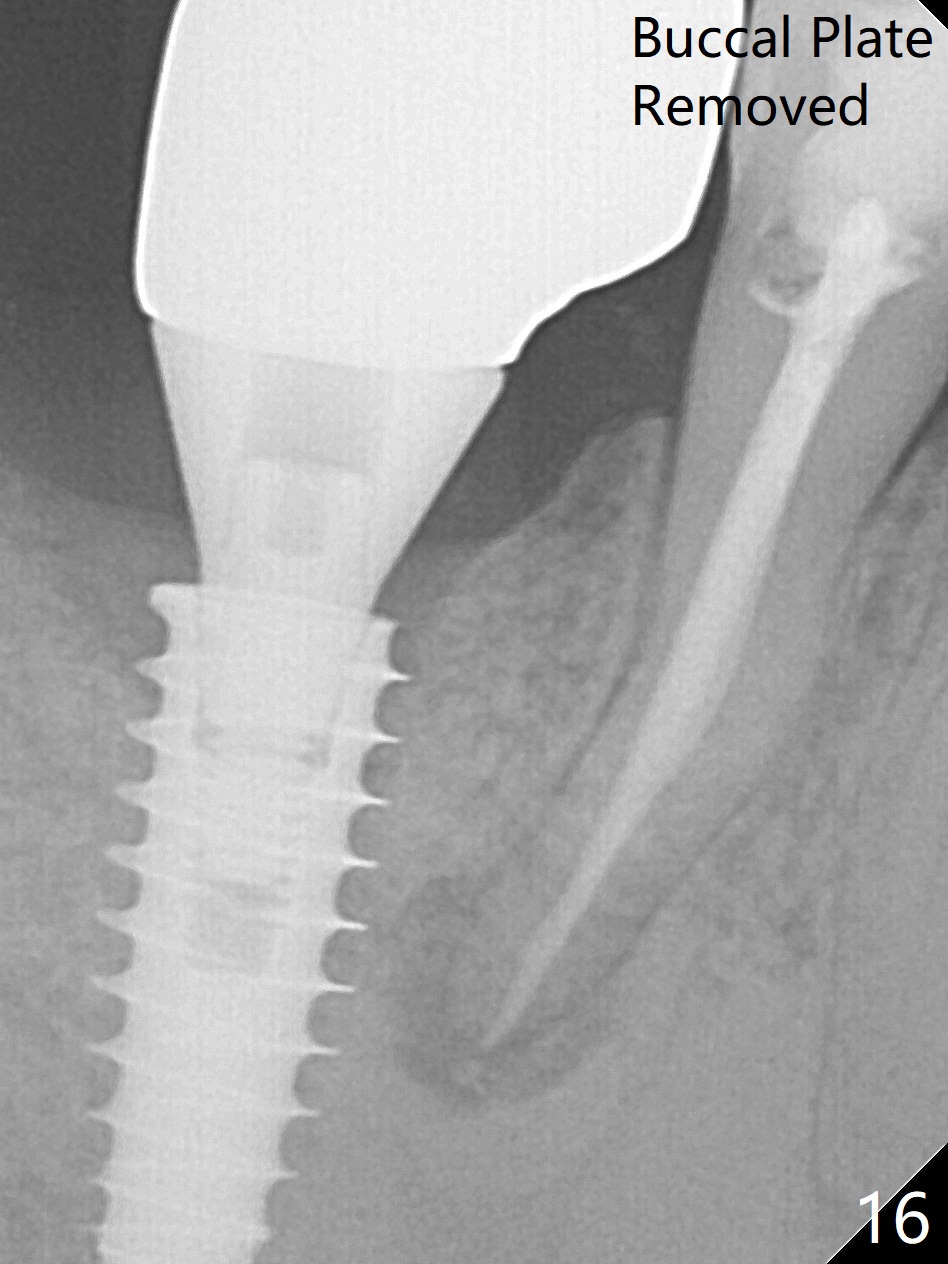
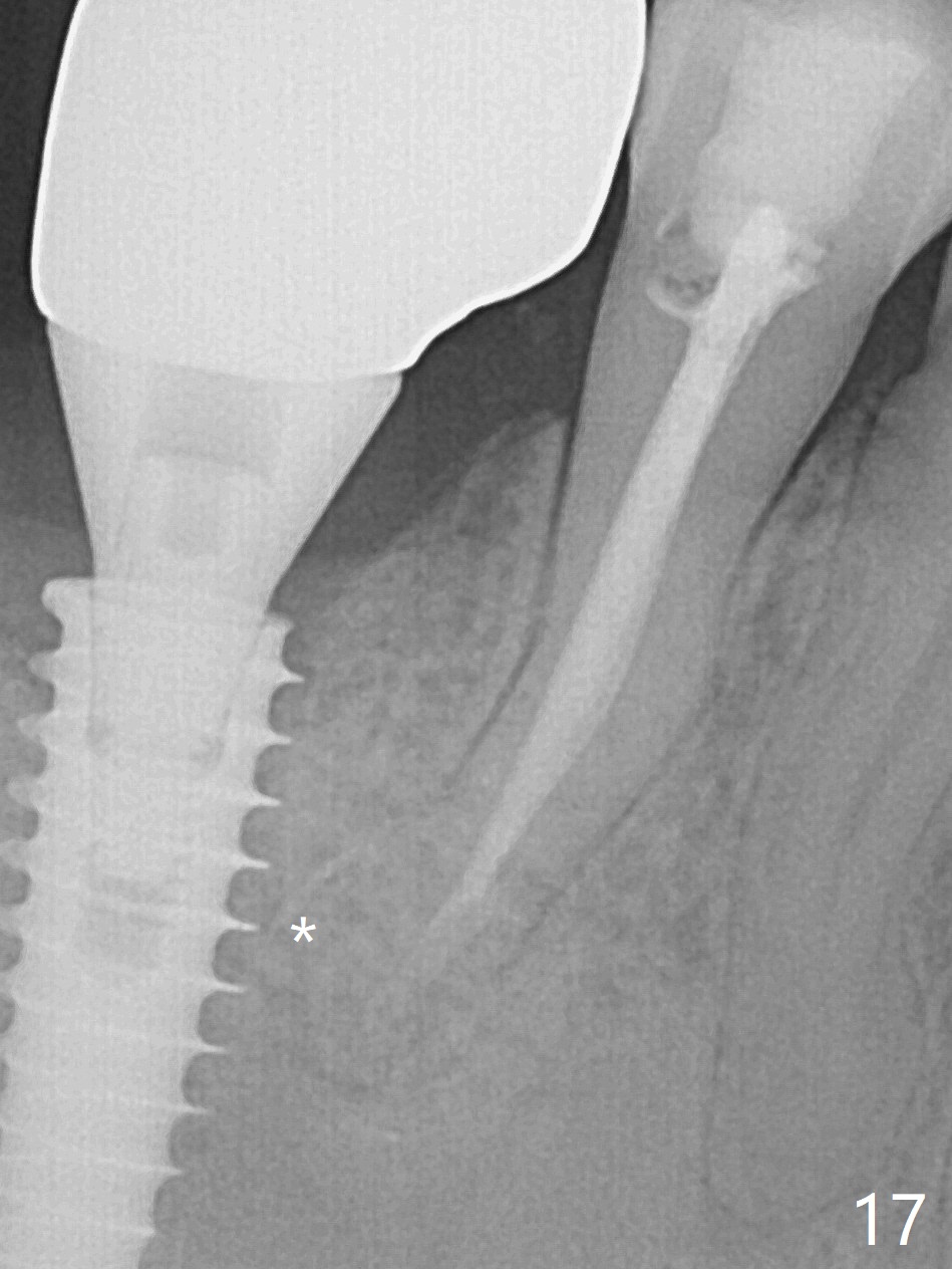
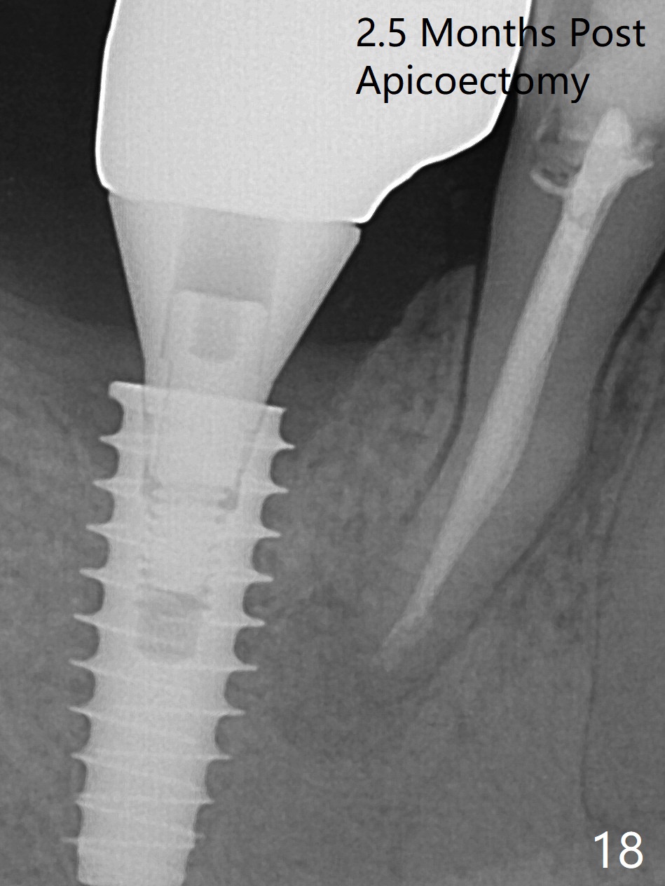
Bone graft seems to sink down and becomes denser 3 months postop (Fig.6 arrow). The bone continues being denser 5 months postop (Fig.7). There is periapical radiolucency of the tooth #29 (^). RCT is done (Fig.8). The pain persists 2 weeks postop (Fig.9,10). There is no missing canal (Fig.9). The apex is close to the implant (Fig.9 *). Apicoectomy will be performed if needed. It appears that the implant is also placed buccal (Fig.10 <) and/or the implant too large for the site. Therefore there should be a 2-3 mm buccal gap before and after implant placement. Separation and reflection of the buccal flap allows better visibility. The pain persists 1 month post RCT and 6 months post implant placement. RCT retreatment is initiated (Fig.11,12) with placement of Calcium Hydroxide paste after redebridement with 30/.04 rotary file at 23.5 mm (.5 mm longer than the earlier RCT, Fig.13). RCT retreatment finishes with apparent transportation and extrusion in 4 weeks (Fig.14,15), followed by apicoetomy (Fig.16,17) (20 days later)). Discomfort remains 2.5 months postop (Fig.18). Keep watching.
Return to
Lower
Molar Immediate Implant, Prevent
Molar Periimplantitis (Protocols,
Table), 1st
Year
Xin Wei, DDS, PhD, MS 1st edition 08/10/2017, last revision 09/07/2018