.jpg)

.jpg) |
 |
After use of the final drill (3 mm), the coronal Incisive Canal is perforated. Following placement of a 3.5x13 mm implant and 4.5x5.5(4) mm abutment, Vanilla Graft is placed (Fig.5 *) to repair the perforation. Retrospectively, the coronal end of the Incisive Canal is revealed at incision (Fig.1 *).
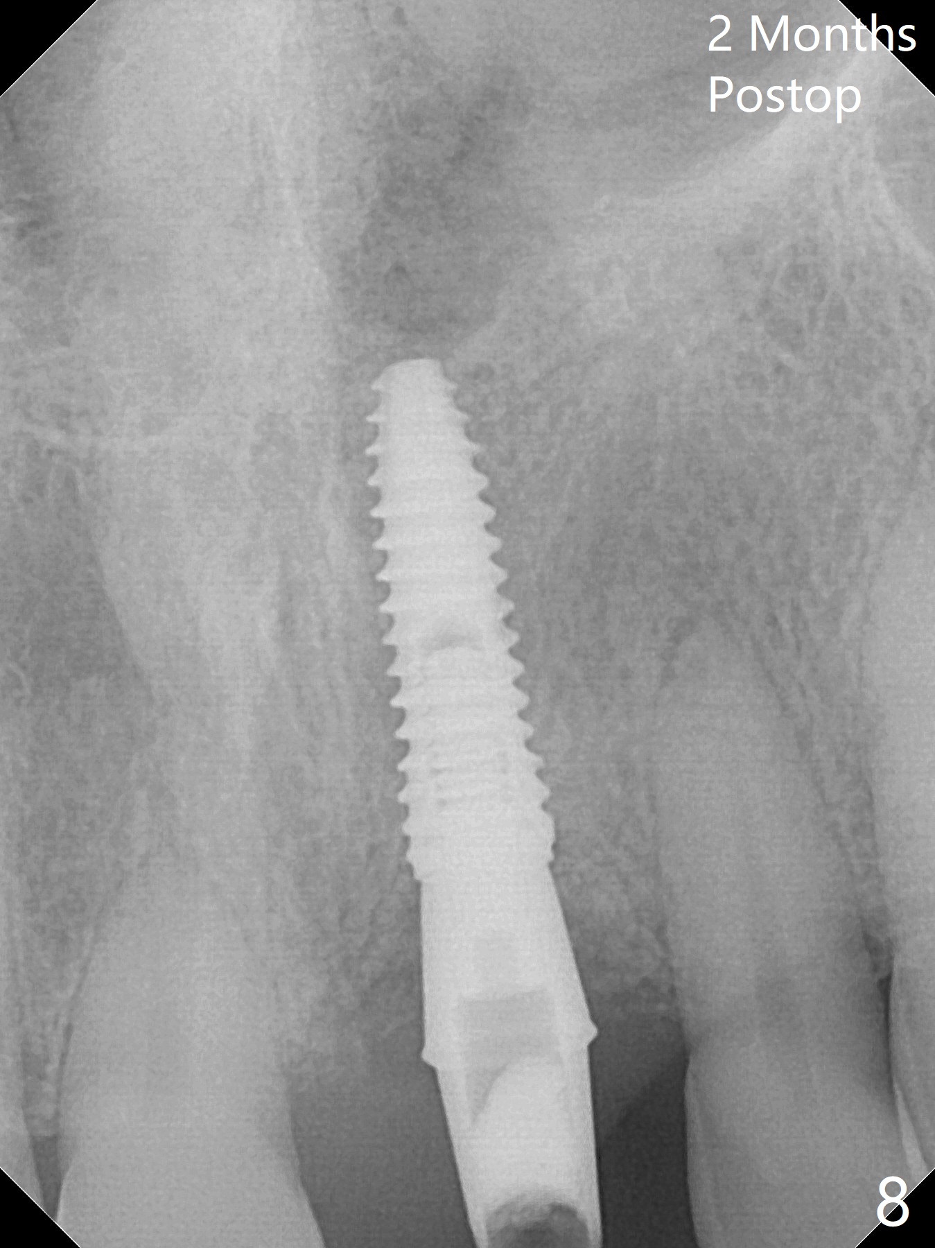
The bone graft appears to remain in place 2 months postop (Fig.8). Impression is taken because of instability of the immediate provisional.
Xin Wei, DDS, PhD, MS 1st edition 12/04/2017, last revision 08/13/2018