|
|
|
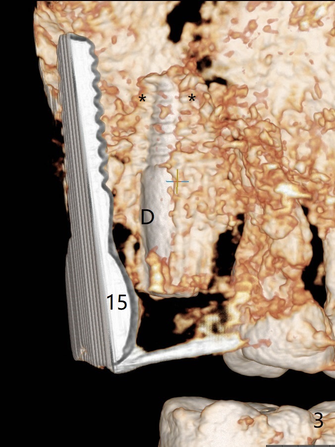
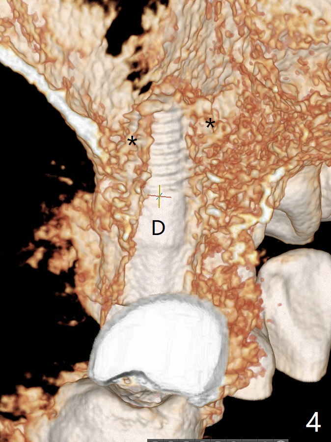
CBCT 3-D images (Fig.3 (palatal view), 4 (distal view) (D: distal)) and coronal section (Fig.5 (P: palatal) with removal of #15 implant image) show the bone graft in the sinus (*).
Xin Wei, DDS, PhD, MS 1st edition 04/22/2019, last revision 04/22/2019