|
|
|
|
|
|
|
|
|
|
|
|
|||
2 Stage
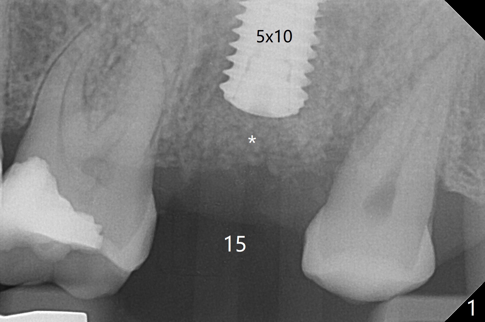
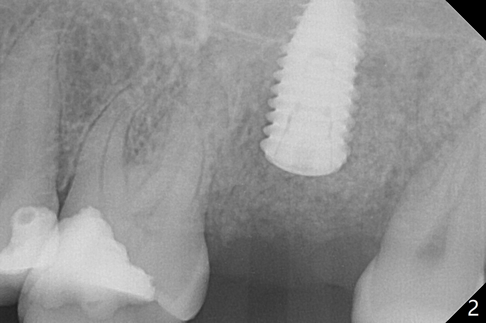
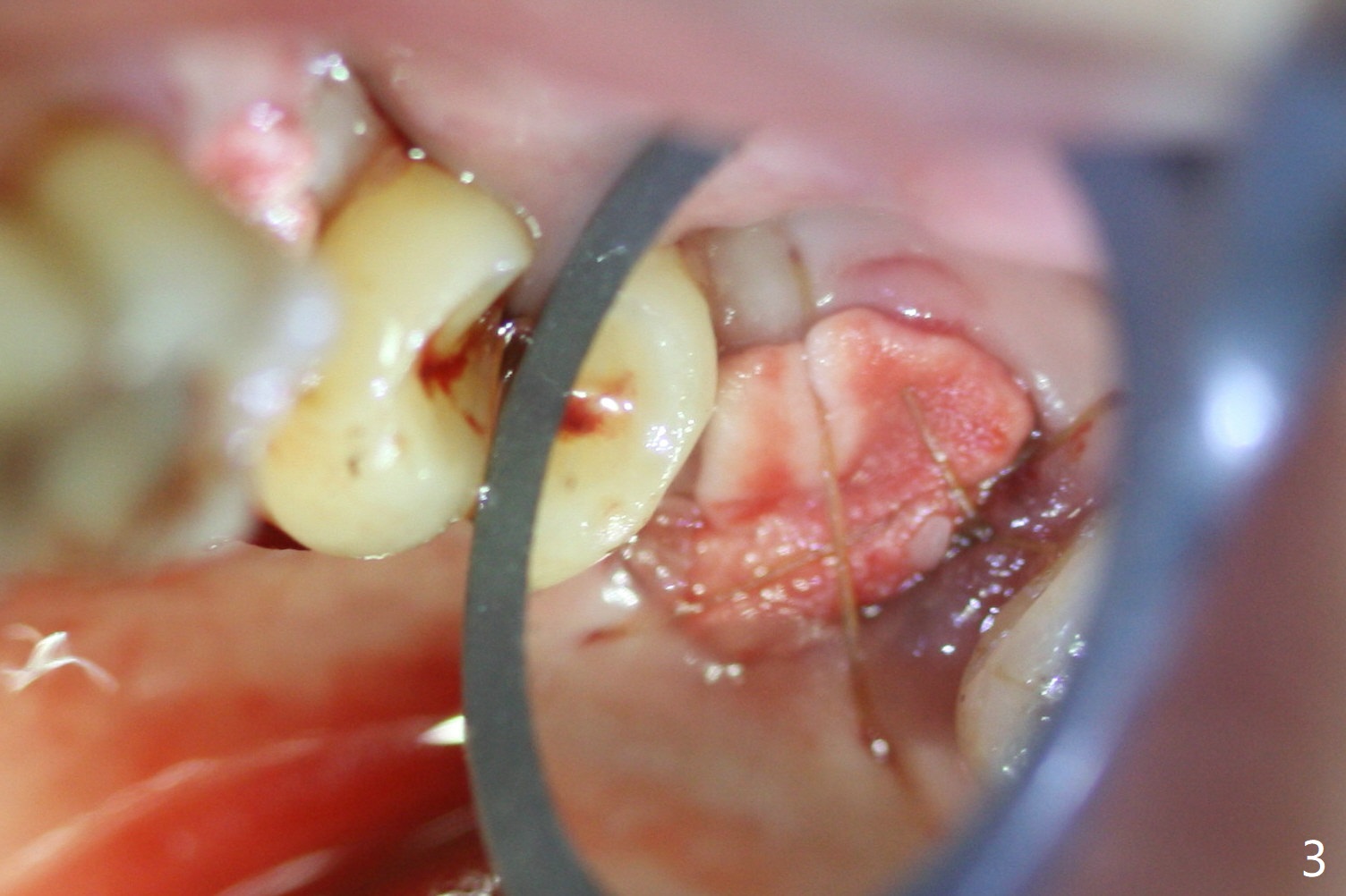
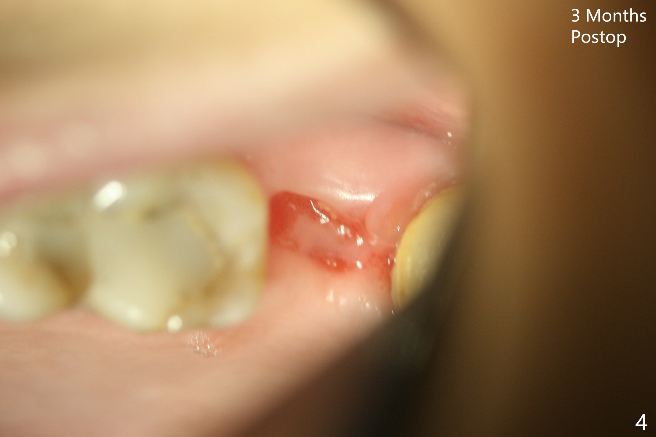
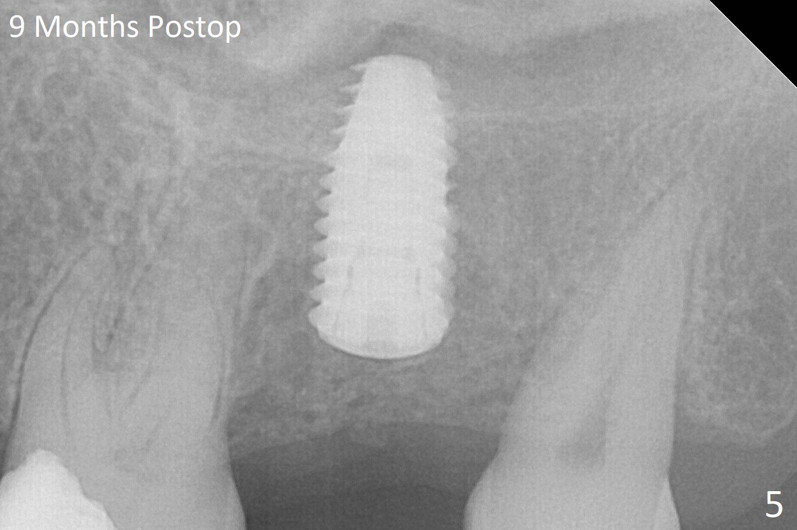
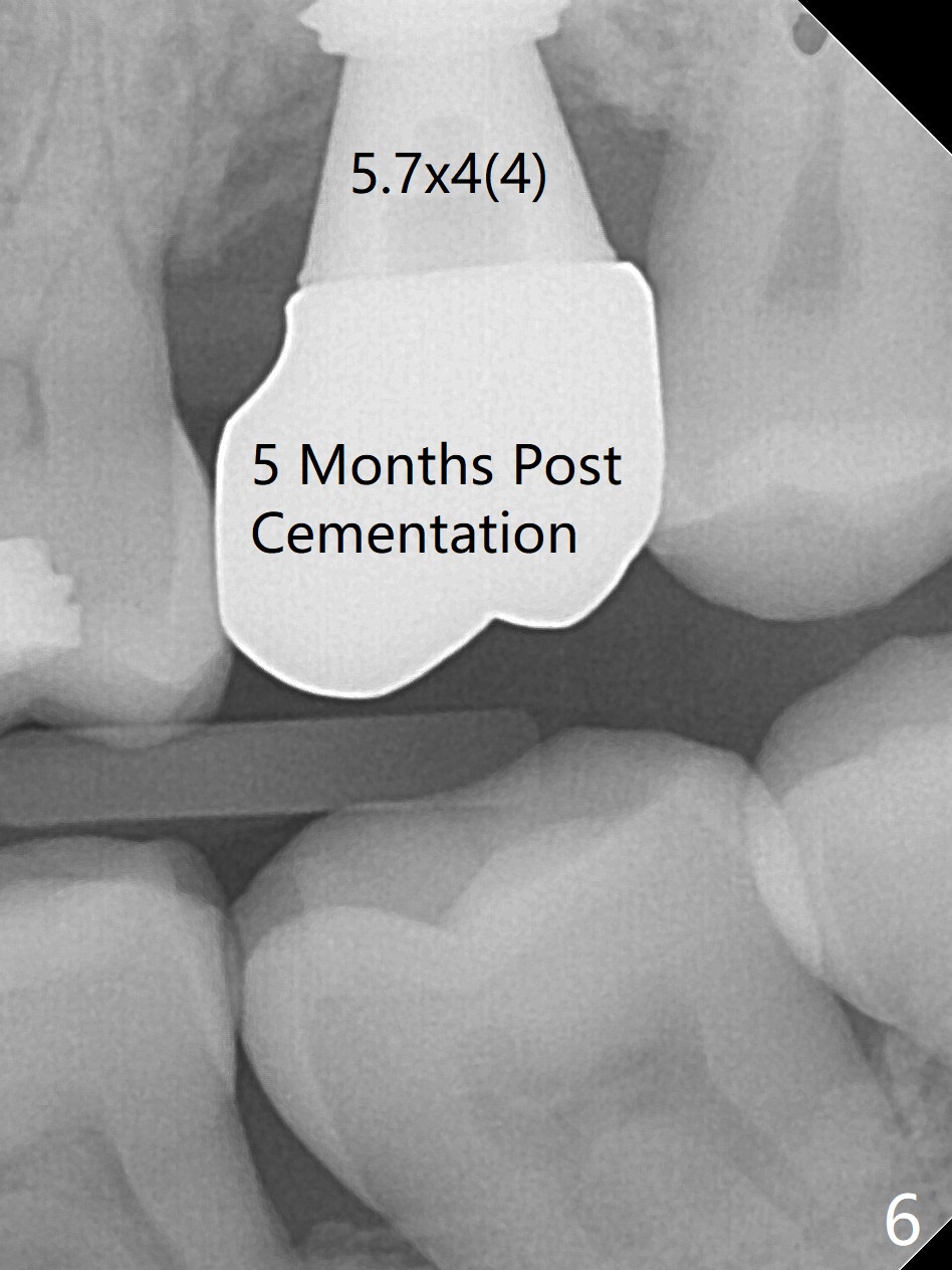
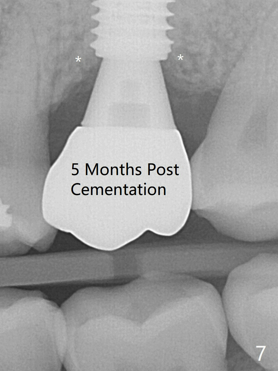
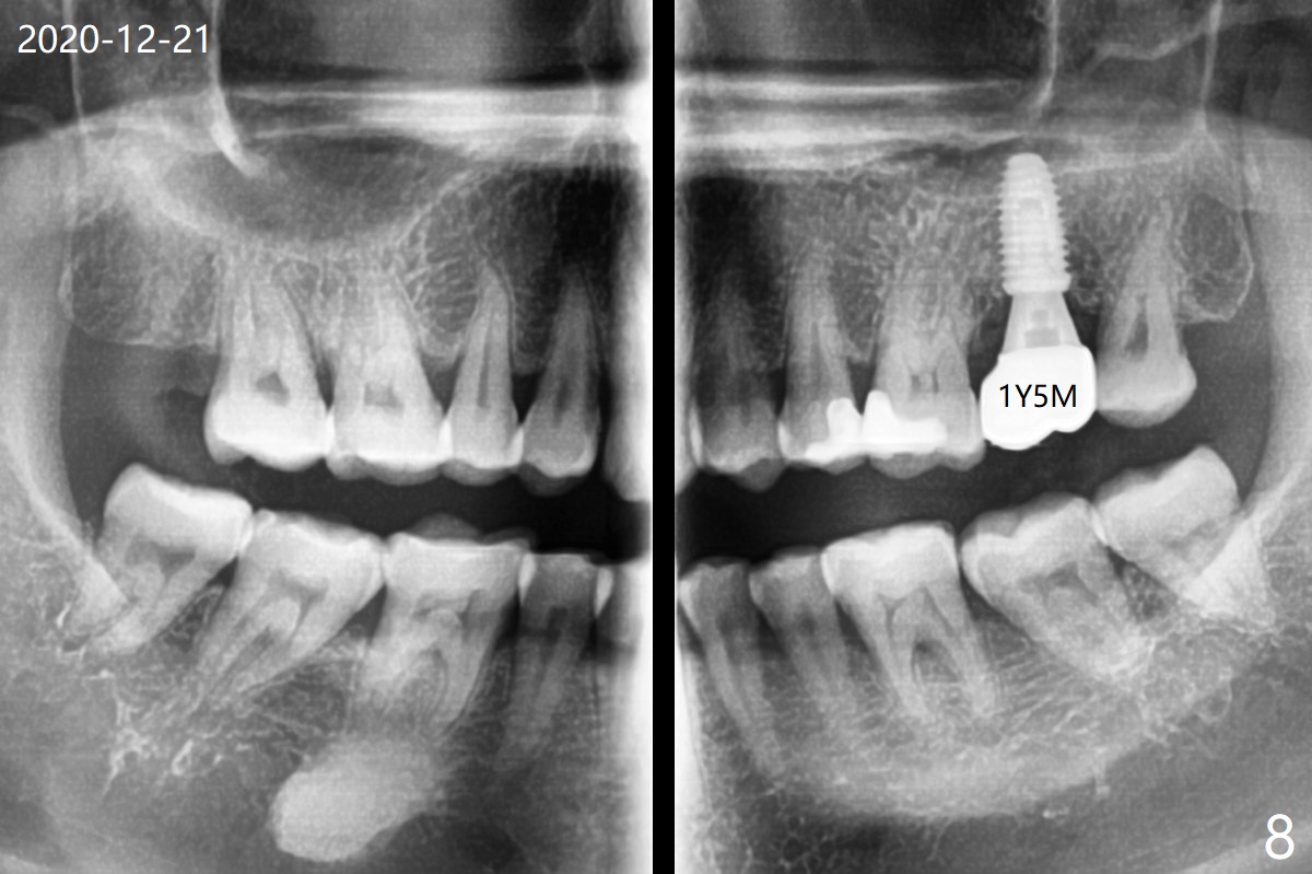
The longest (12 mm) bone trimmer seems not to work over the guide at #15. When osteotomy finishes, the guide is removed. The sinus floor happens to have been removed, whereas the sinus membrane remains intact. After partial insertion of a piece of PRF membrane (due to hemorrhage) and Vanilla graft for sinus lift, a 5x10 mm implant is placed with 15 Ncm. The implant appears to be short and placed deep (Fig.1,2). When an implant is not too large, it can be placed ~ 1 mm subcrestal (vs. 2-3 mm for this case) in spite of the palatal wall defect. A cover screw is used, followed by allograft (Fig.1 *). The latter is covered with another piece of PRF membrane, collagen membrane (Fig.3) and periodontal dressing. There is no nasal hemorrhage postop. PRF membrane and collagen membrane dislodge a few days postop, although the bone graft remain coronal to the implant. The healed socket appears to have been shrunken buccopalatally 3 months postop (Fig.4), probably due to the absence of an abutment and a provisional. The mechanics (abutment and provisional) seems to be more important than the chemical (membrane). Bone forms overlying the implant plateau 9 months postop (Fig.5), which is confirmed in implant uncover. The implant plateau remains to be covered by the bone 5 months post cementation (Fig.6,7 *). No threads are exposed 1 year 5 months post cementation (Fig.8).
Return to Upper Molar Immediate Implant, Prevent Molar Periimplantitis (Protocols, Table), Trajectory, Guided Surgery 1/2 Application
Xin Wei, DDS, PhD, MS 1st edition 09/28/2018, last revision 01/31/2021