.jpg)
|
|
|
|
|
|
|
|
|
|
|
|
||
|
|
|
|
|
||
The Larger Implant, the More Thread Exposure M
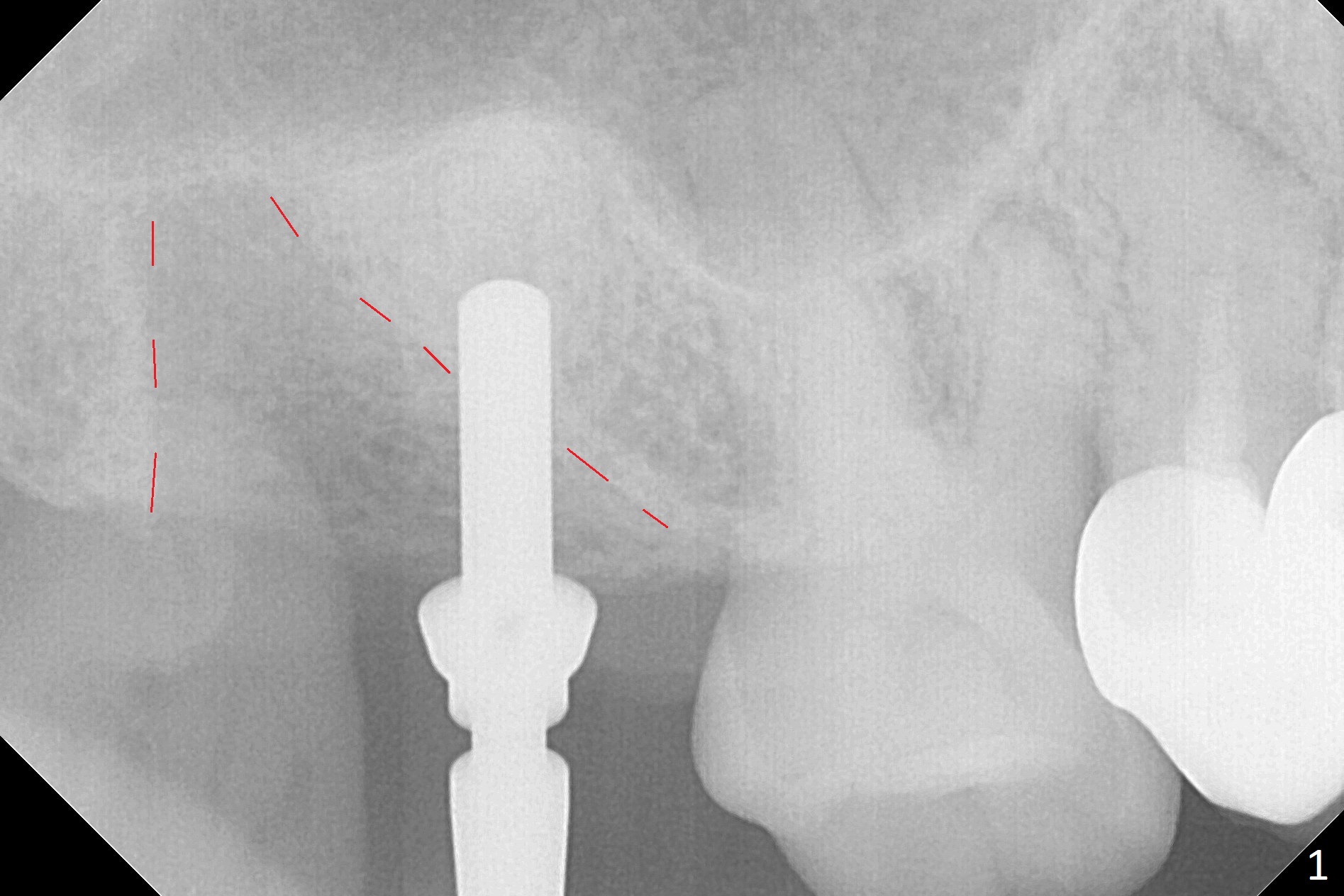
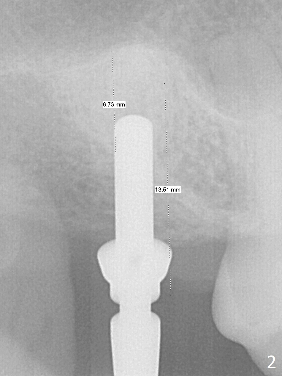
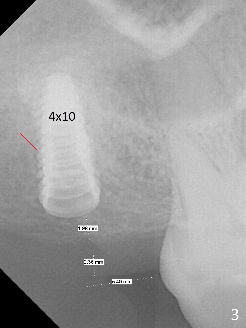
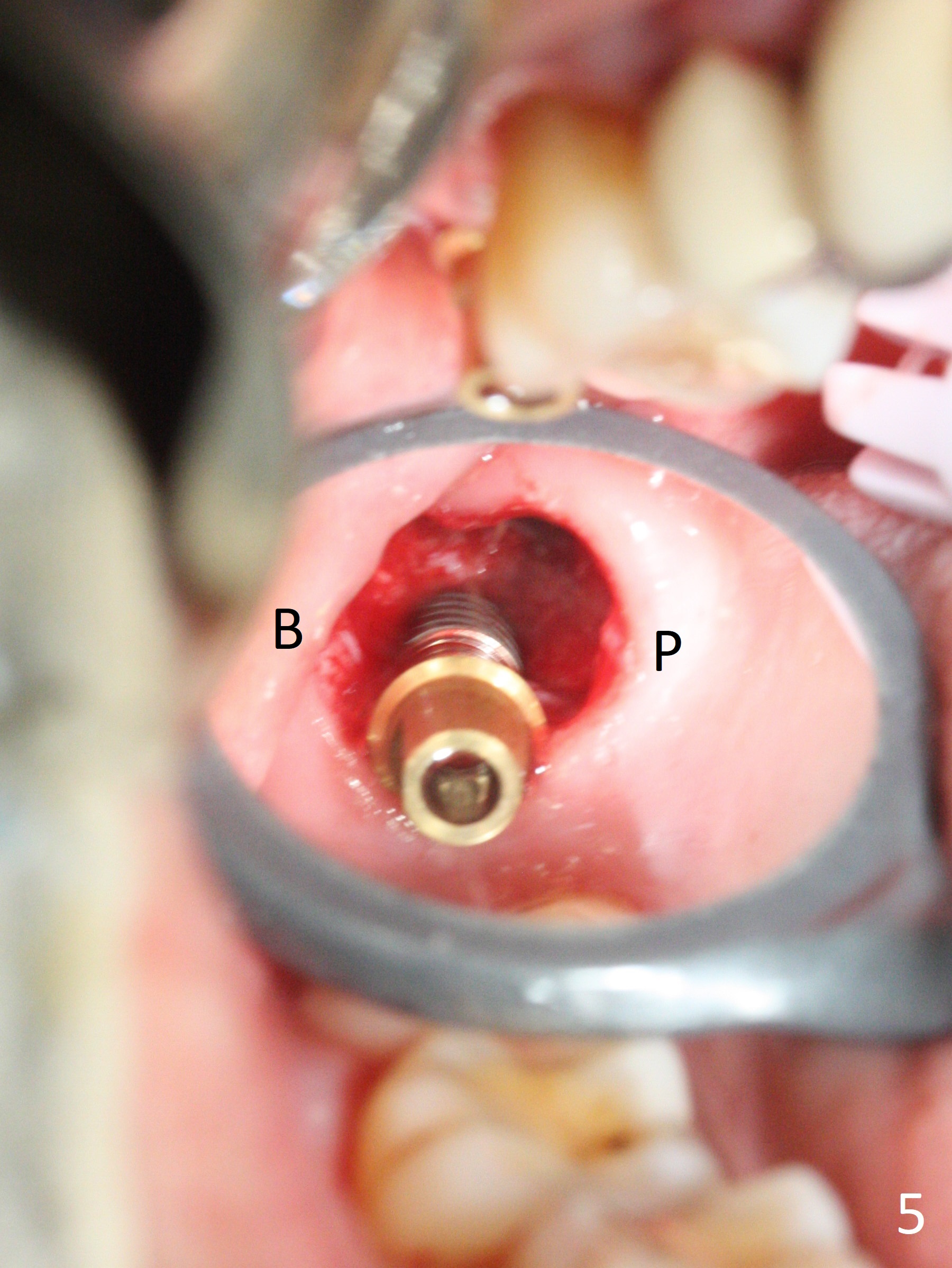
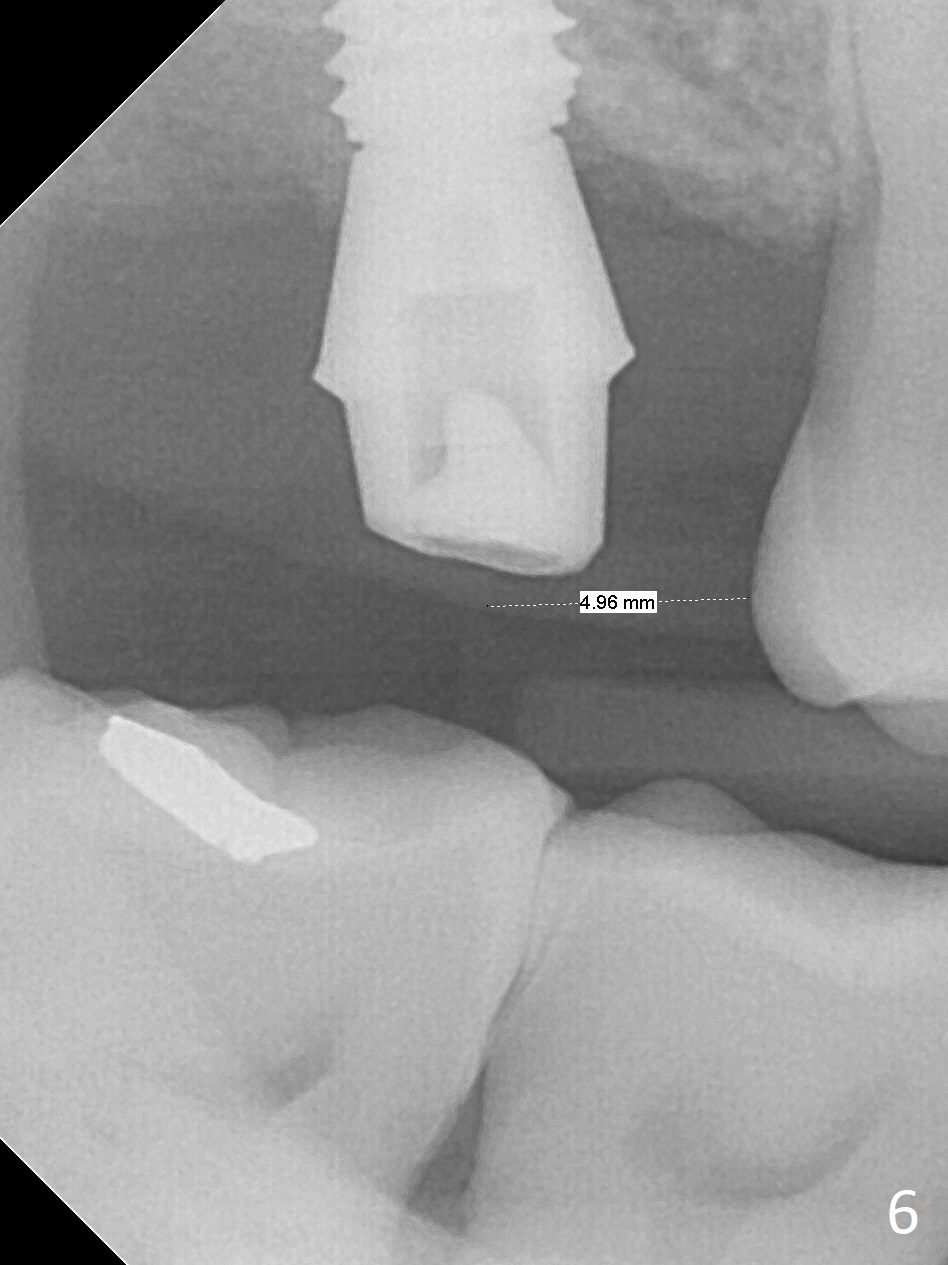
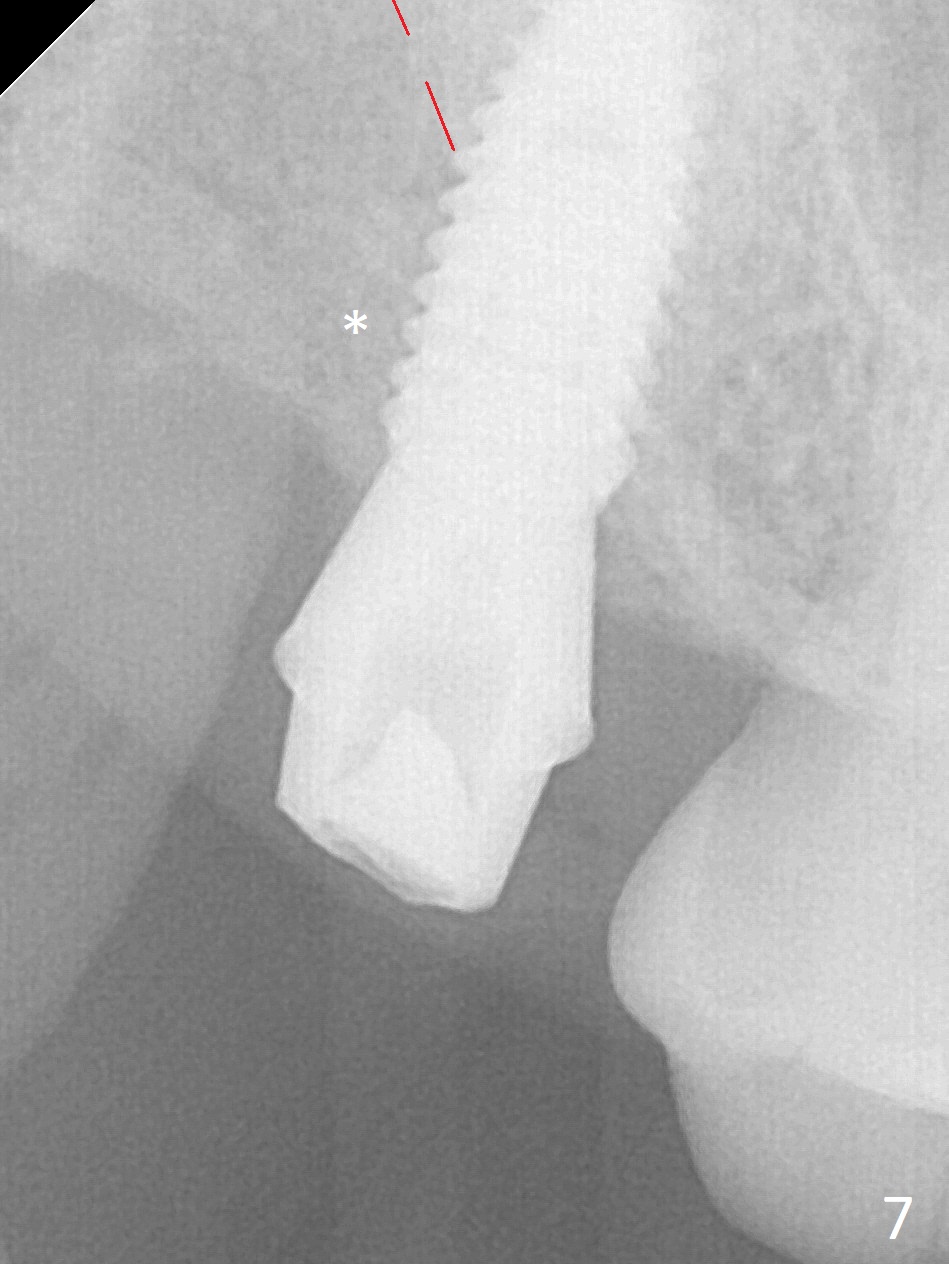
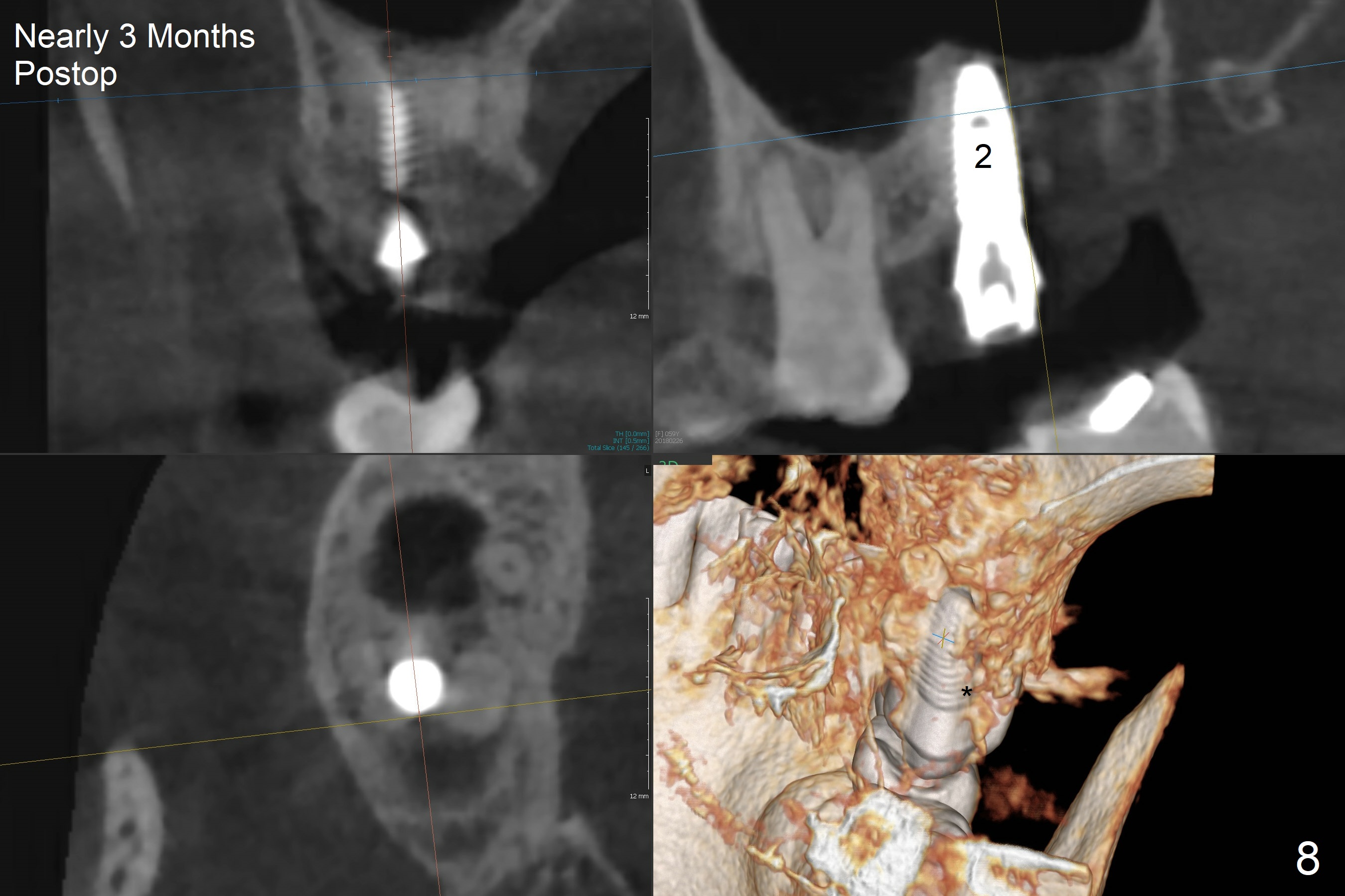
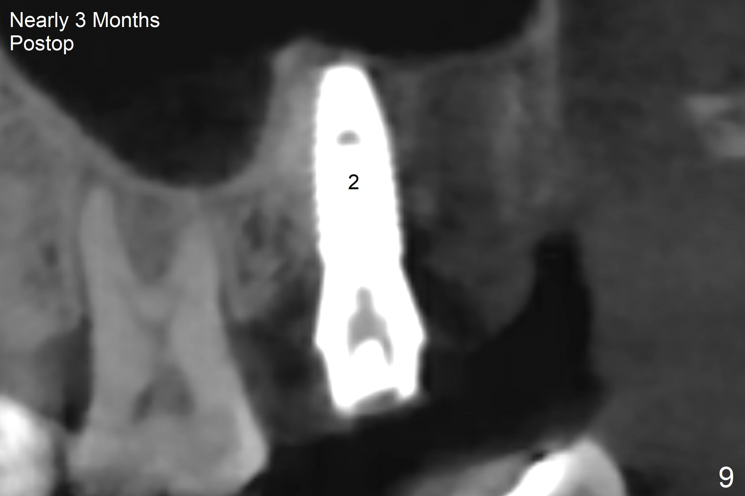
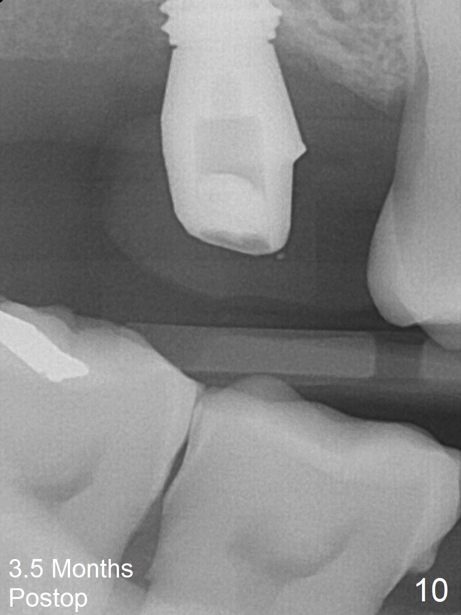
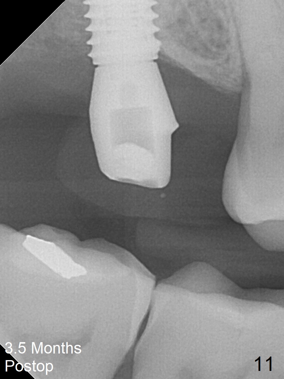
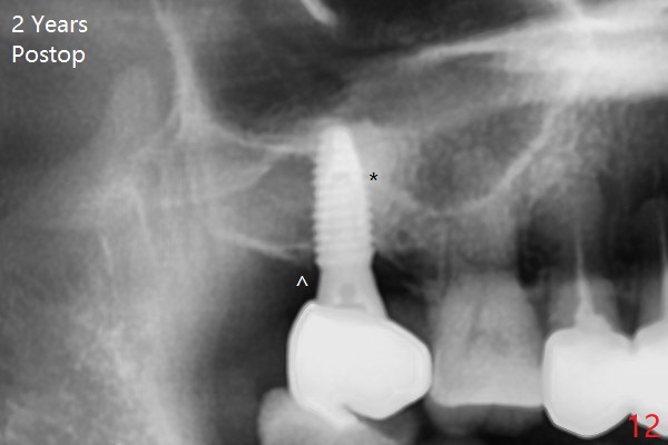
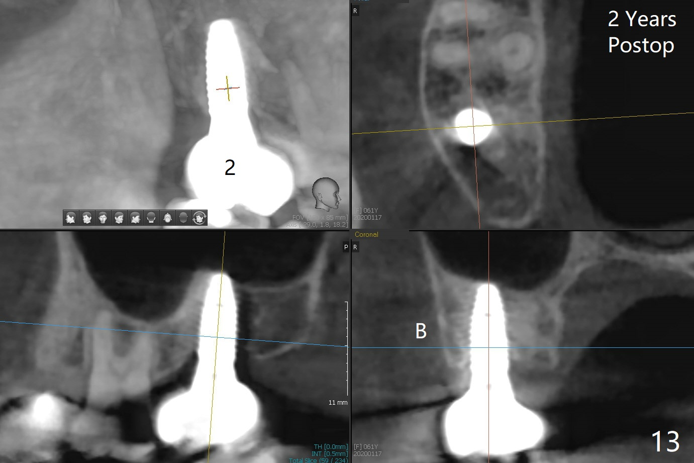
After extraction of the 3rd molar with mesial periodontal defect, osteotomy for 2nd molar implant is initiated in the mesial slope of the 3rd molar socket (Fig.1 red dashed line). When osteotomy is finished with IS drills and sinus lift with Magic Sinus Lifter (S-reamer with 11 mm stopper being short), a 4x10 mm dummy implant is placed with ~ 4 implant thread exposure (Fig.3). Following use of Lindamann bur to move osteotomy mesial and larger drill, a 4.5x10 mm implant is placed with 5-7 implant threads exposed distally (Fig.4,7 (~ 50 Ncm)). The bucco(B)-palatal(P) extent of the implant thread exposure is larger (Fig.5) than that associated with the 4 mm dummy implant (data not shown). The exposed implant surface is covered with Vera Graft (Fig.7*), Collagen plug and an immediate provisional after adjustment of abutment height (Fig.6,7). The bone density distal to the implant is low 3 months postop (Fig.8,9 CBCT) and 3.5 months postop (Fig.10,11). The permanent crown is cemented nearly 4 months postop. The distal cortical bone contacts the implant (Fig.12 ^), while the mesial bone increases in density (*) 2 years postop.
Return to
Upper
Molar Immediate Implant, Prevent
Molar Periimplantitis (Protocols,
Table),
Armaments,
#6
(final)
15
Xin Wei, DDS, PhD, MS 1st edition 12/01/2017, last revision 01/19/2020