|
|
|
|
|
|
|
|
|
|
|
|
Remove Implant to Prevent
Neighboring Implant from Periimplantitis
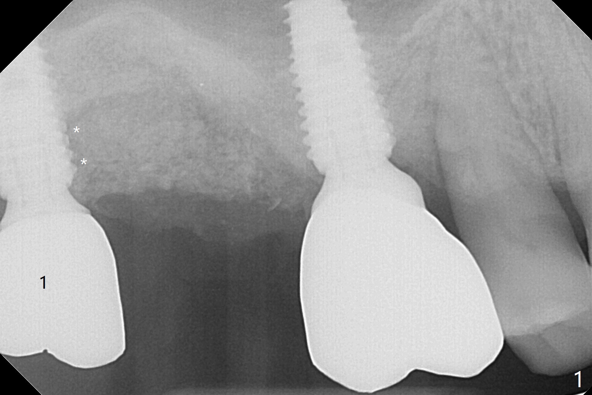
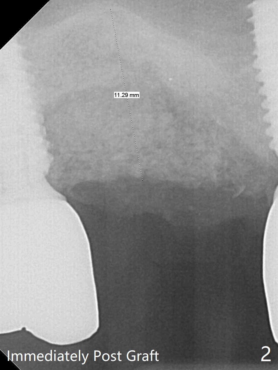
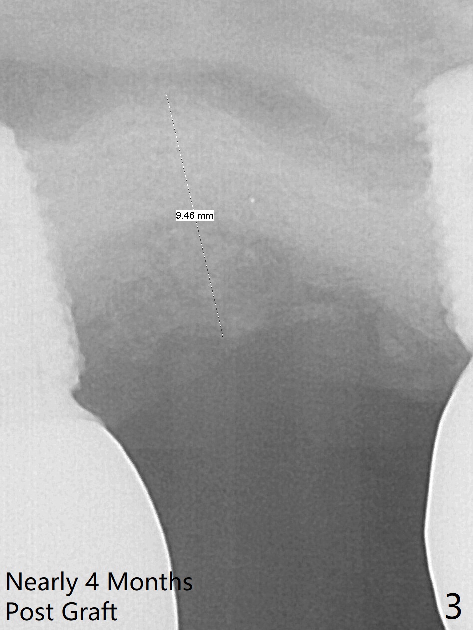
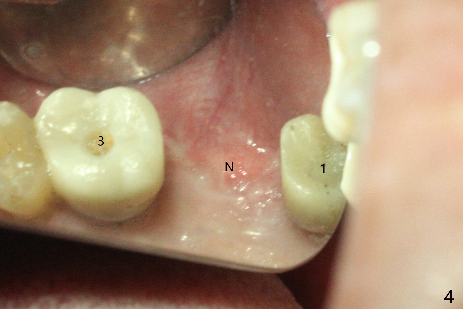
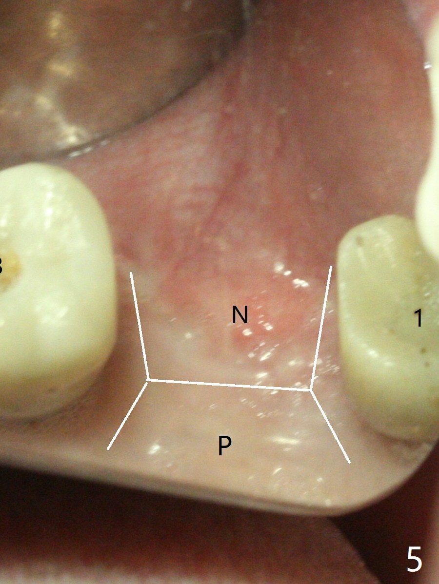
Removal of the implant at #2 is extremely difficult because of fusion of the abutment with the implant. Buccal and palatal incisions are made to remove the bone buccomesiodistal to the implant. After implant removal, the exposed mesial coronal implant threads at #1 are cleaned with Titanium brush; allograft is placed against the exposed threads (Fig.1 *), followed by 2 pieces of PRF membrane and 6-month collagen membrane. Periodontal dressing is applied after suturing. Partially due to traumatic implant removal and partially due to easy and repeated loss of periodontal dressing, bone height reduces nearly 4 months postop (compare Fig.2,3). More discouraging is the invasion of loose nonkeratinized buccal gingiva into the healed socket nearly 4 months post graft (Fig.4 N). Tissue punch thorough a guide should expose the implant site to the nonkeratinized tissue. Can we fabricate a guide with buccal clearance underneath so that a flap can be raised buccally, i.e., to push the palatal keratinized tissue buccally?
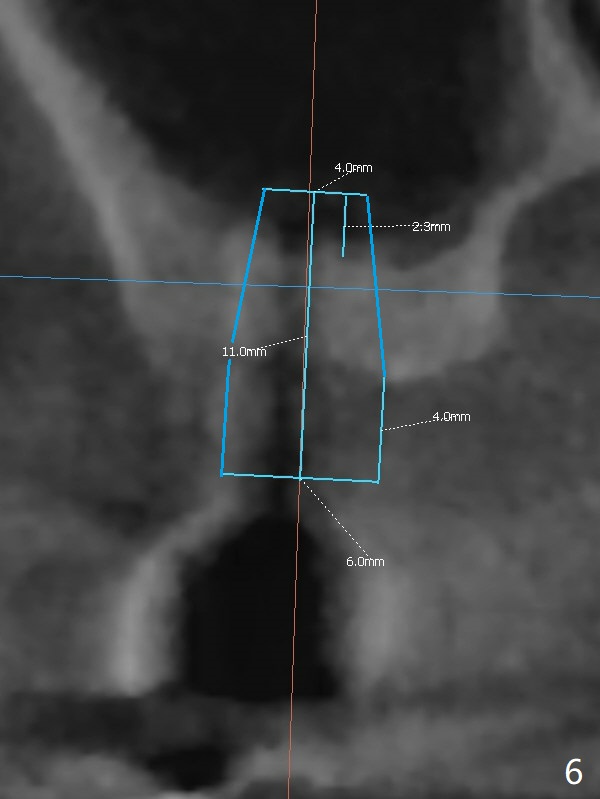
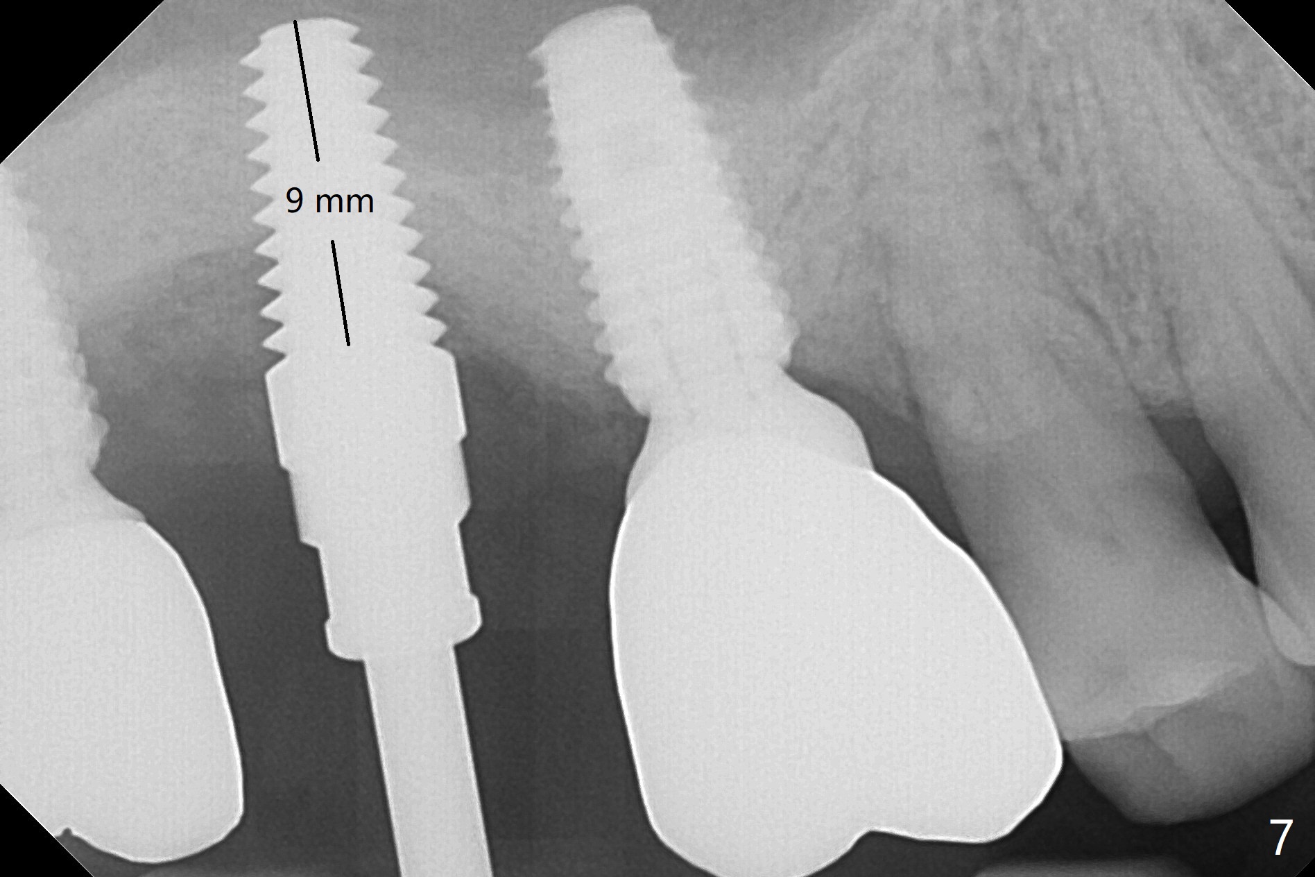
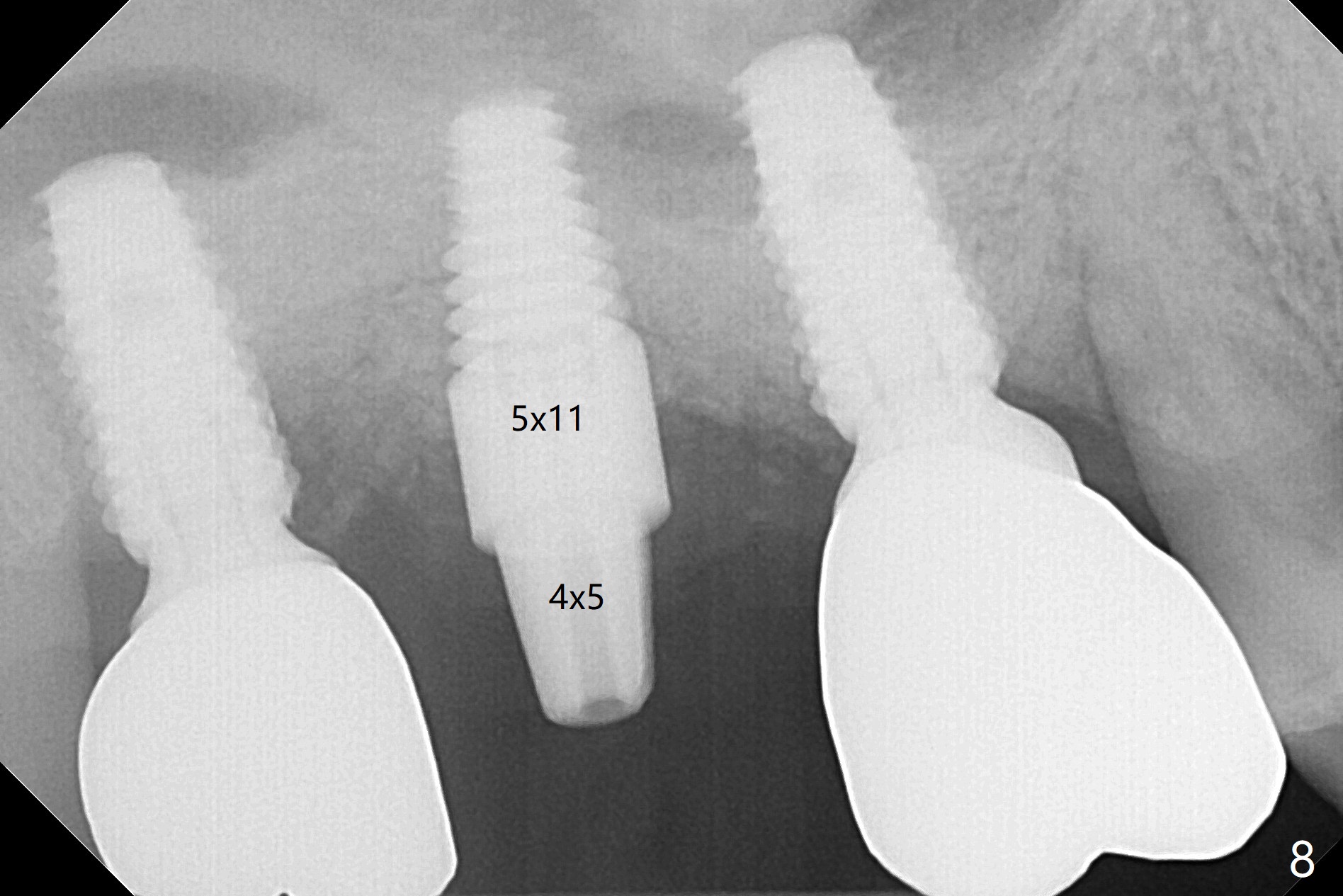
Hi Dr. WeiDue to time constraint (he may move out of state), implant placement will be free hand with palatal incision (Fig.5 P). Surgical stent will be ready. Since the patient is a heavy bruxer, a 5 or 6 x14 mm tissue-level implant will be placed using DIO Sinus Lift Master Kit and Bone Expander Kit (Fig.6). Prepare water pump syringe. The first 6 mm osteotomy with regular drills (1.2 and 2.0 mm) is easy and soft, whereas the last 1-2 mm is hard. In the end, the sinus membrane is perforated. After use of 3.5 mm reamer for 8 mm, a 5x14 mm tap is inserted for ~ 8 mm (Fig.7). A 5x11 mm implant (9 mm in bone) is placed ~ 35 Ncm; following placement of bone graft around the junction of thread and the unpolished portions of the implant, a 4x5 mm abutment is placed for retention of periodontal dressing (Fig.8). Since #2 implant removal, #3 implant crown/abutment has rotated twice (screw not loose). On the 2nd occasion (2 months post #2 implant placement), a provisional is fabricated to stabilize #3 crown. During #2 temporization, the crown of #1 dislodges and recements. Return to Upper Molar Immediate Implant, Trajectory Similar Case Xin Wei, DDS, PhD, MS 1st edition 01/08/2019, last revision 08/18/2019