|
|
|
|
|
|
|
|
|
|
|
Sinus Mucocele
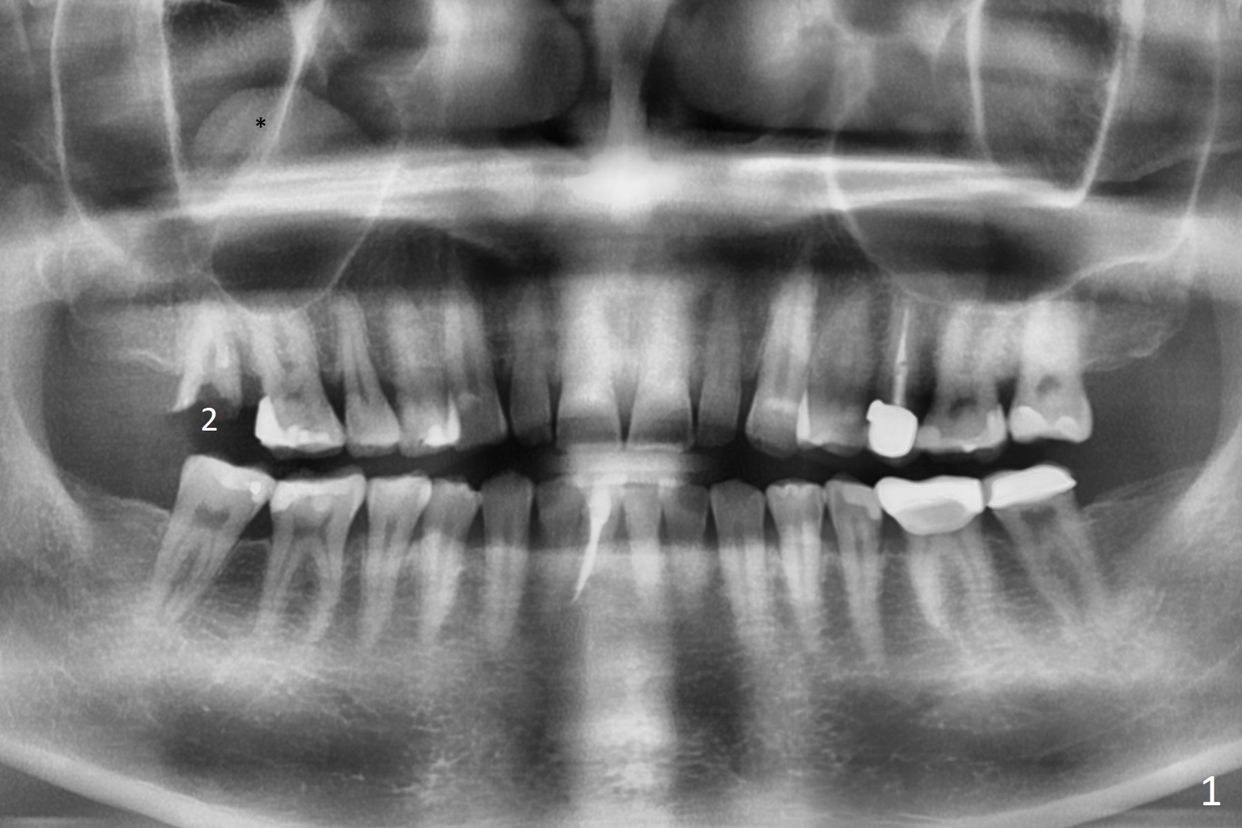
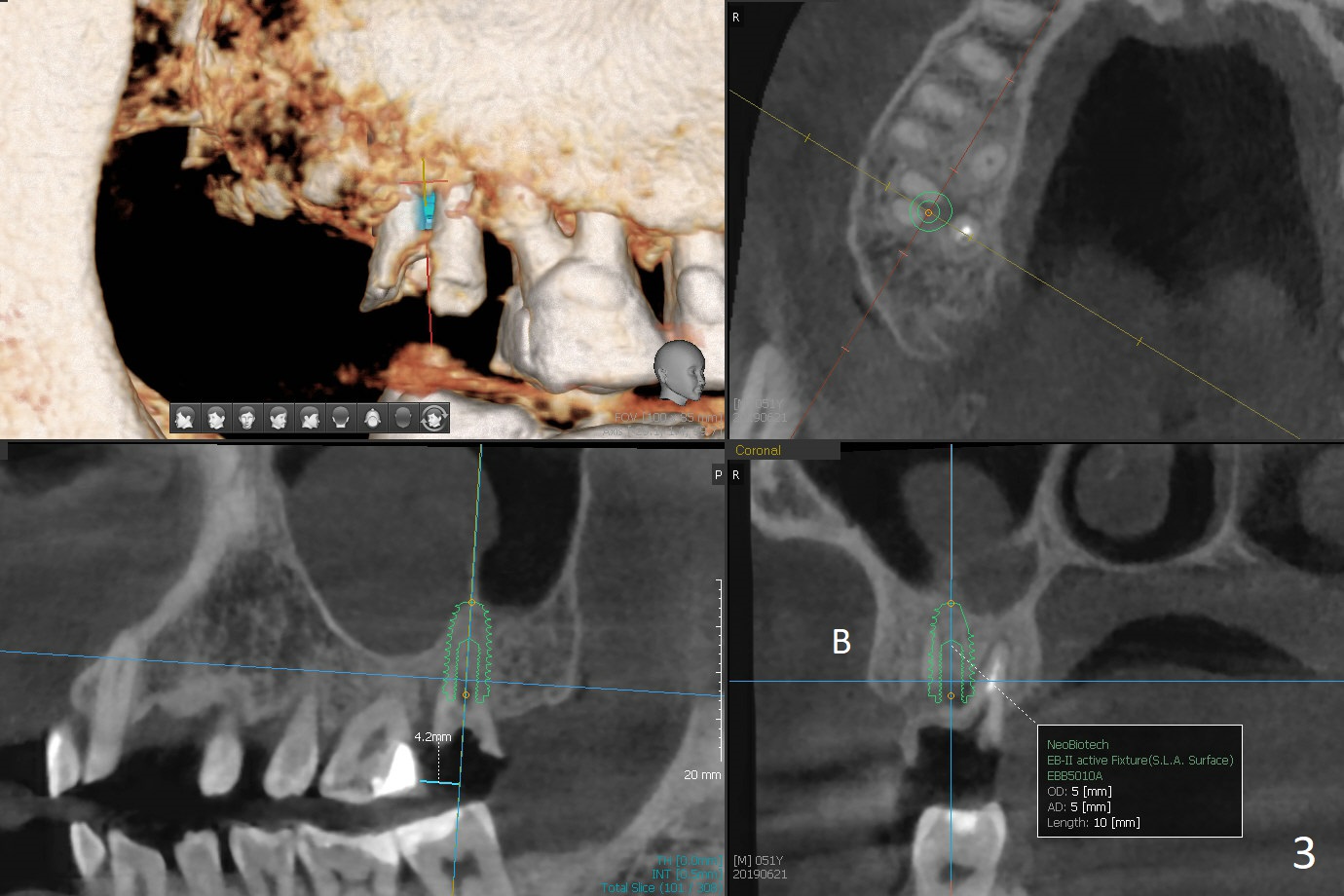
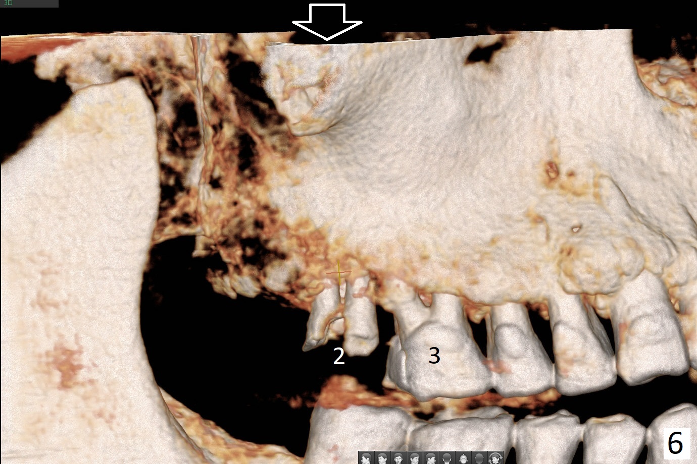
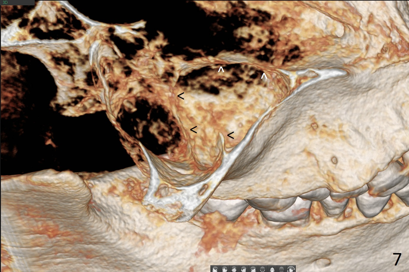
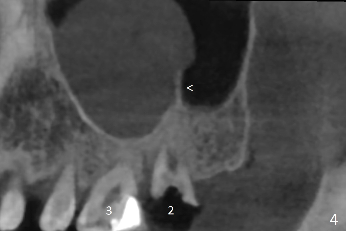
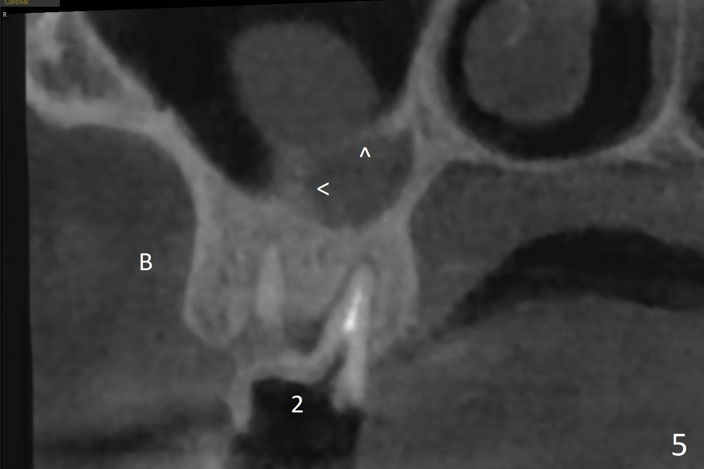
A 51-year-old man agrees to have #2 residual root to be extracted for implant. Panoramic X-ray (Fig.1) and CT coronal section (Fig.2) show maxillary sinus mucocele (*). A 5x10 mm implant will be placed not to intrude into the sinus (Fig.2,3). In case sinus membrane perforation, prepare PRF membranes for repair. The apex of the implant will be engaged to an apparent sinus septum for stability (Fig.4,5,7 arrowheads). Fig.4,5,6 are sagittal and coronal sections and 3-D image of Fig.3 without an implant at #2, while Fig.7 is the inferior view of Fig.6 (arrow).
Return to
Upper
Molar Immediate Implant,
Prevent Molar Periimplantitis (Protocols,
Table),
Trajectory,
Clindamycin
第二磨牙即种
IBS
Xin Wei, DDS, PhD, MS 1st edition
06/21/2019, last revision
05/23/2021