%20to%205.8x4(1)%20with%20plastic%20sleeve_1y7m%20postop.jpg)
|
|
|
|
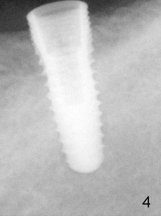
A 4.5x12 mm implant is placed as planned (Fig.4). Dashed line: the upper border of the inferior alveolar canal. Then autogenous bone (harvested from reamers) is placed around the most coronal portion of the implant (with microthreads).
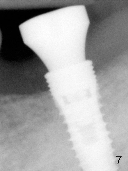
The 1st 4 coronal threads remains outside the bone 3.5 months postop (Fig.7). It appears that less thread exposure (3 instead) 1 year 7 months postop (Fig.8), suggesting bone growth.
Return to Mission Accomplished
Xin Wei, DDS, PhD, MS 1st edition 08/10/2014, last revision 01/19/2018