|
|
|
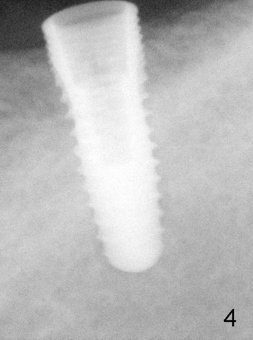
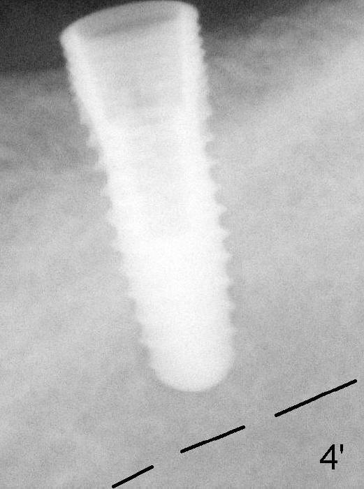
A 4.5x12 mm implant is placed as planned (Fig.4). Dashed line: the upper border of the inferior alveolar canal. Then autogenous bone (harvested from reamers) is placed around the most coronal portion of the implant (with microthreads).
Return to Mission Accomplished
Xin Wei, DDS, PhD, MS 1st edition 08/10/2014, last revision 01/19/2018