|
|
|
|
|
|
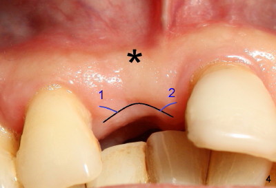
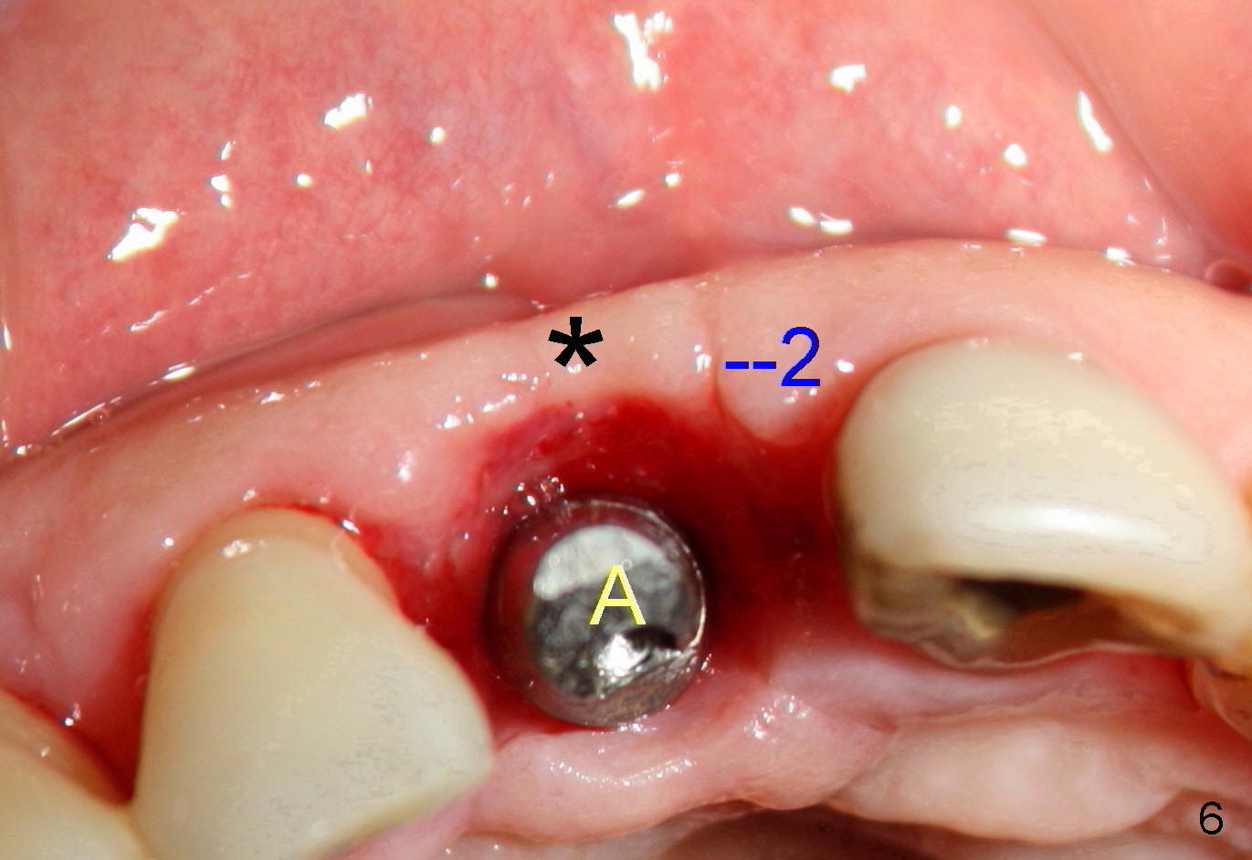
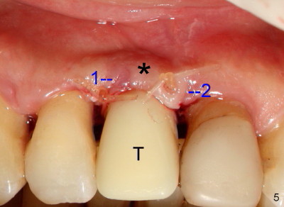
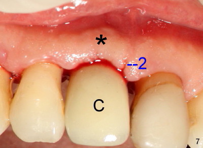
Fig.4-7 show modified semilunar incision helps form cosmetically-pleasing attached gingiva. Prior to uncover, the buccal plate of #8 (*) is concave, as compared to those of the neighboring teeth. The main incision is indicated by black line, lingual to the crest so that the buccal flap is more bulky. It is curved and festooned to create the future cosmetic buccal gingival margin. If the main incision is placed too lingual to expose the implant, one or two accessory incisions (red) can be made. After insertion of 4x6.5 mm non-shouldered abutment 3 mm post, PVS impression is taken. Fig.5 shows temporary crown in place (T) with two vertical mattress suture in the mesial and distal papillae. Two accessory incisions are approximated. The attached gingiva over the temporary crown immediately becomes bulky (*). Two weeks later, the temporary crown is removed. The accessory incision #1 has healed, while #2 is healing (Fig.6). The temporary crown helps form a nice socket around the abutment (A). The buccal attached gingiva (*) is leveling with that of the neighboring teeth, as compared to concavity before uncover (Fig.4). Fig.7 shows the gingiva around the crown (to be cemented) with healing incision #2. The buccal attached gingiva(*) is as bulky as immediately after uncover (Fig.5). Return to original article
Xin Wei, DDS, PhD, MS 1st edition 05/1/2011, last revision 05/15/2011