|
|
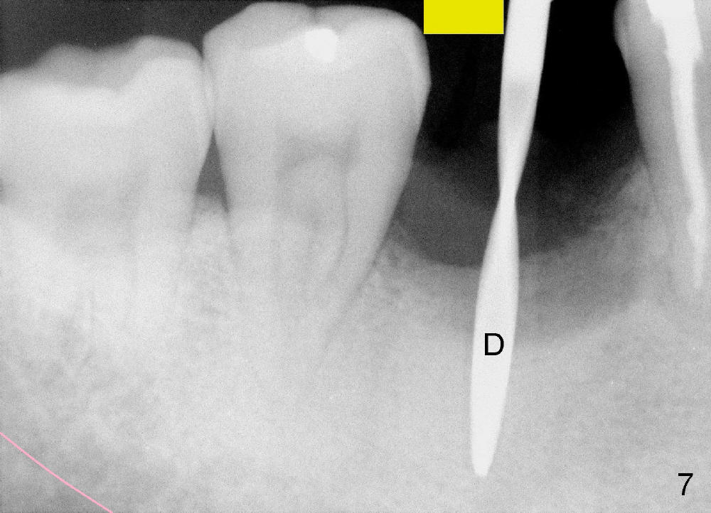
Fig.7: When the pilot drill is inserted into the initial osteotomy site for X-ray confirmation of its depth (D), the vertical block of the endo device (yellow rectangle) may be blocked due to the narrow divided edentulous space. The crowns of the molars are shown, whereas the inferior alveolar nerve is not shown except a small portion in the left lower corner of Fig.7 (pink line). Return to original article
Xin Wei, DDS, PhD, MS 1st edition 12/09/2012, last revision 12/09/2012