|
|
|
|
|
|
|
|
|
|
|
|
|
|
|
|
|
|
|
|
|
|
|
|
|
|
|
|||
Immediate Provisional
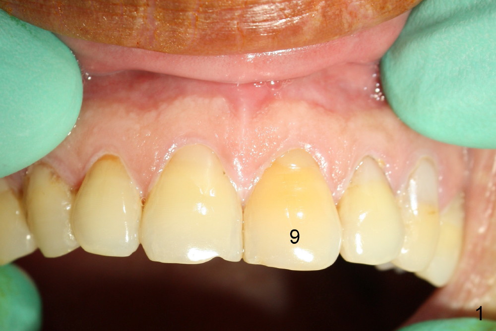
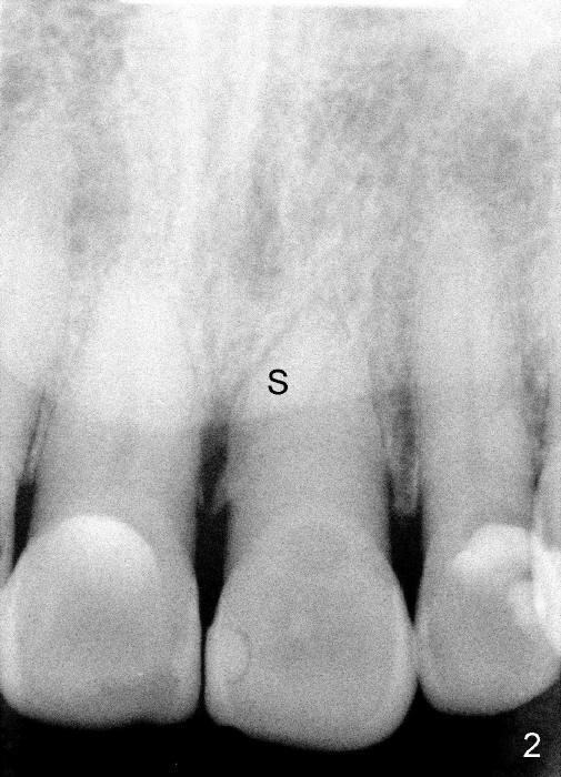
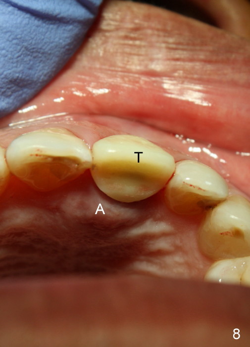
A 65-year-old man had injury to the tooth #9 20 years ago. The tooth has been discolored since (Fig.1). The tooth remained asymptomatic until last year. It has been loose since then. X-ray shows short root (Fig.2 S), sclerosed canal and bone loss mesially. There is a lingual abscess (Fig.8 A) with deep pockets.
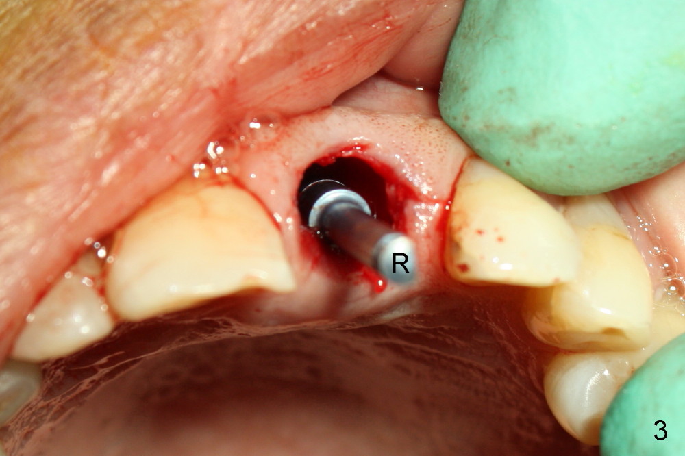
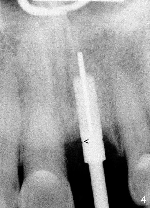
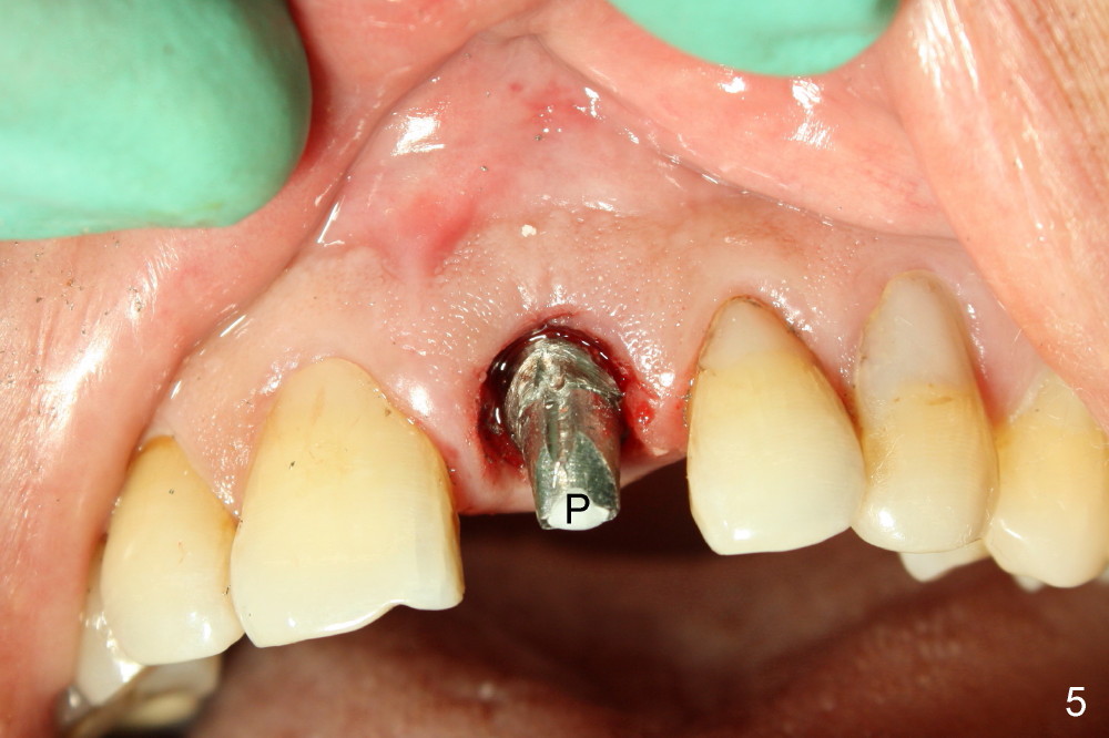
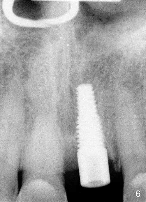
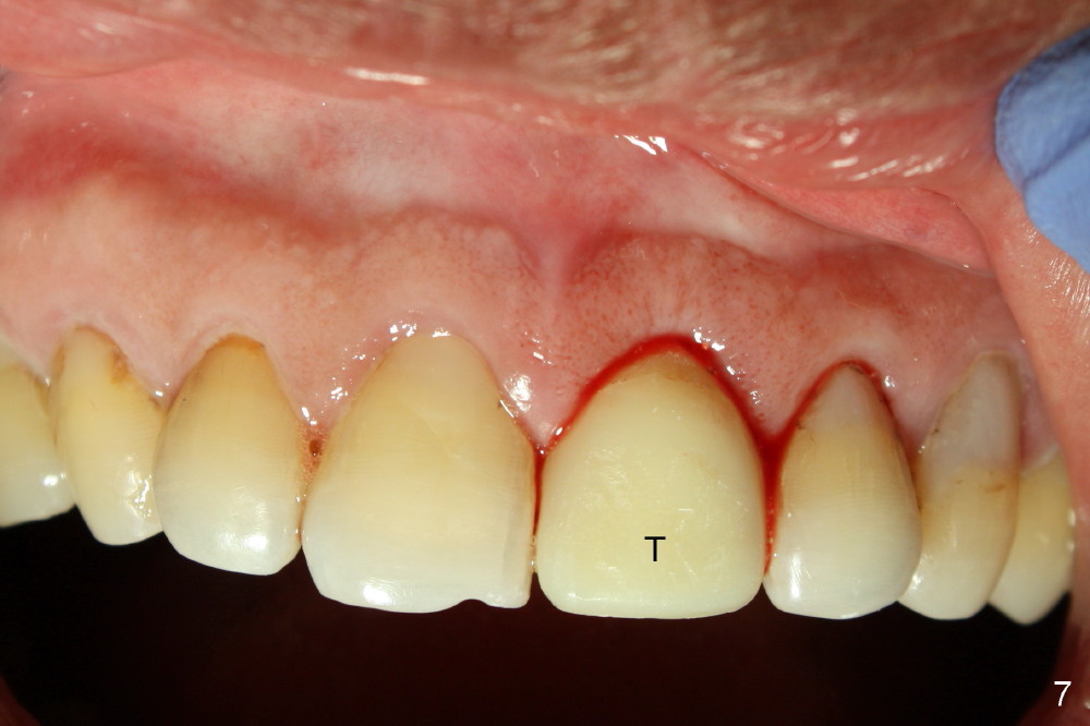
After extraction, osteotomy is made as palatally as possible so that the end of a reamer/drill (Fig.3 R) in the line of the incisal edge of the neighboring teeth. The depth of the 3.5 mm reamer is 14 mm from the crest (Fig.4 <). Next, a 5x17 mm tapered implant is placed with insertion torque > 60 Ncm (Fig.6). A 4 mm 0° unipost is permanently cemented (Fig.5: P). Finally a provisional is cemented on a temporary basis (Fig.7,8: T). There is no contact in centric or any lateral excursions.
In case the implant needs to be removed, how can it be done considering permanent cementation of the unipost? Use of a post guide as a temporary abutment appears to have advantage of retrievability. Are there any precaution about this method? I may try it next time.
The root dimension of the extracted tooth is 6x7 mm. Is it necessary to place a 6 mm implant to close the socket opening more tightly?
Dr Wei, Nicely done ! If not integrated it could be removed by unscrewing with pedo forceps. If integrated you will need to use trephine after shortening post. Does not appear to be a concern. Dr Borgner Tuesday, June 25, 2013 5:29 AM
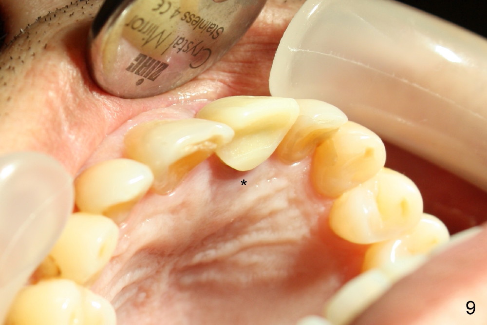
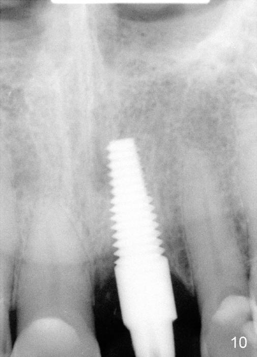
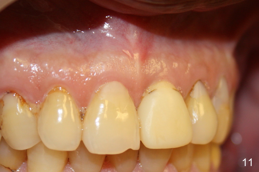
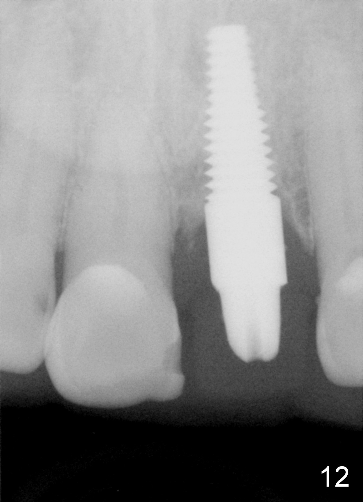
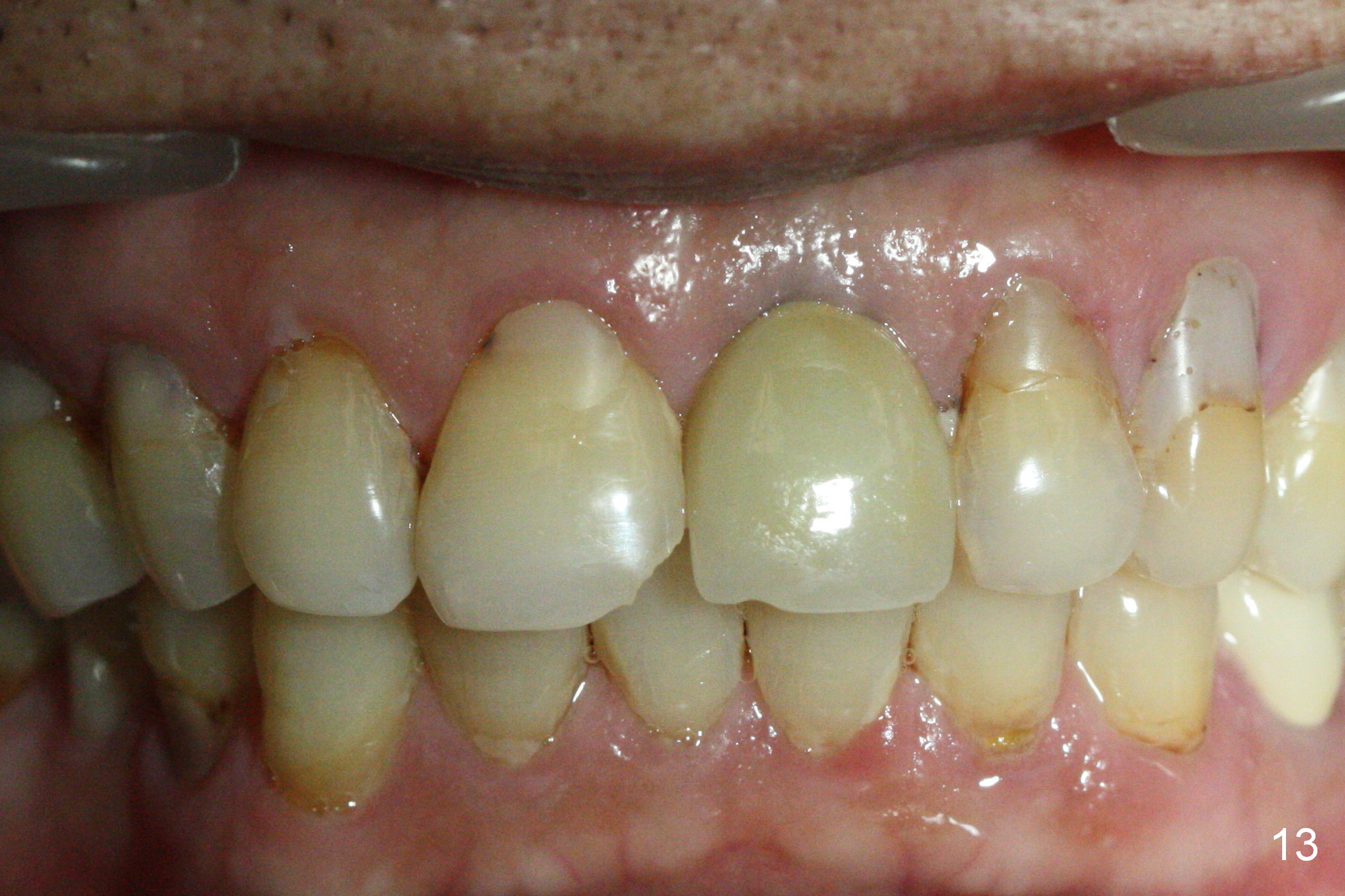
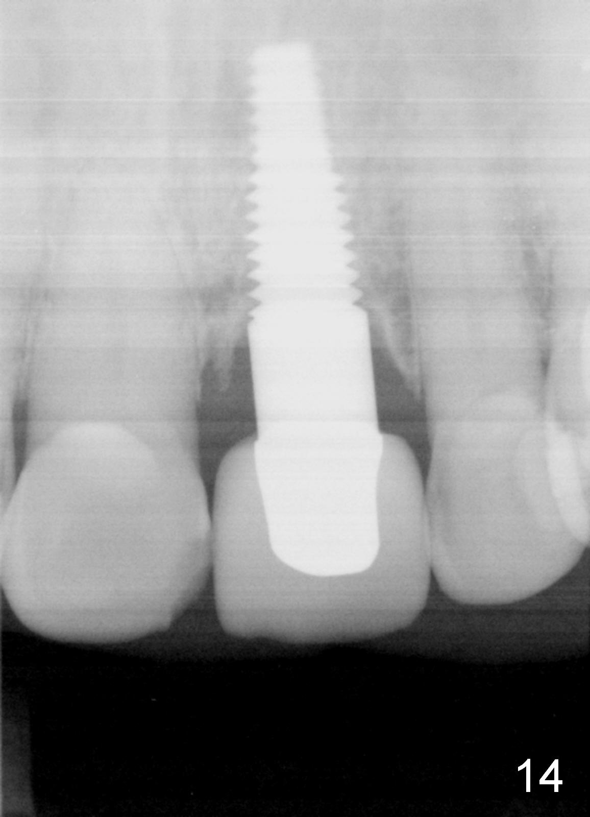
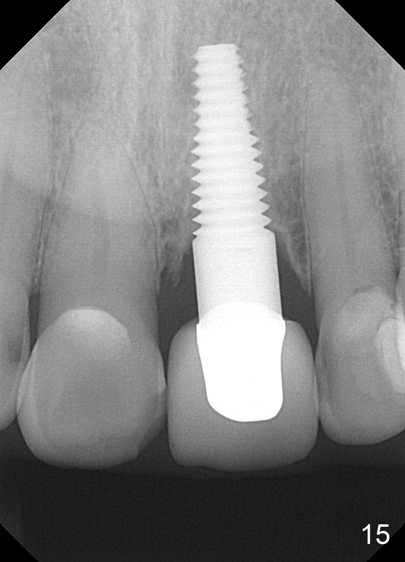
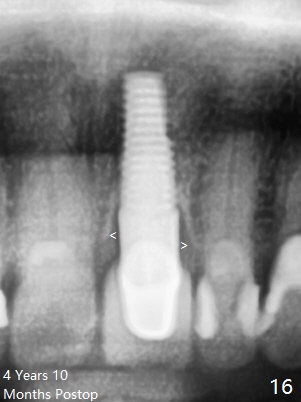
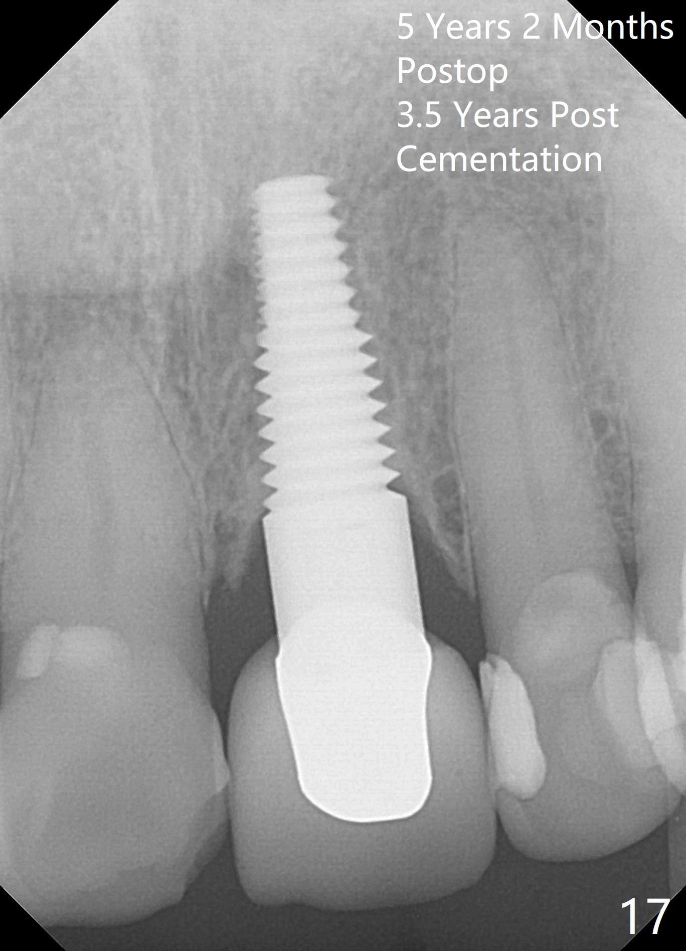
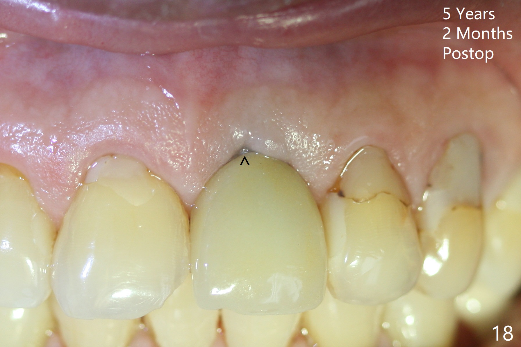
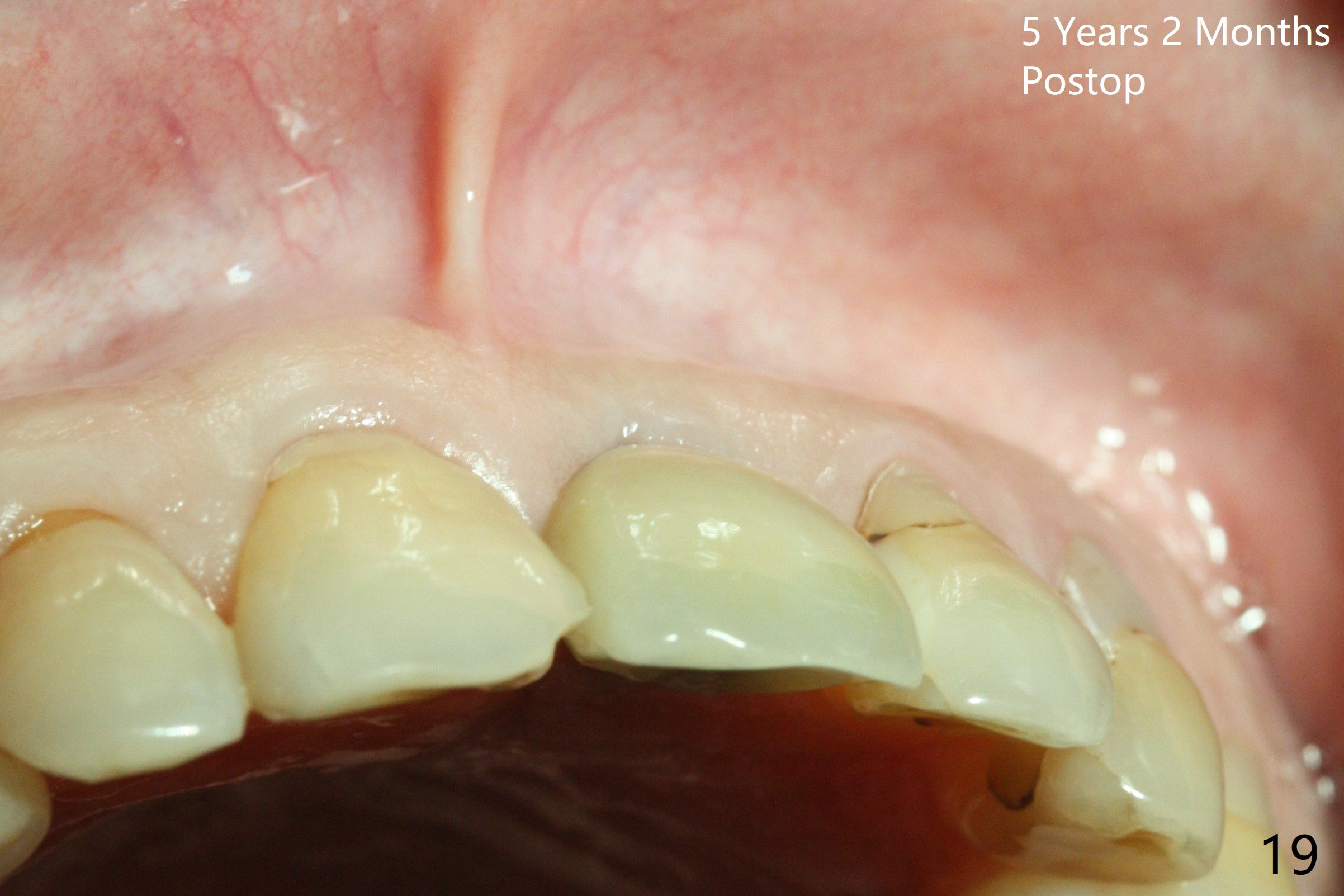
Postop tenderness lasts for two days. The palatal swelling is unnoticeable 7 days postop (Fig.9 *, as compared to A in Fig.8). The bone remains stable around the implant (Fig.10), while the gingiva healthy around the immediate provisional (Fig.11) 5 months postop. Due to insurance limit, the permanent restoration is delayed (16 months postop, Fig.12). The patient is satisfied with the function and appearance 3 years 8 months postop (21 months post cementation, Fig.13,14). PA is taken 4 years 1 month postop (2.5 years post cementation, Fig.15). The lamina dura forms from the most coronal threads (Fig.16). Although there is no bone loss around the implant (Fig.17), metal starts to show 5 years 2 months postop (Fig.18 ^), probably related to the buccal placement, too large the implant for the site or buccal plate atrophy (Fig.19).
Upper Incisor Immediate Implant
Xin Wei, DDS, PhD, MS 1st edition 06/24/2013, last revision 08/29/2018