,%20bone%20graft.jpg)
|
|
|
|
|
|
|
|
|
|
|
|
|
|
|
|
||
|
|
|
|
||
|
|
|
|||
The Labial Margin Should be More Apical
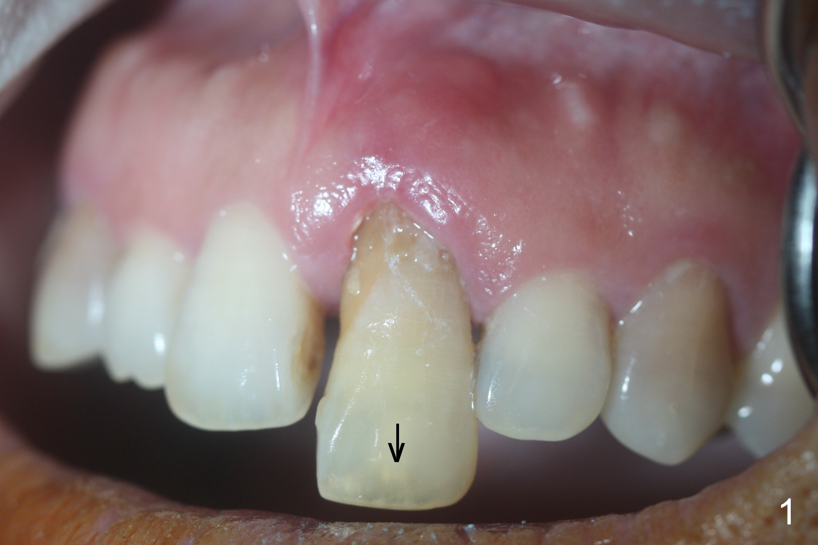
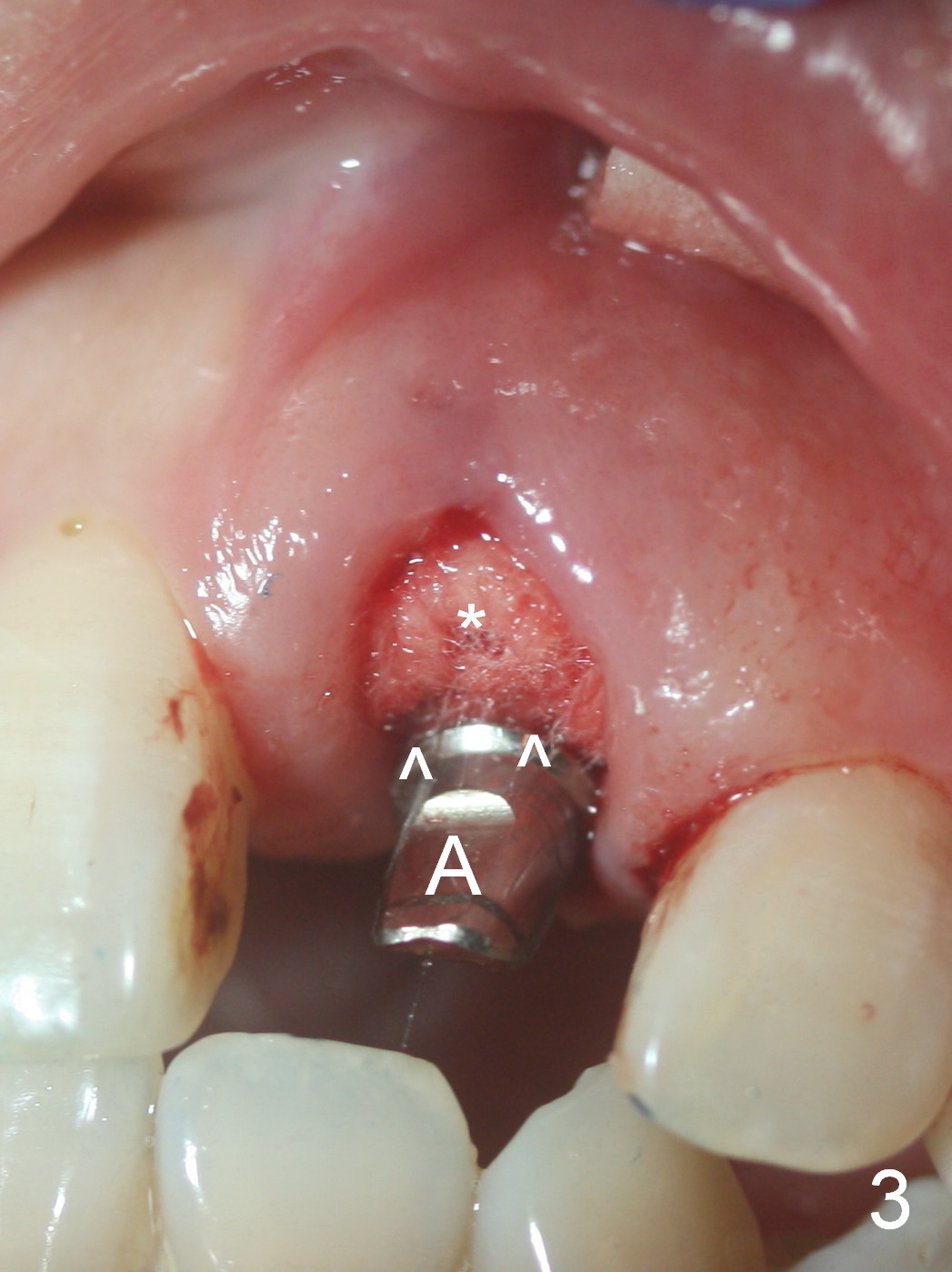
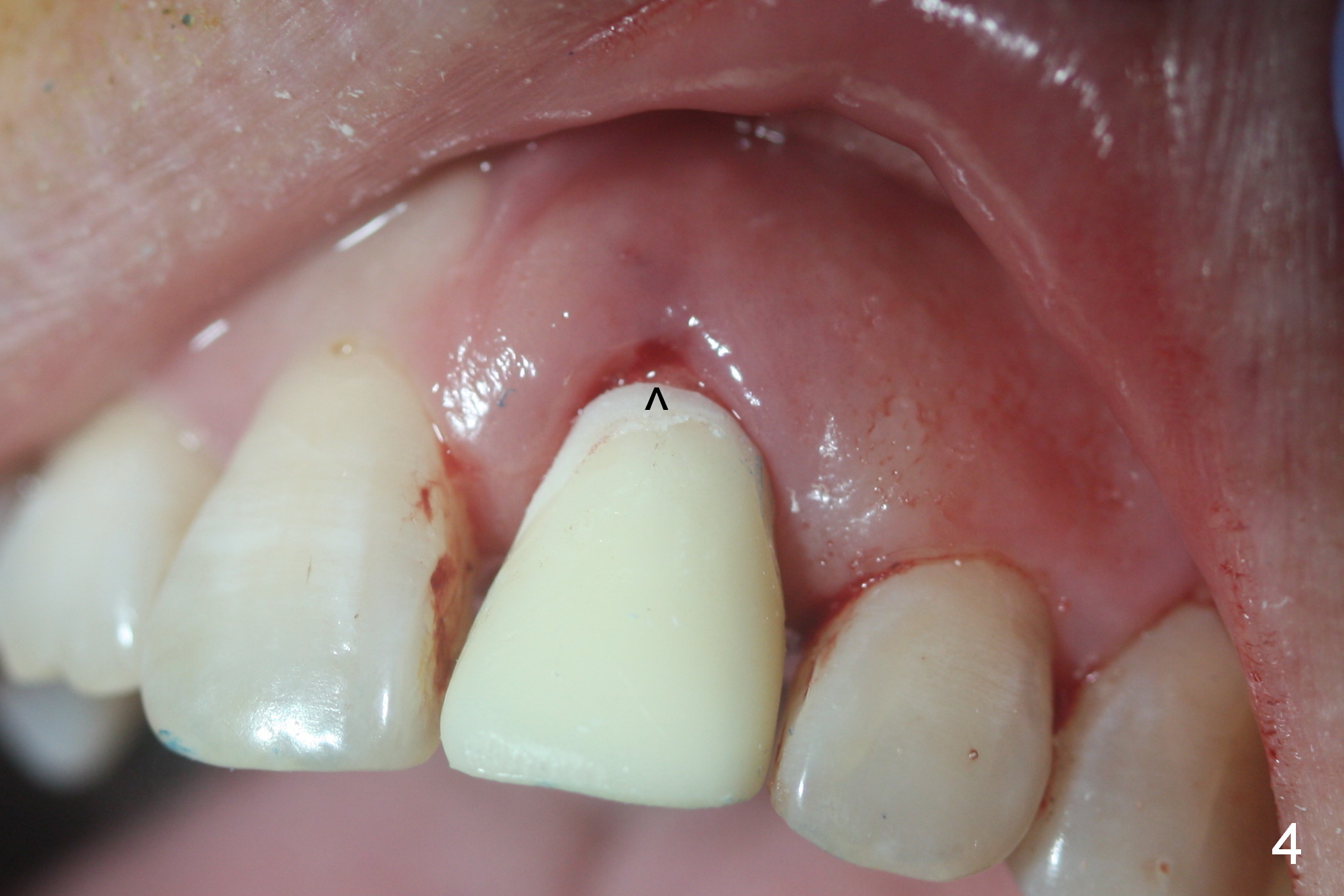
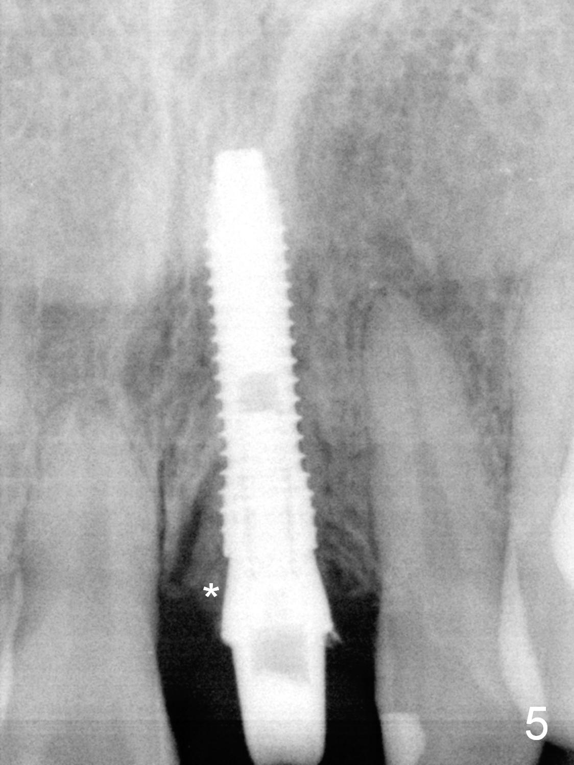
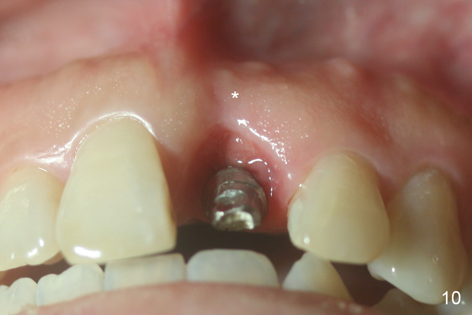
The tooth #9 is severely extruded prior to extraction (Fig.1 arrow). As expected, the labial plate is absent with granulation tissue. After 2 and 3 mm drills, a 3.8x16 mm implant is placed, followed by collagen membrane/allograft (Fig.2 < (buccally)) and a cemented abutment (A: 4.5x5(3) mm). In fact, the implant is placed too palatal so that there is heavy palatal reduction in the abutment (Fig.3 A). With a piece of gauze in the buccal gap to cover the bone graft (Fig.3 *), an immediate provisional is fabricated; attention is paid to making sure that the labial margin (Fig.4 ^) is more coronal than the counterpart so that there will be no pressure on the gingival margin. The gap between the gingival and provisional margins is to be closed by collagen plug. Prior to temporary cementation of the provisional, the gauze is removed and more bone graft is placed (Fig.5 *).
For the best cosmetic result, the labial margin of the abutment (Fig.3 ^) should have been prepared more apically. Because of the palatal placement of the implant, the lingual cervical portion of the provisional is bulky. Probably an angled abutment should be placed once the wound heals. The 2nd drawback of this procedure is failure to use the crown shell as a provisional.
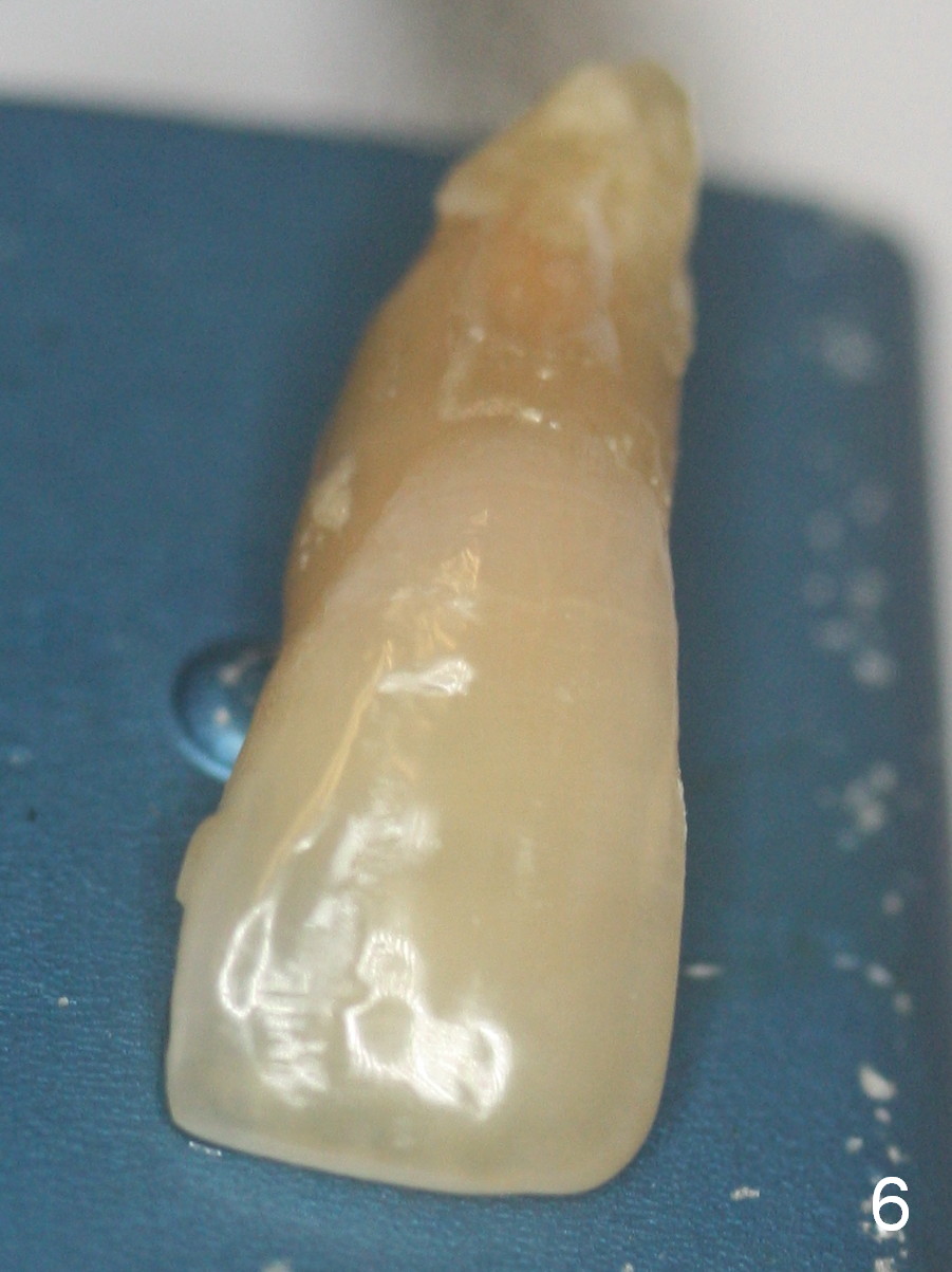
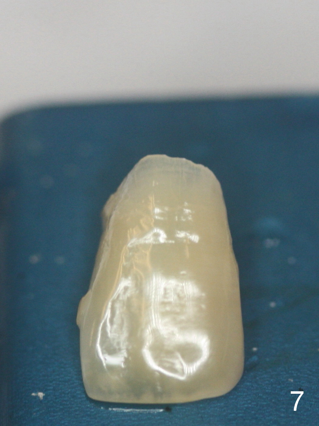
Nine days postop, the patient returns for follow up. She is pleased with the result except the shade of the provisional. The extracted tooth is removed from a jar with diluted bleaching solution (Fig.6). After removing the root and making crown shell, the tooth looks brighter (Fig.7). The tooth is placed in the diluted bleaching solution. It will be used as a provisional after choosing a right shade for acrylic.
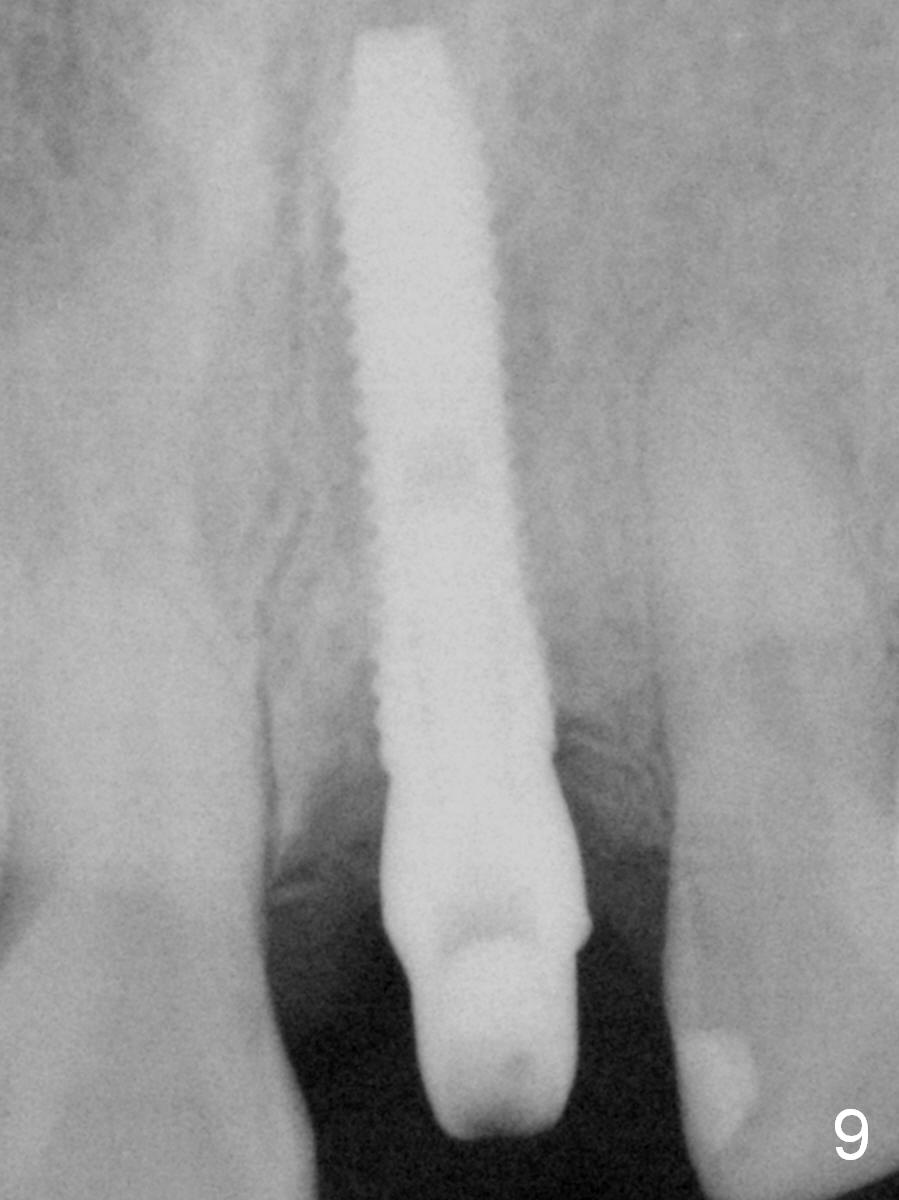
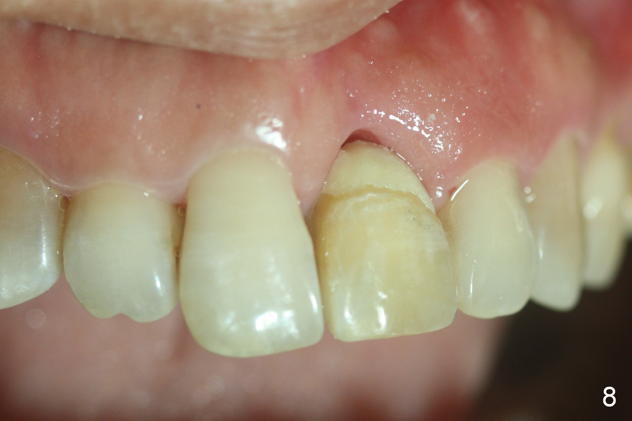
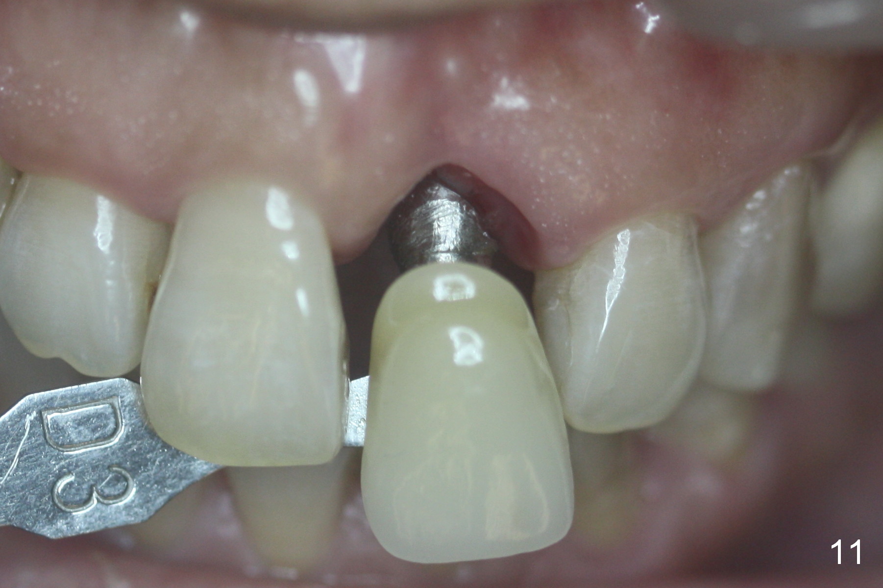
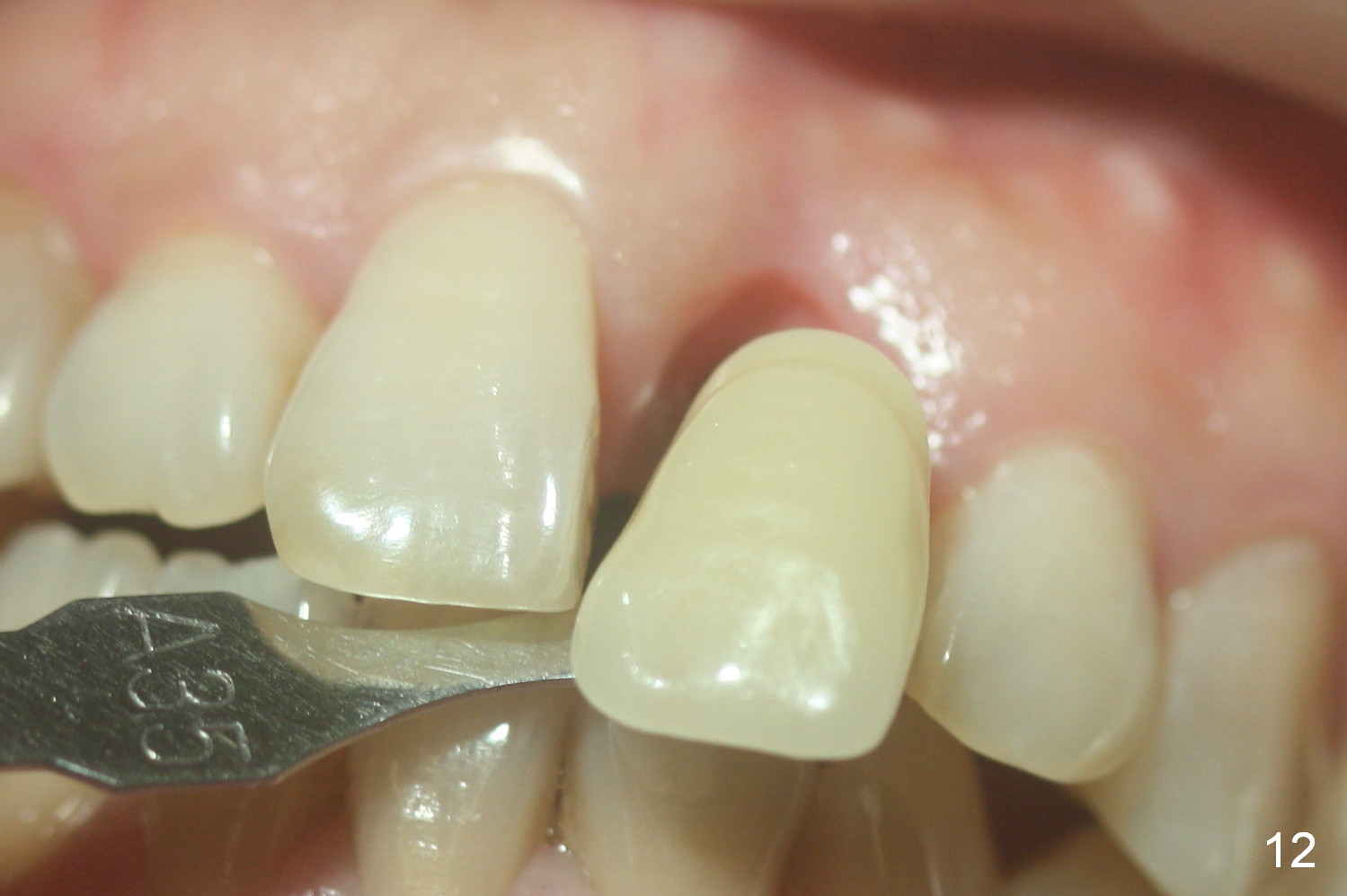
The crown shell has been used for a provisional, which is relined once. More modification is done (Fig.8) 1 week prior to impression (<3 months postop, Fig.9-12). There is no torque prior to impression. Apply sufficient amount of cement. After cementation, remove the crown/abutment complex to remove residual cement. Torque will be applied. The crown needs reshade and reshape.
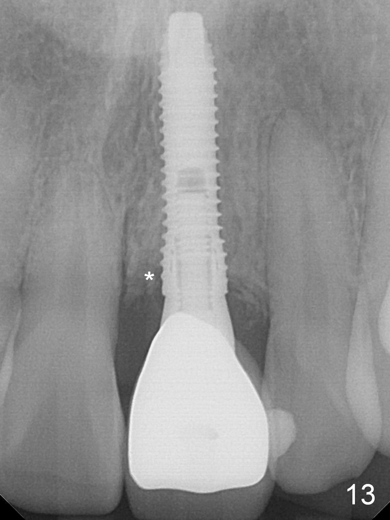
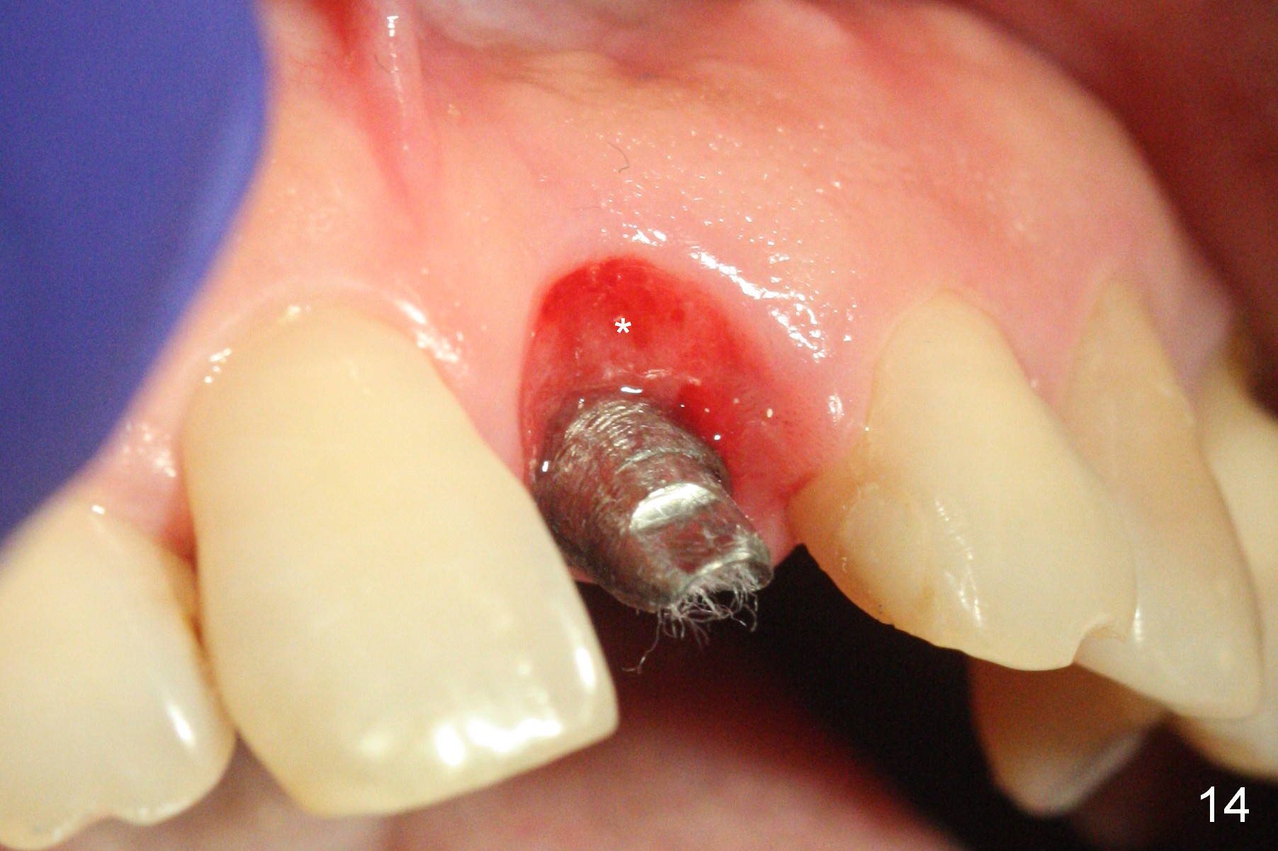
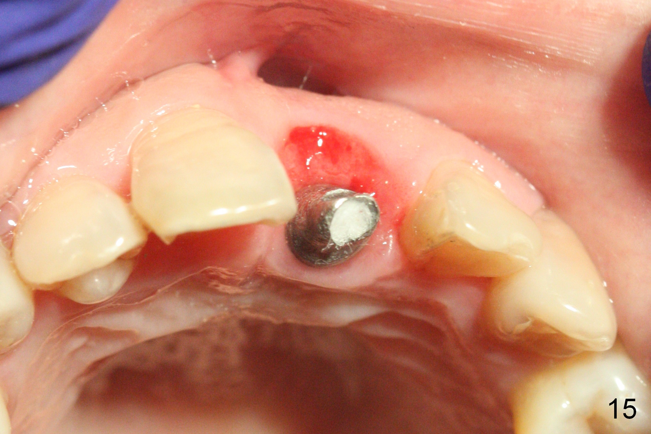
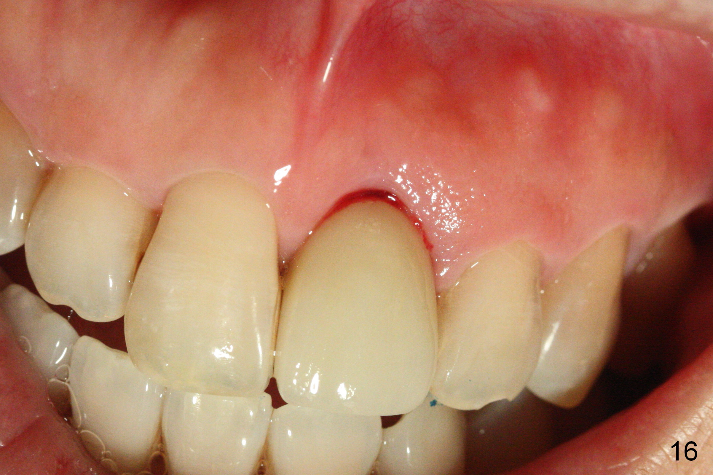
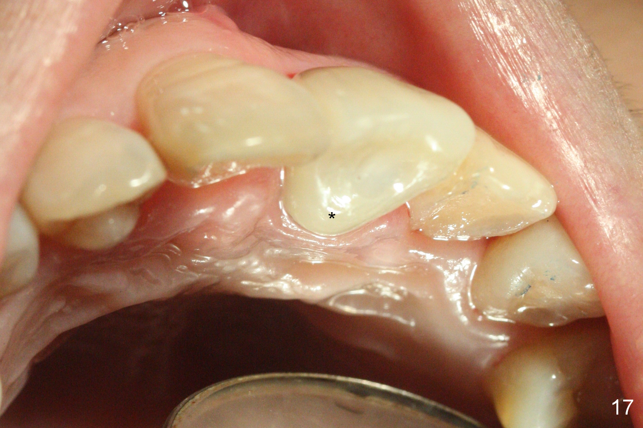
A PA is taken after crown try in (8 months postop); the mesial socket heals (Fig.13 *). The gingiva inside the socket is slightly erythematous (Fig.14). The implant/abutment is palatal (Fig.15). The patient is satisfied with the crown shade and form immediately post cementation (Fig.16). She does not note the palatal position of the cingulum (Fig.17 *). She then requests implants at #3 and 4.
Return to Upper Incisor Immediate Implant
Xin Wei, DDS, PhD, MS 1st edition 05/23/2016, last revision 01/28/2017