|
|
|
|
|
|
|
|
Palatal Placement
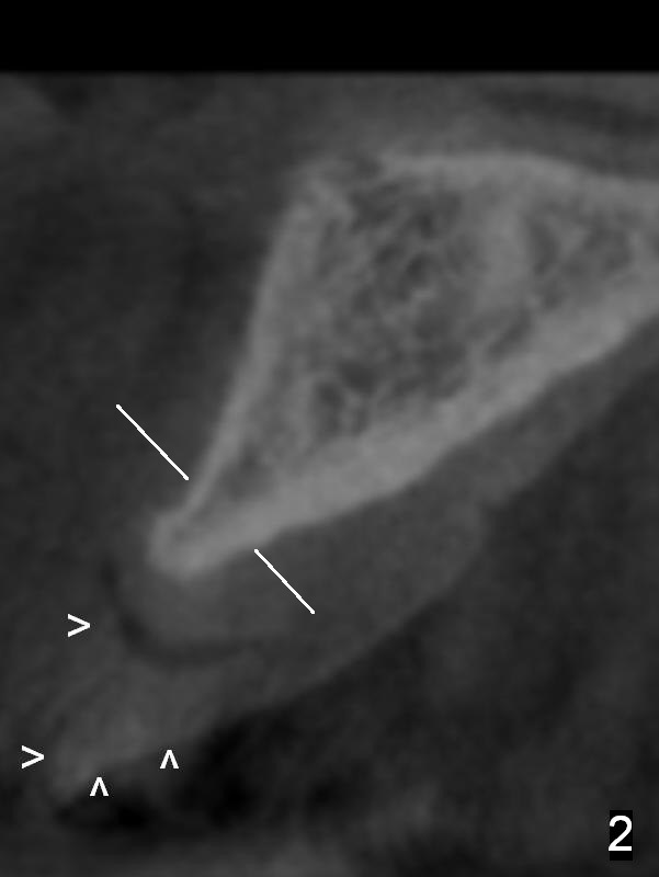
A 48-year-old man requests implant at the site of #9, 4 years after extraction (due to accident). The buccal plate is atrophic (Fig.1 CT axial section (section level as indicated by white lines in Fig.2). Arrowheads in Fig.2 show the denture tooth at the site of #9.
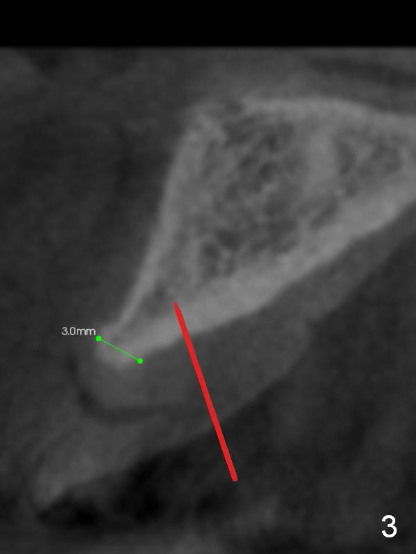
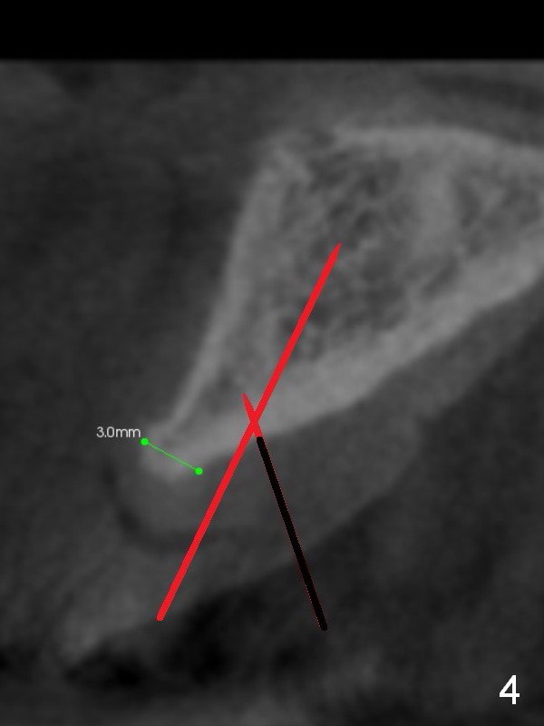
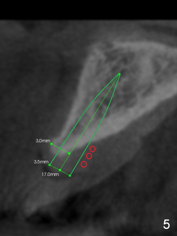
After incision, osteotomy starts in the palatal wall, ~ 5 mm from the crest (Fig.3). Once penetrating the thick palatal cortex, the 1.5 mm pilot drill changes the trajectory, more or less parallel to the buccal wall (by palpation, Fig.4). The depth will be 17 mm from the gingival margin. The depth of the 2nd (2 mm) drill will be 8 mm. The implant will be 3x17 mm one piece (Fig.5). The osteotomy is initiated intentionally more palatal. Since the resistance of the palatal wall is greater than that of the buccal one, the implant may be deviated slightly buccally, hopefully in the center of the crown. The palatal exposure threads will be covered by allograft/Osteogen and Osteotape (red circles).
If the implant is placed too buccally, but stable, bone graft will be placed buccally, probably from chin. If necessary, gingival graft will be harvested from the palate. A surgical stent is in the implant shelf (not in a box).
Return to
Upper Incisor Immediate Implant
CT/前牙植牙
Xin Wei, DDS, PhD, MS 1st edition 08/10/2015, last revision 10/17/2020