
|
|
|
|
|
|
Bone Expansion
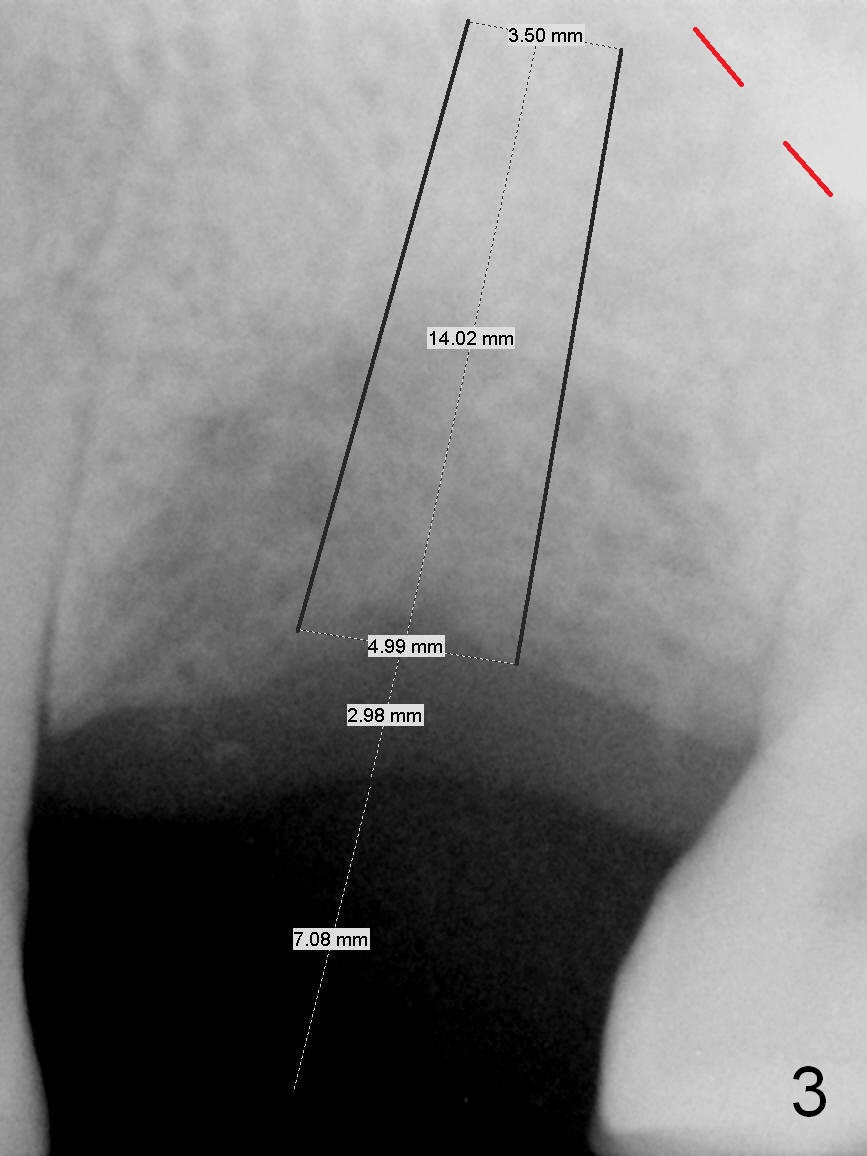
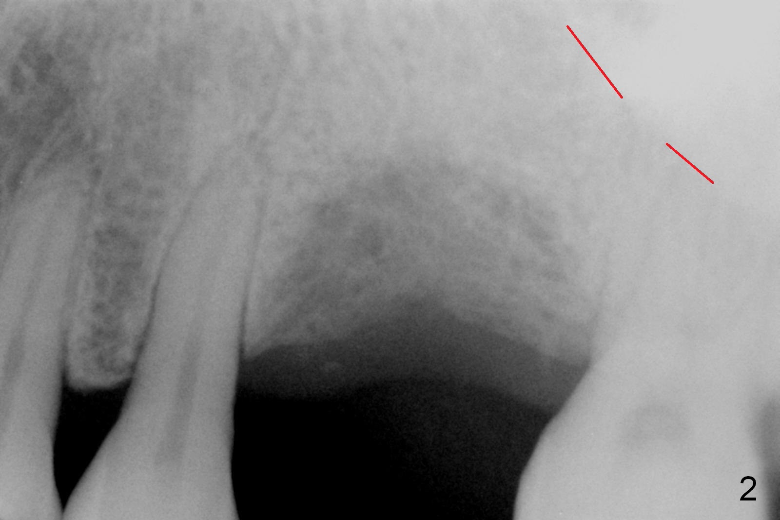
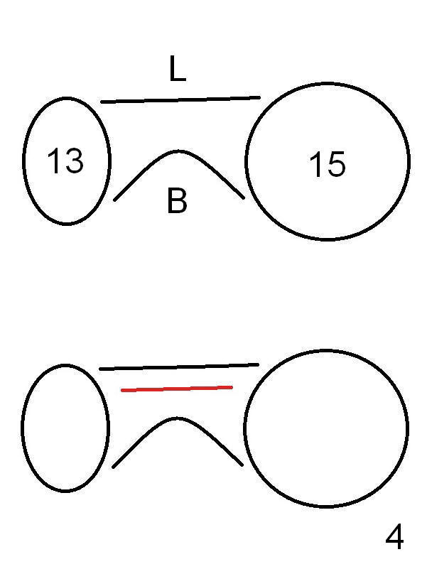
The tooth #14 of a 52-year-old lady had severe bone resorption 6 years ago (Fig.1 (red dashed line: sinus floor)). The bone resorption persists after the tooth exfoliated (Fig.2). Clinically, the buccal plate (Fig.4 (occlusal view) upper panel: B) is concave (take preop photos). #15 scalpel will be used for incision (red line in Fig.4 lower panel, near the lingual plate (L)) and initiation of bone expansion. The latter is followed by bone scalpels, bone blades and RTs at the depth of 14 mm. A 4.5x17 mm Tatum tapered tap is used for 14 mm. PA is taken. If the bone is dense, the depth is less than 14 mm in bone. Use RT2,3 to reach 17 mm. A 5x14 mm bone-level implant is placed (Fig.3). Crown cementation will be done for the tooth #3 before surgery.
Return to
Upper Molar Immediate Implant
Xin Wei, DDS, PhD, MS 1st edition 10/04/2015, last revision 01/19/2019