|
|
|
|
|
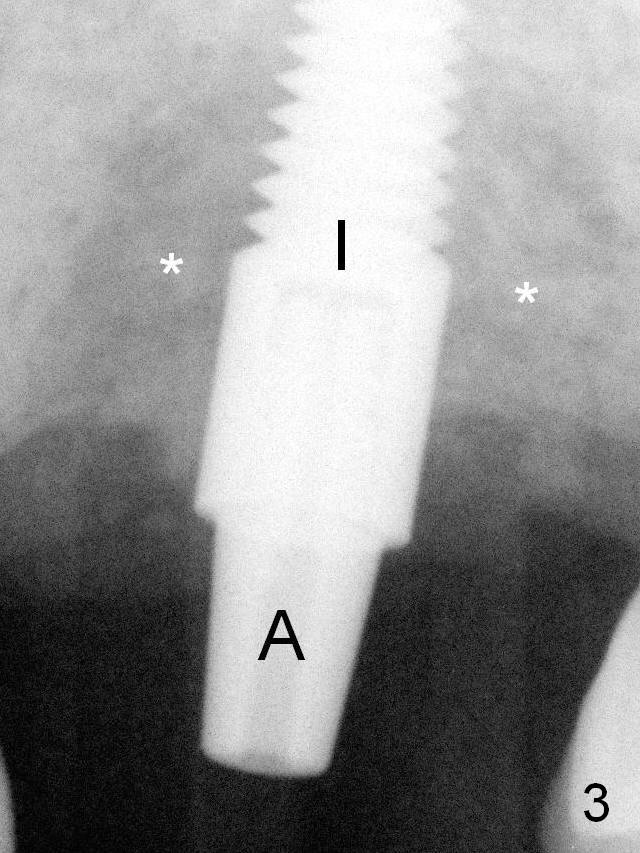
A 4.5x17 mm tissue-level implant is placed (Fig.3 I) with insertion torque >60 Ncm. Allograft and Osteogen are placed around the bulging buccal and lingual plates (*). Collagen membrane with a hole in the center is fixed around a 3.5x5 mm abutment (A). An immediate provisional is fabricated mainly for wound protection and perio dressing fixation.
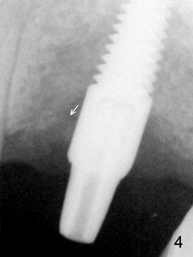
The patient returns 3 months postop; the bone graft appears to have been displaced mesioocclusally (Fig.4 arrow).
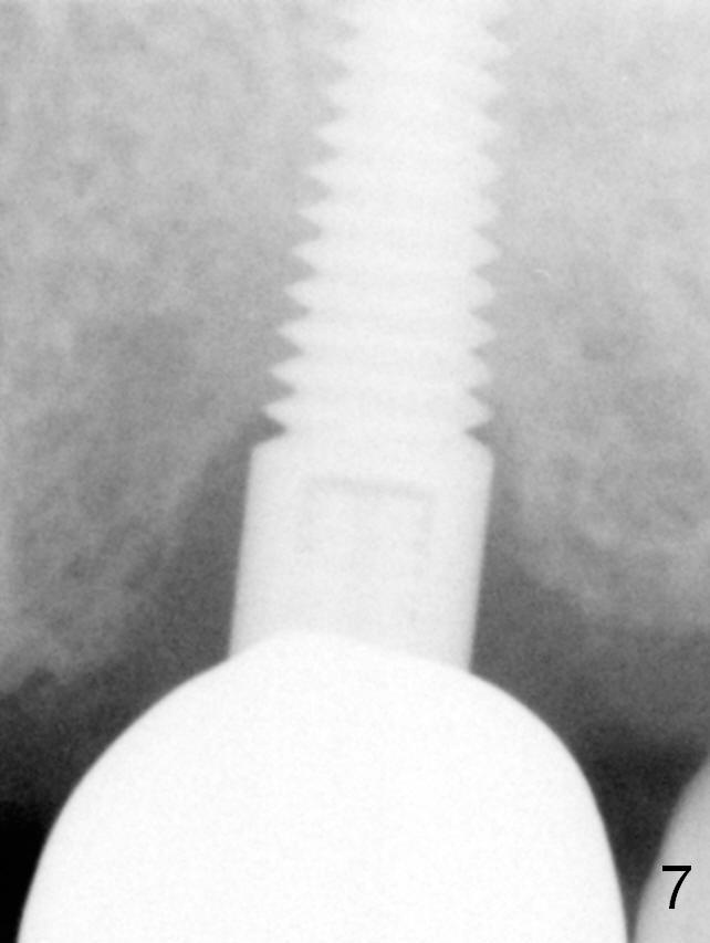
Fig.7: 7 months post cementation.
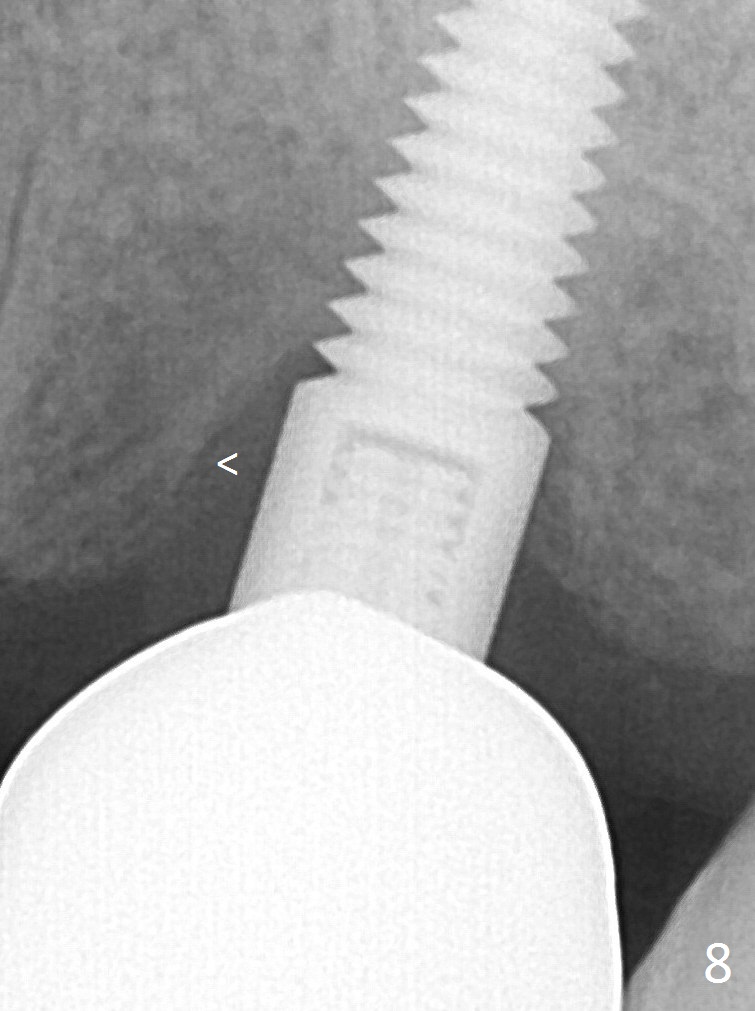
The chief complaint remains the same 1 year post cementation; mesial bone appears to form lamina dura-like structure (Fig.8 <). Is this the earliest sign of periimplantitis?
Dense Bone Last Next Xin Wei, DDS, PhD, MS 1st edition 01/17/2016, last revision 01/19/2019