|
|
|
||
|
|
|
|
|
3rd Molar Immediate Implant
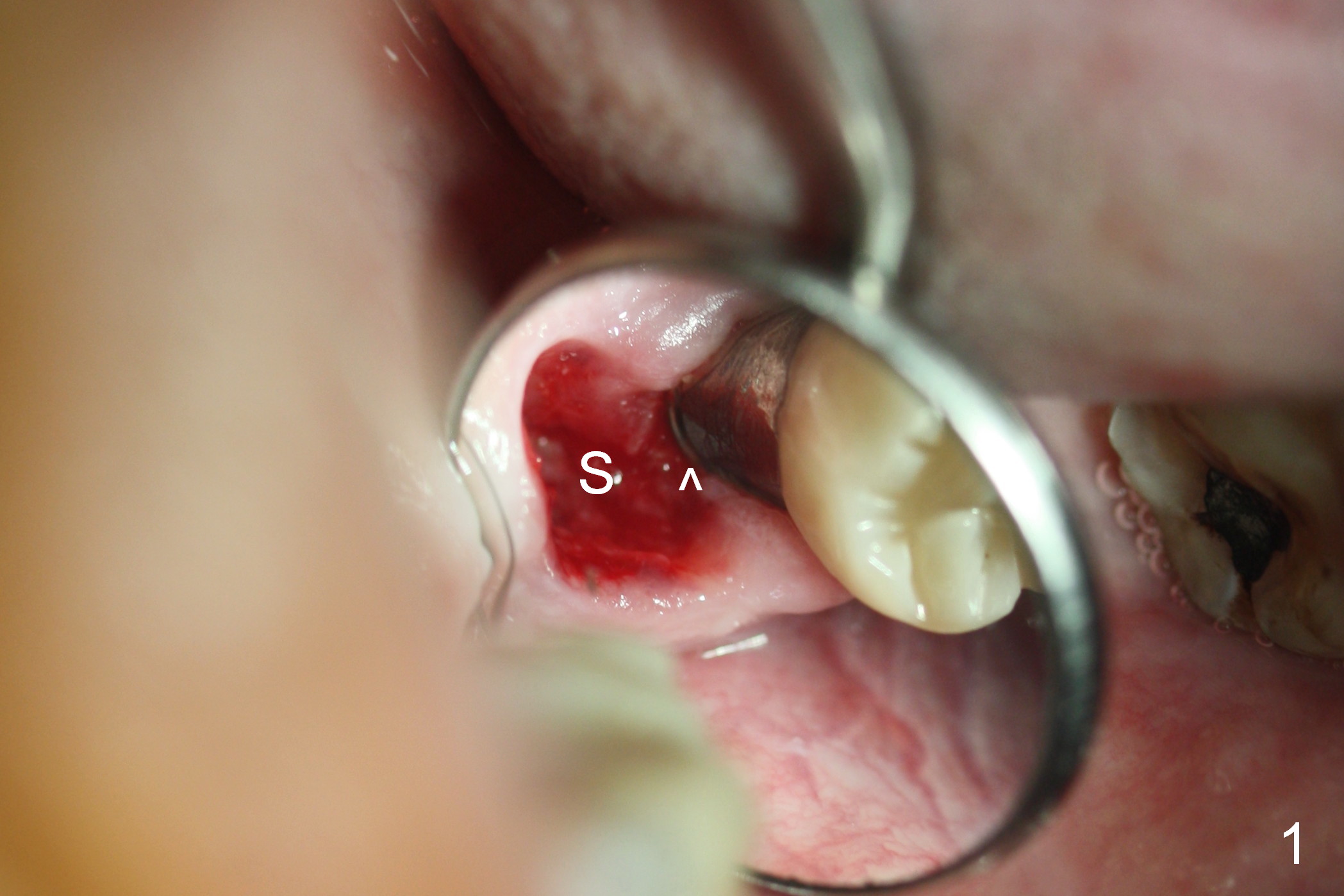
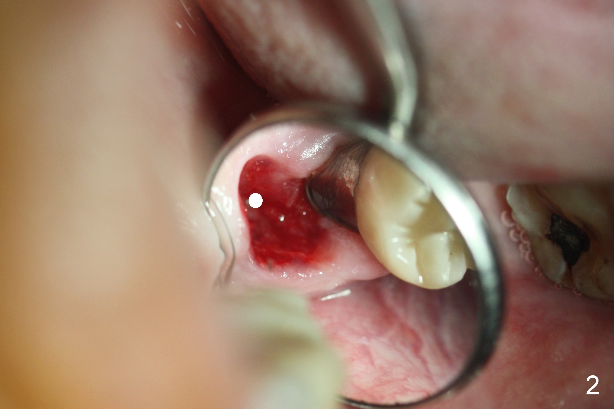
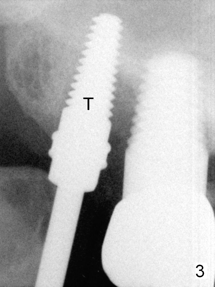
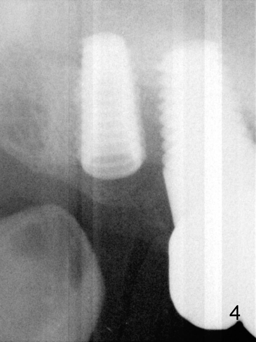
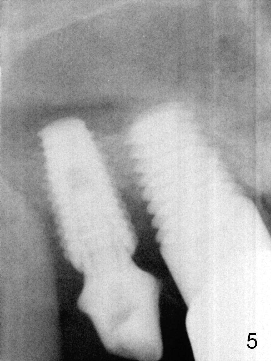
In fact there is no sign of peri-implantitis at #2 except exposure of 1 implant thread (Fig.1 ^) after extraction of the tooth #1. There is minimal or no granulation tissue in the socket (S). Osteotomy is initiated as distopalatal as possible (Fig.2 white circle). After using 3 mm reamer at 11 mm, a 4.5x14 mm tap is intended to be inserted at 11 mm. It becomes borderline stable when the tap is inserted at the full length (Fig.3). The sinus membrane is perforated when bone graft is pushed upward. Finally a 5x10 mm bone-level implant is placed (Fig.4) with insertion torque of <50 Ncm. A 5.5x5(2) mm abutment is placed for an immediate provisional. It is challenge to place the abutment due to limited space and near to the Stensen's duct (saliva). Bone graft is then placed between the implants. In fact the margin of the abutment prevents sufficient placement of the bone graft, which is evident by possible mesial coronal threads not covered by bone 4 months postop (Fig.5). Six months post #1 implant placement, the tooth #3 is deemed non salvageable by the patient. Panoramic X-ray is taken 2.5 years post cementation.
Upper Molar Immediate Implant 2 3 14
Xin Wei, DDS, PhD, MS 1st edition 11/20/2015, last revision 12/09/2018