,%20bone%20graft.jpg)
|
|
|
|
|
|
|
|
|
|
|
|
|
|
|
|
|
|
|
Palatal Socket Placement
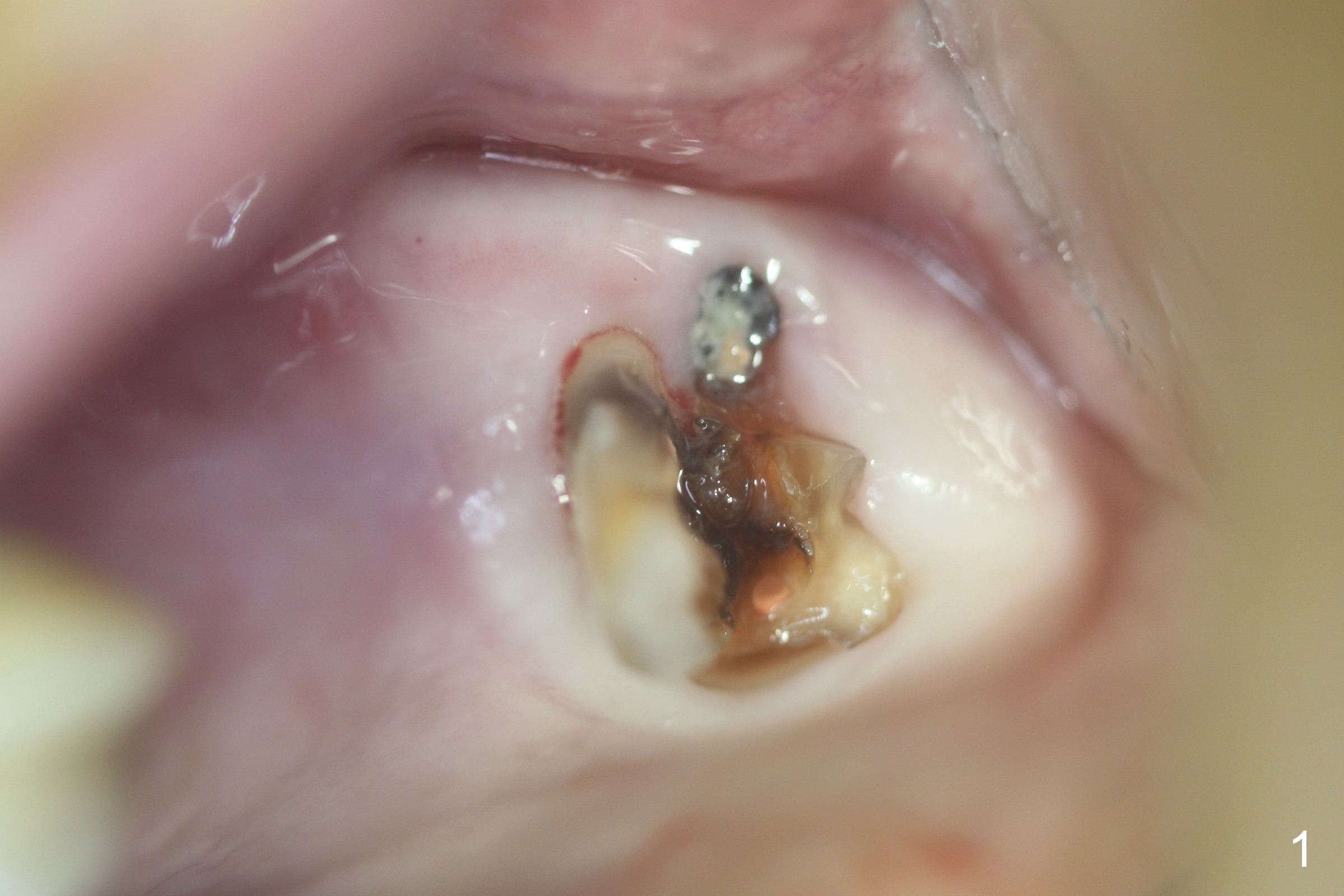
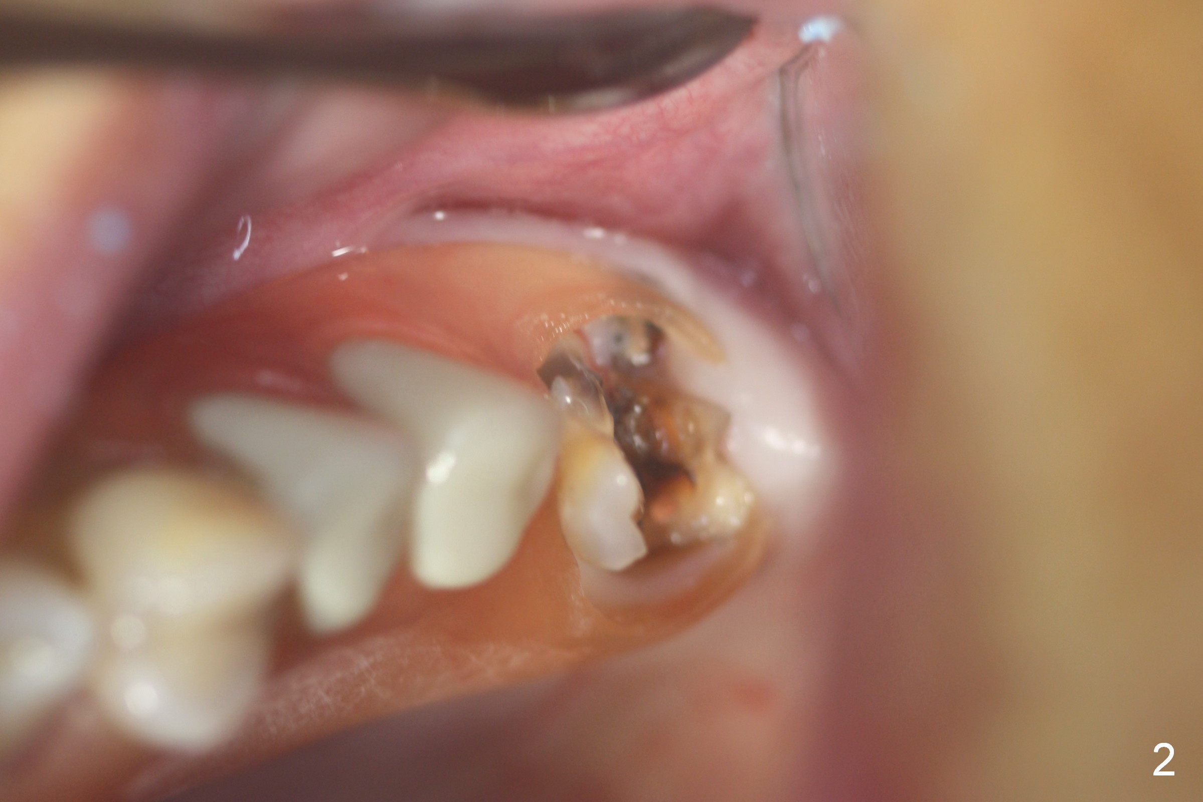
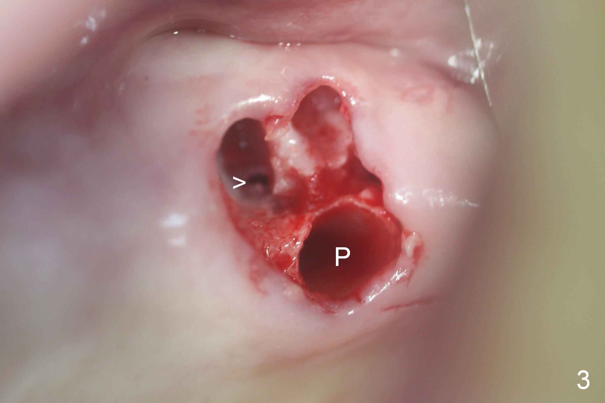
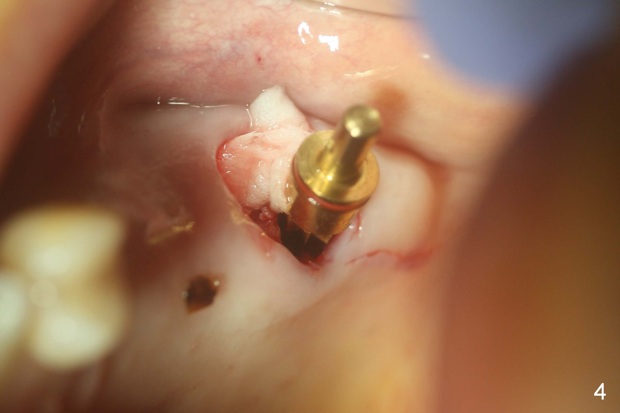
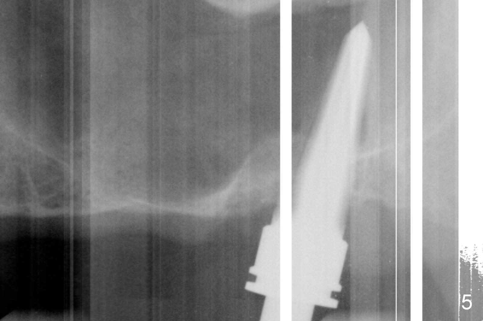
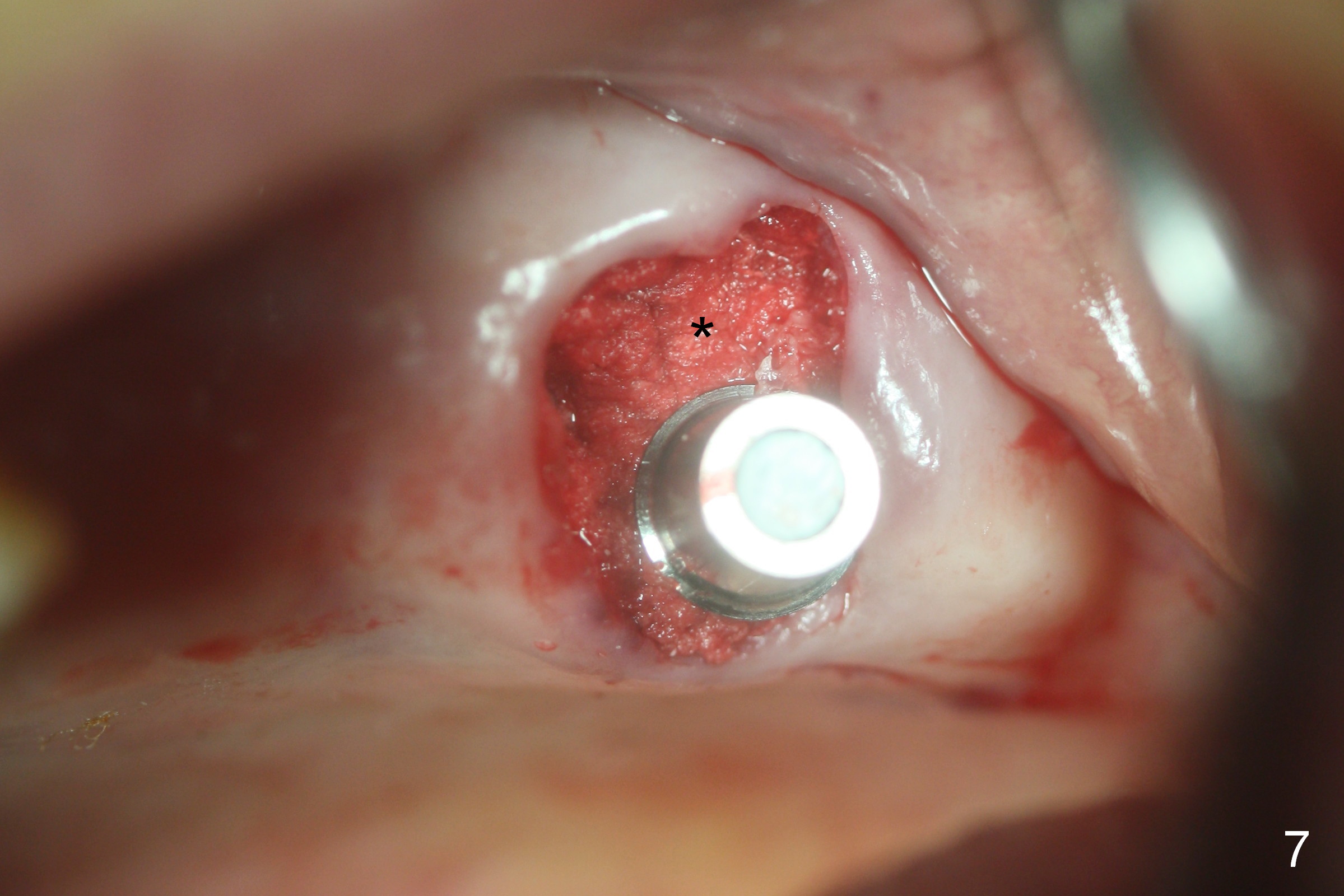
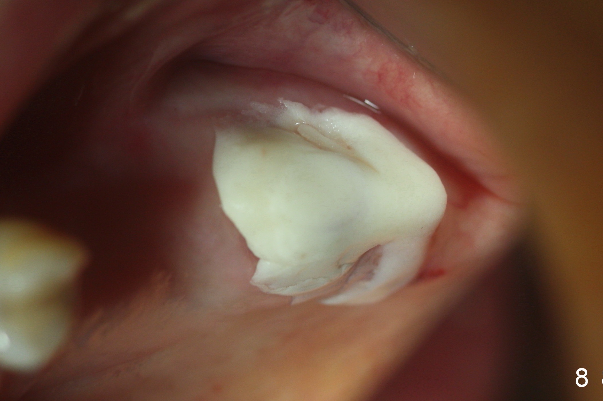
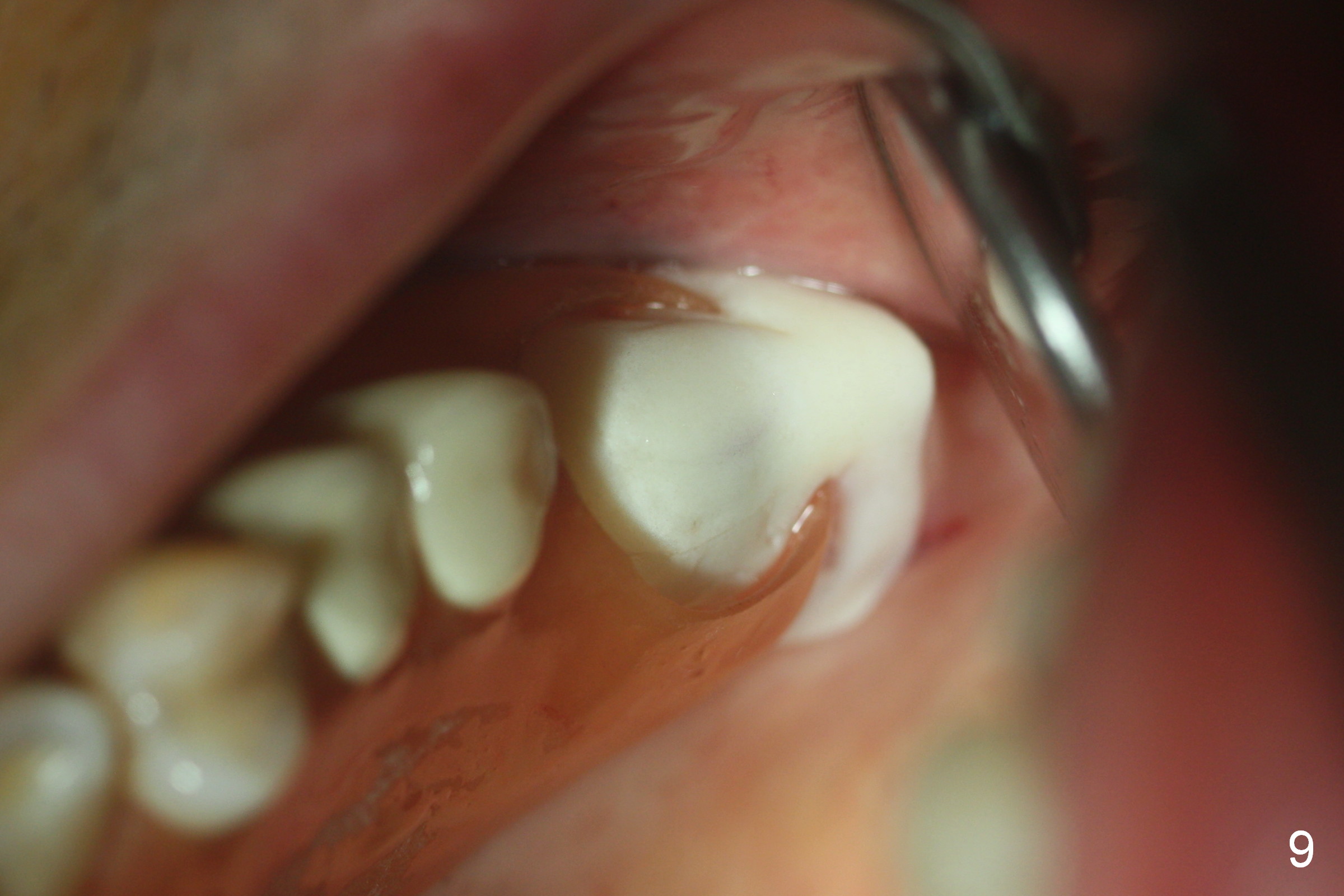
Fig.1,2 show the close relationship of the upper flipper and the residual roots at #15. Atraumatic extraction using periotomes and surgical sectioning still results in perforation of the mesiobuccal socket (Fig.3 >). The distobuccal one is shallow, while the septum is small. It appears that the palatal (Fig.3 P) socket is the most ideal recipient site for the immediate implant and is expanded with Magic Osteotomes until 4.3 mm (Fig.4,5) with the coronal end pushed as buccal as possible. After placing allograft for sinus lift (Fig.6 >), a 5x11 mm IBS implant is placed with insertion torque ~ 50 Ncm. A 6x4(3) mm pair abutment is placed, followed by bone graft in the remaining sockets (Fig.6 *) and by Osteogen plug (Fig.7 *). Finally the socket is sealed by applying acrylic over the abutment (Fig.8). While the acrylic is setting, the flipper is seated and excess acrylic is removed and pushed away from the flipper (Fig.9). Advise the patient not to wear the flipper. If it is being worn, there will be minimal contact between the flipper and the immediate provisional.
The patient will return for provisional revision 3 weeks postop. There will be a gap between the provisional and the gingiva. Remove the former, possibly using high-speed handpiece. Make a new and smaller one (big enough to close the socket and keep the graft in place) so that there is no interference when the flipper is on and off.
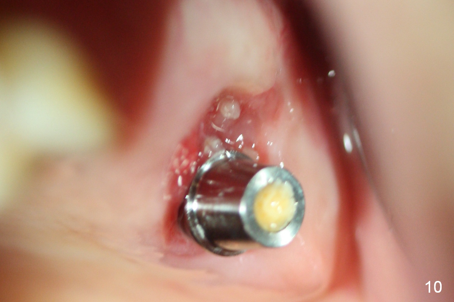
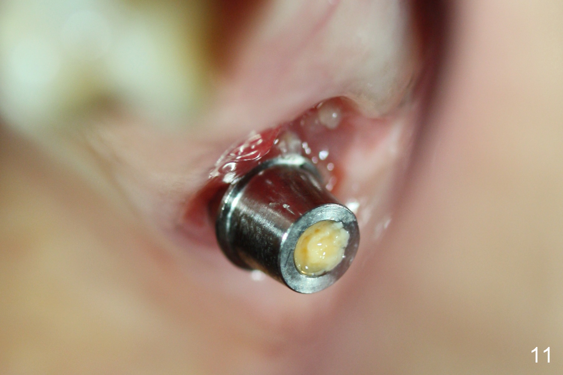
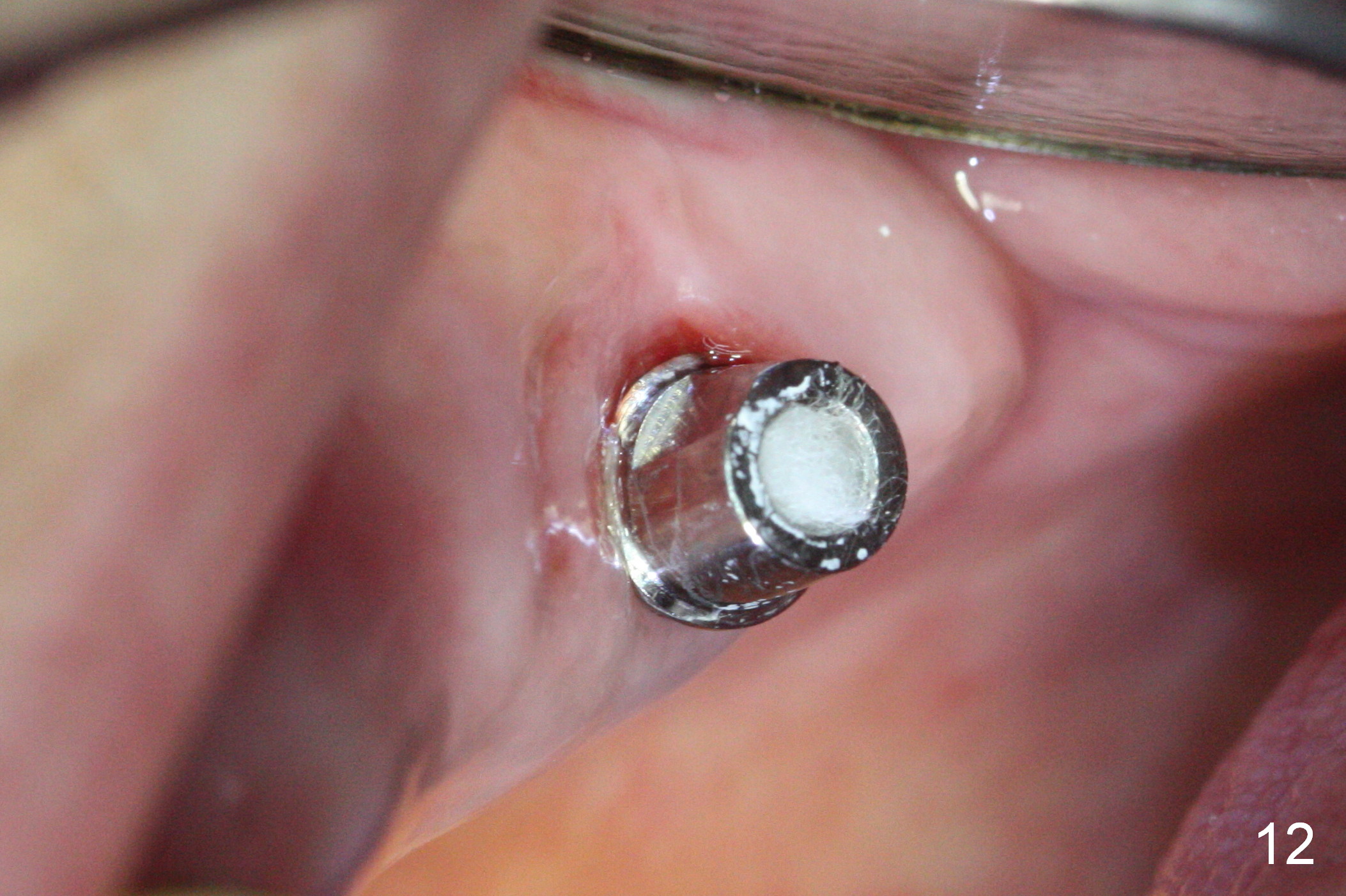
When the provisional is removed for revision 3 weeks postop, there is no gap as mentioned above. The provisional is easily removed. The socket is healing with the bone graft apparently incorporated (Fig.10,11). The provisional is modified to cover the abutment margin. The provisional is removed because of abutment screw loosening; the socket has healed (Fig.12; 1.5 months postop).
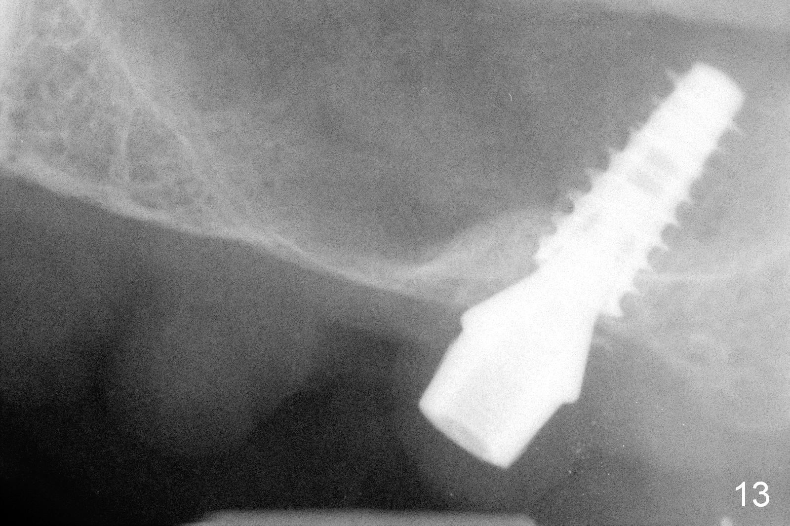
The bone density in the mesial socket appears to increase and becomes more homogenous 3 months postop (Fig.13). Impression is taken for final restoration.
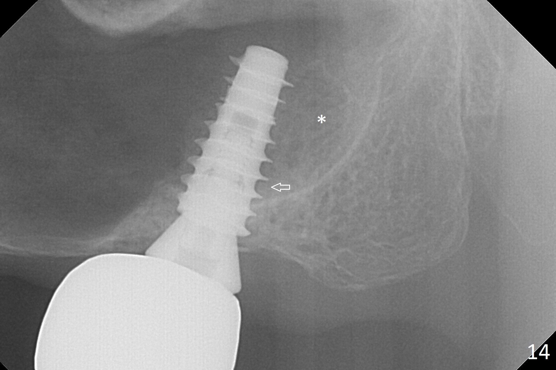
There is bone formation in the sinus distal to the implant 6 months post cementation (Fig.14 *) with the osseous tissue having grown into between the threads (arrow).
Return to
Upper
Molar Immediate Implant,
Prevent Molar Periimplantitis (Protocols,
Table), #10,
1st Year
Xin Wei, DDS, PhD, MS 1st edition 08/17/2016, last revision 06/03/2018