,%205x13,%206.5x4(3).jpg)
|
|
|
|
|
|
|
|
|
|
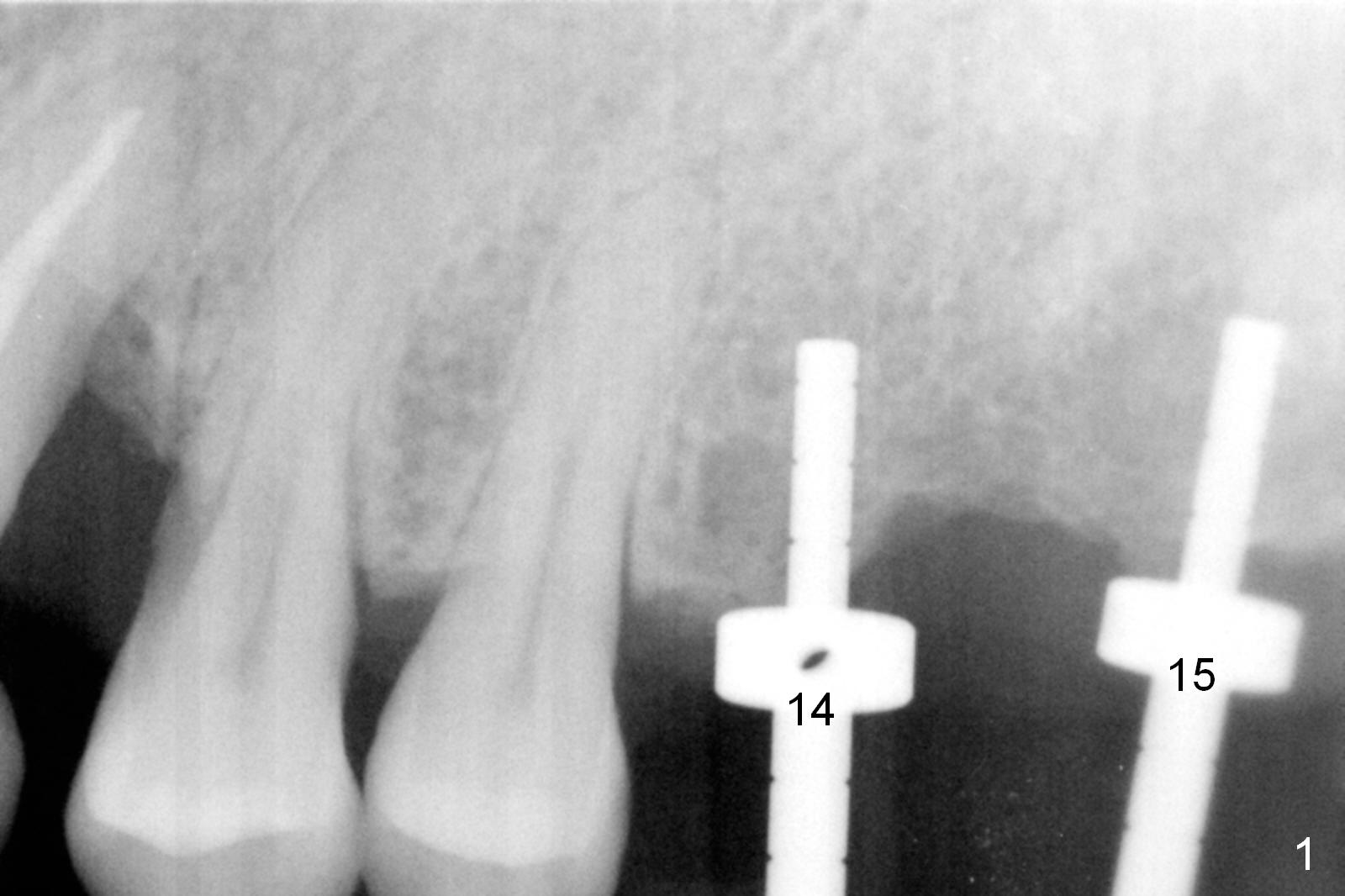
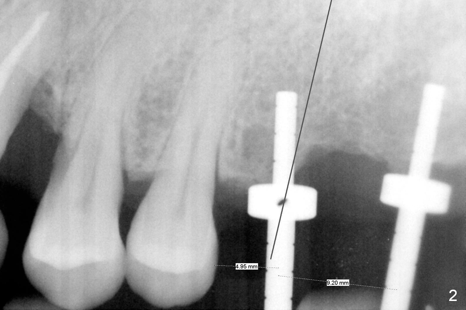
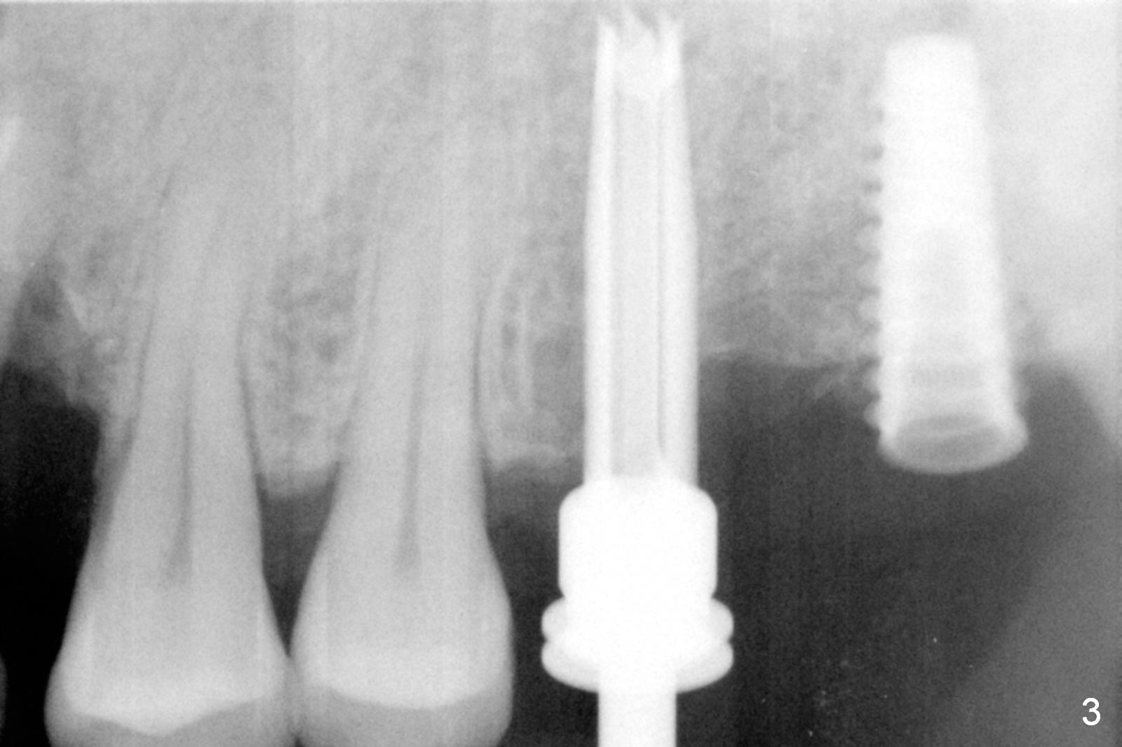
Combination of Osteotomes and Drills
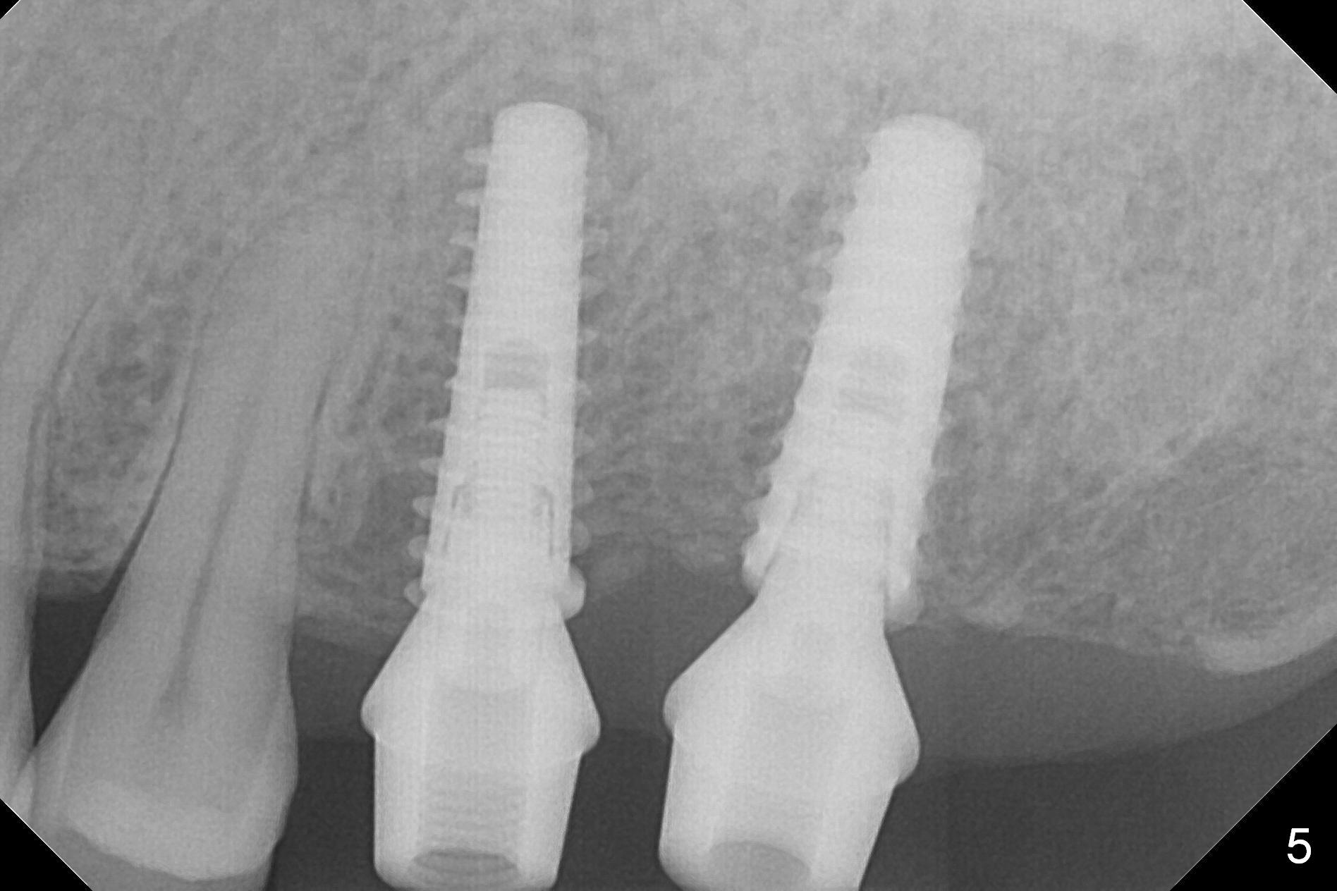
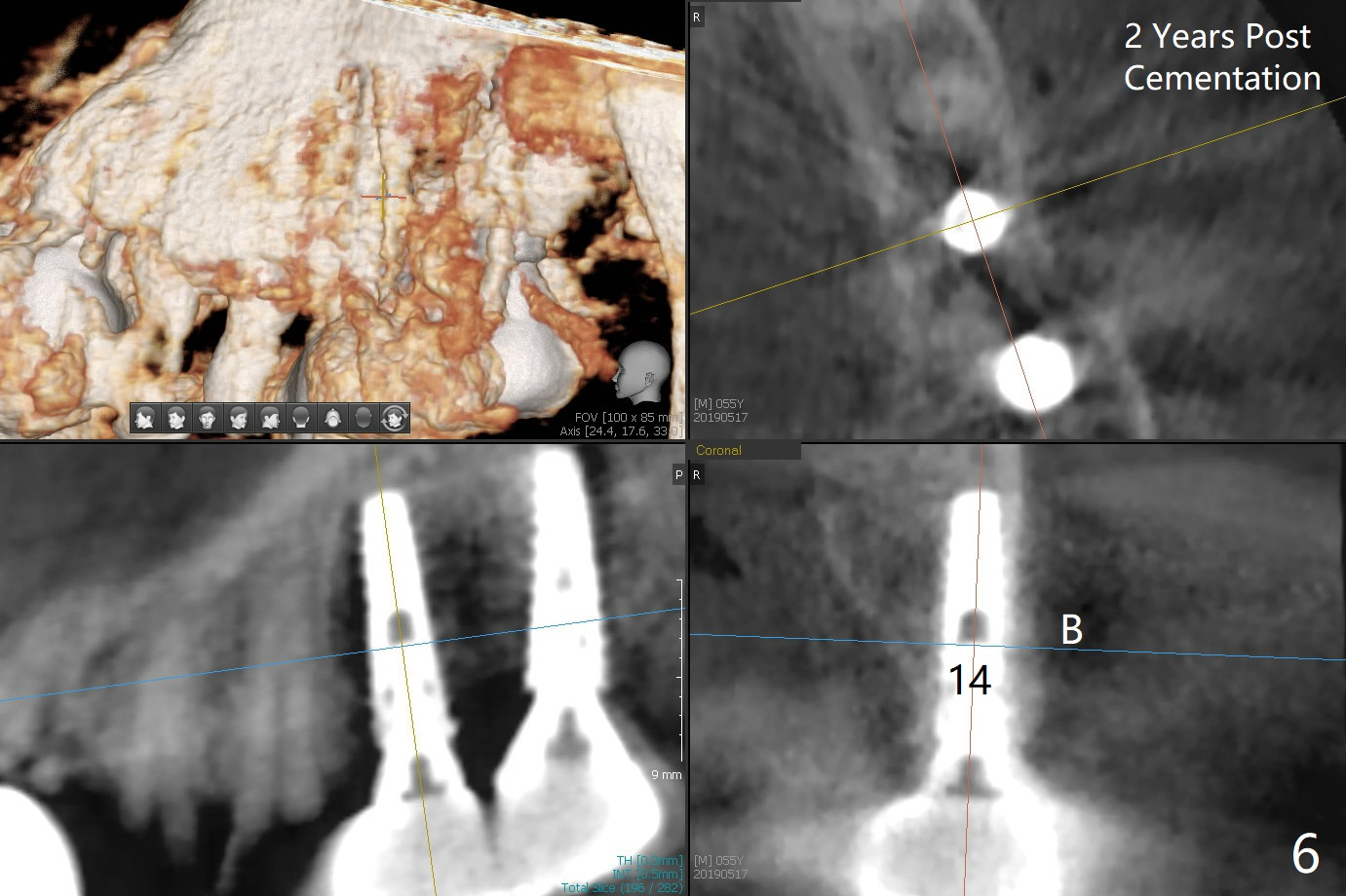
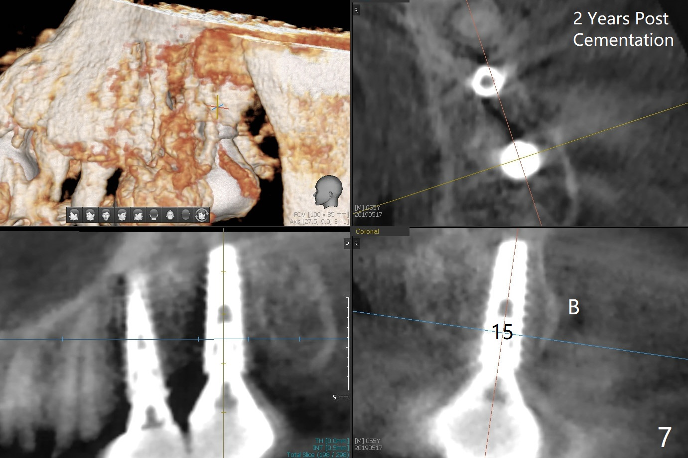
As shown by CT, the ridge at #14 is narrower than that of #15 (confirmed after incision), whereas the density at #14 is higher than that of #15. The osteotomies are established by combination of magic osteotomes and drills after change in trajectory (Fig.1-3). Following placement of 4.5x13 and 5x13 mm IBS implants and 6x4(3) and 6.5x4(3) mm pair abutments at #14 and 15 (Fig.4), flaps are sutured for hemostasis. Since the patient does not tolerate the surgery too well (unstable hypertension and oozing), immediate provisional is delayed. Periodontal dressing is applied instead. While the implants are healing, porcelain chips at the upper anterior bridge. There appears no bone resorption 6 months postop (Fig.5). Impression is taken following changing abutment to 5x4(2) mm at #14 and Diode gingivectomy. A panoramic film is taken 1 year 2 months post cementation. CT taken 2 years post cementation shows relatively good trajectory of these 2 implants (Fig.6,7).
Return to Upper Molar, Arch Immediate Implant, #10 24/25 19/21/30 Xin Wei, DDS, PhD, MS 1st edition 09/08/2016, last revision 05/24/2019