.jpg)
|
|
|
|
|
|||
|
|
|
|
|
|||
|
|
|
|
|
|
||
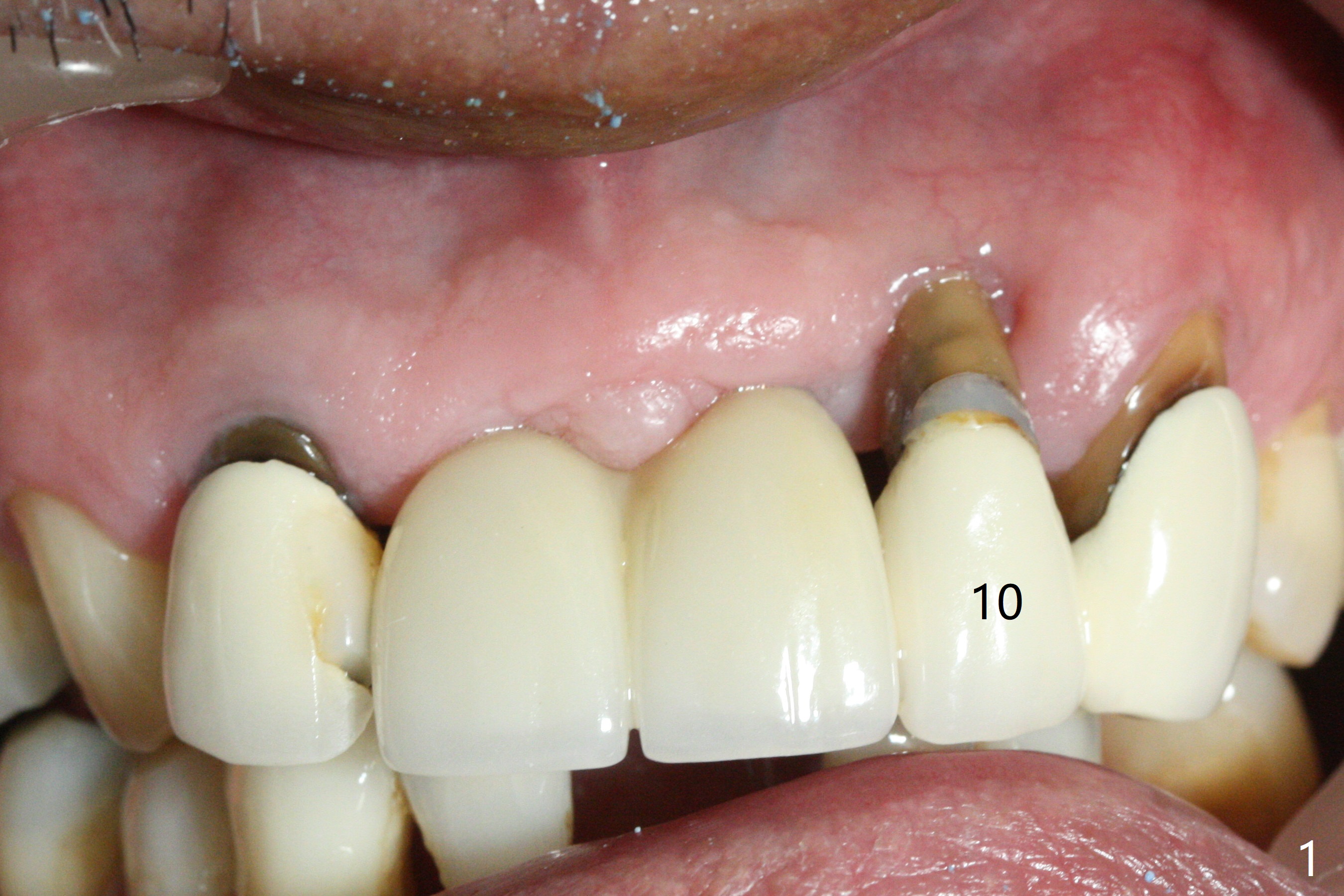
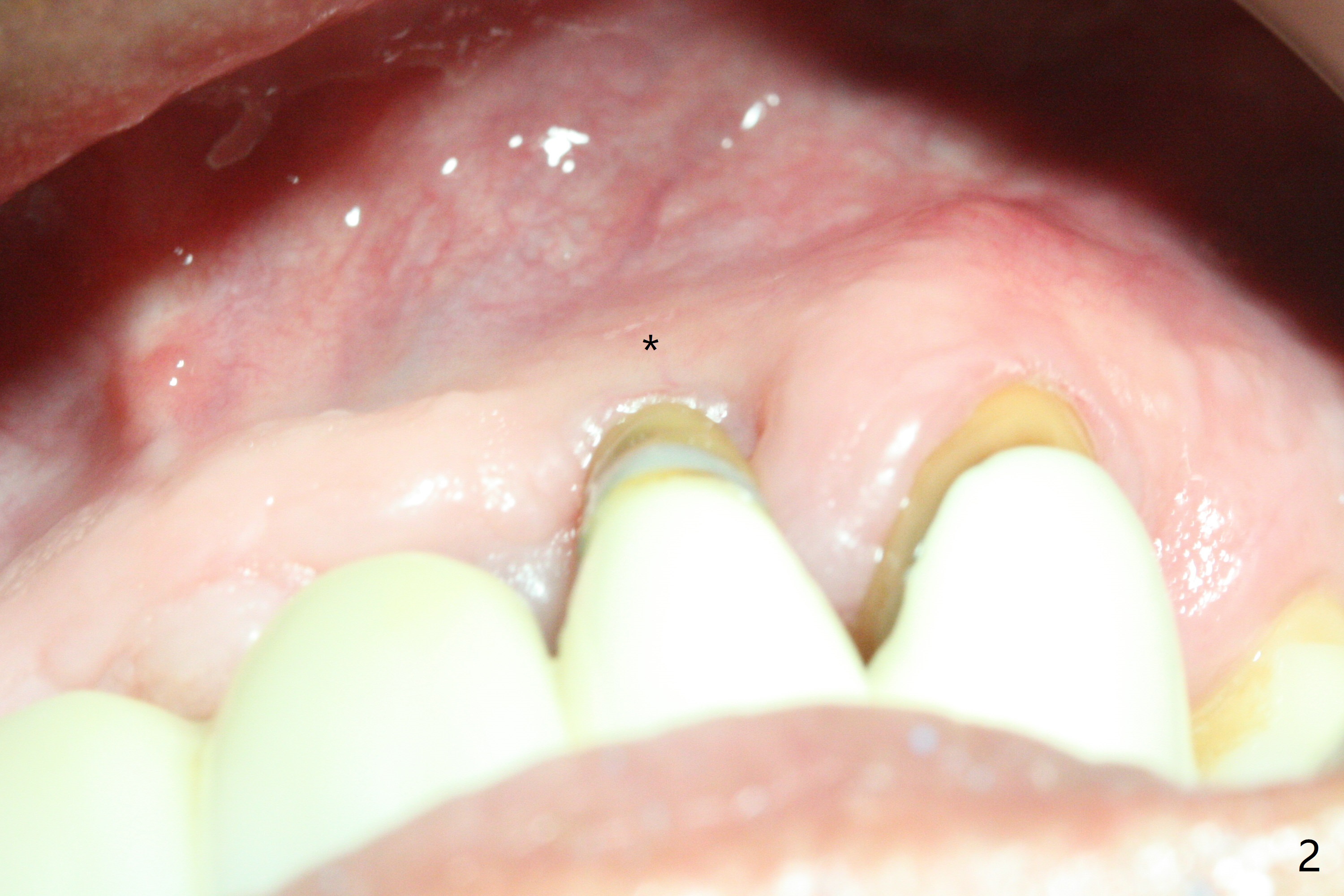
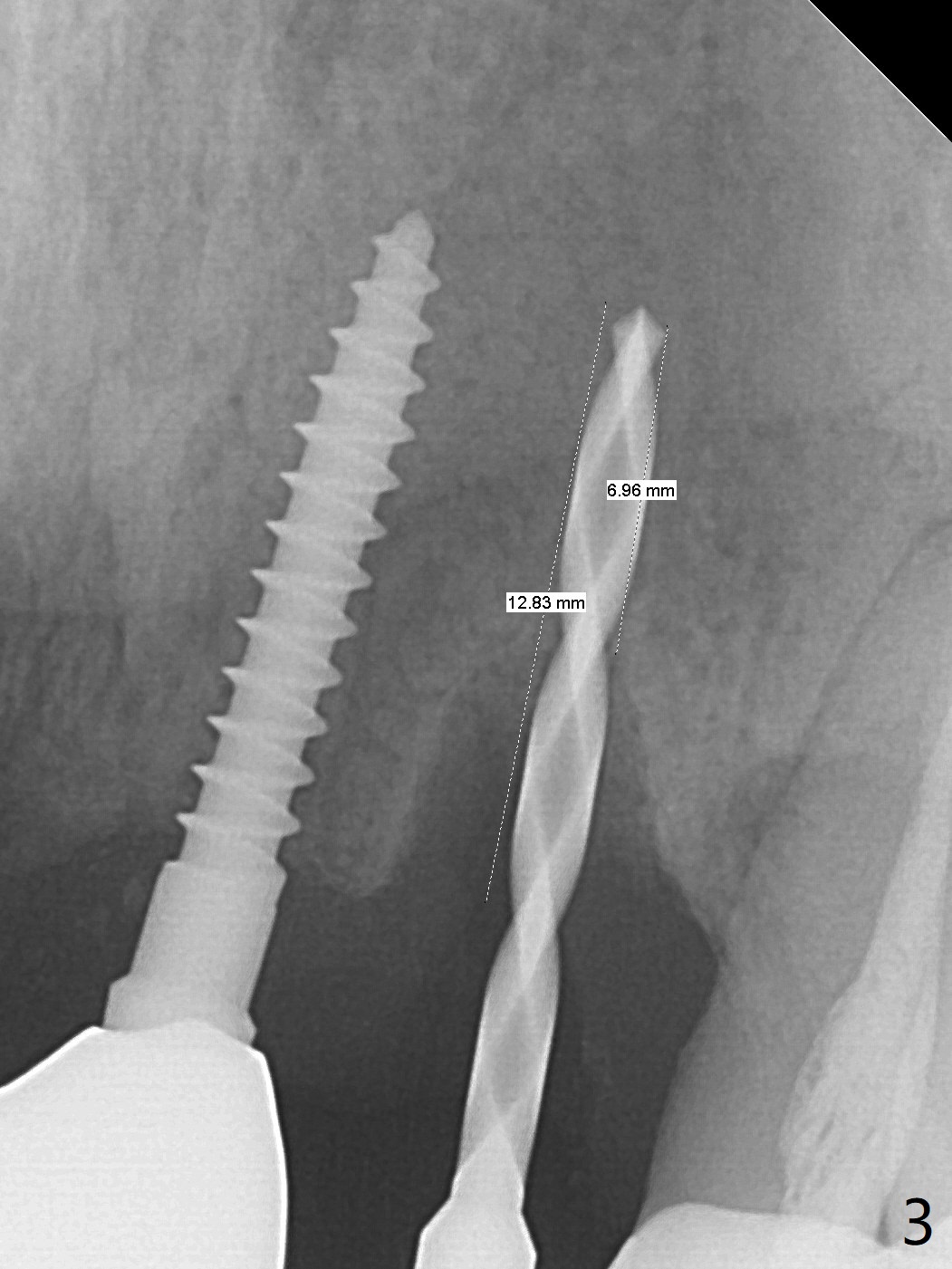
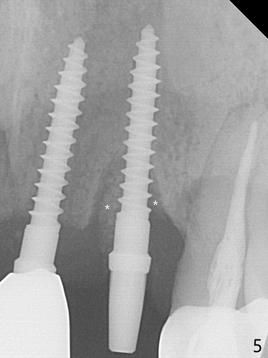
Native Bone Wider than CT Indicates
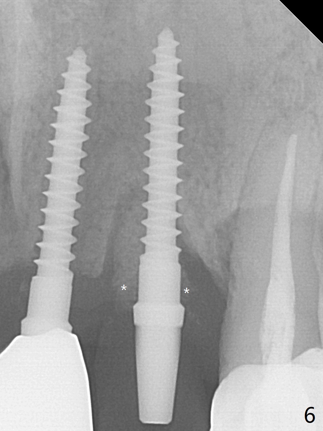
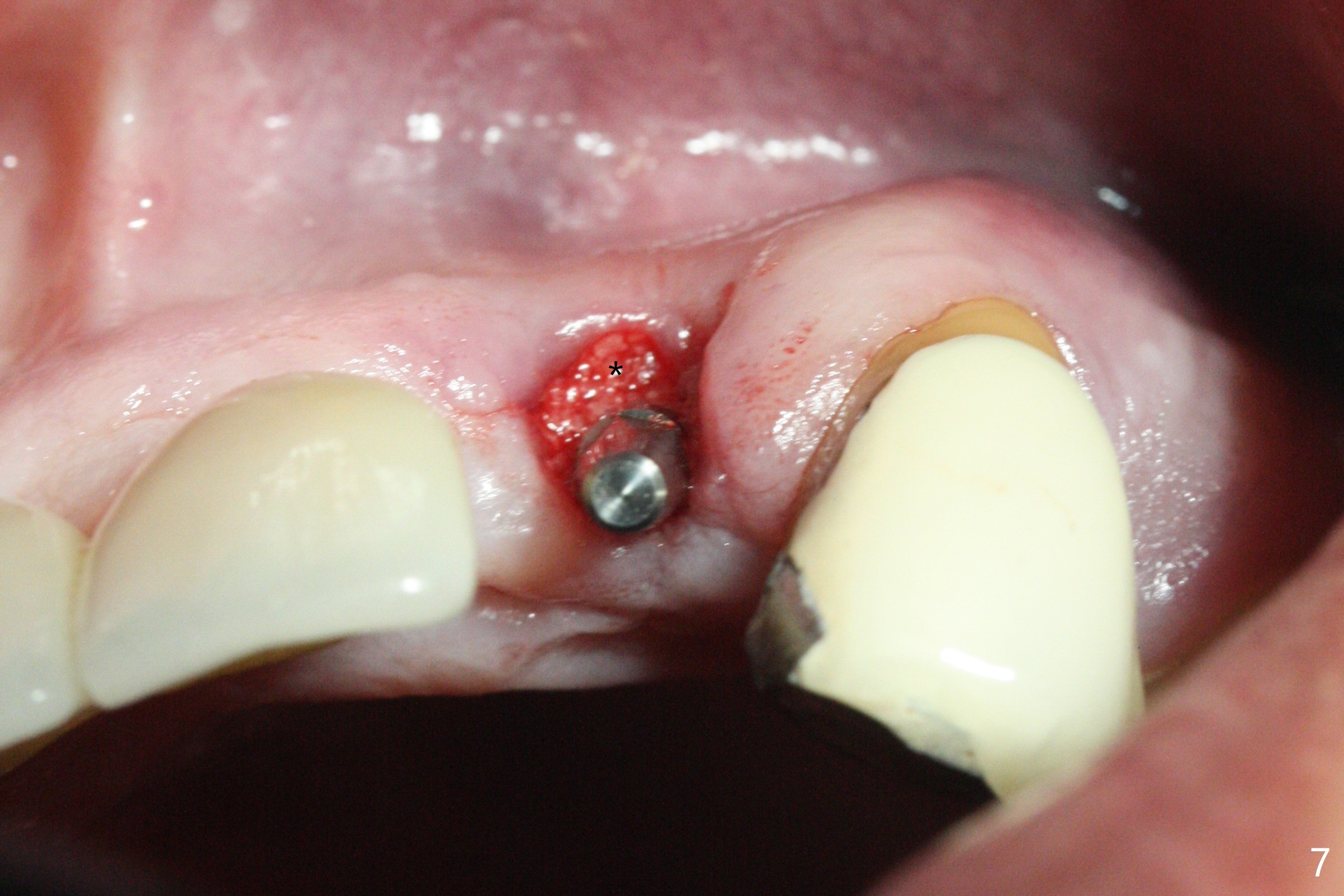
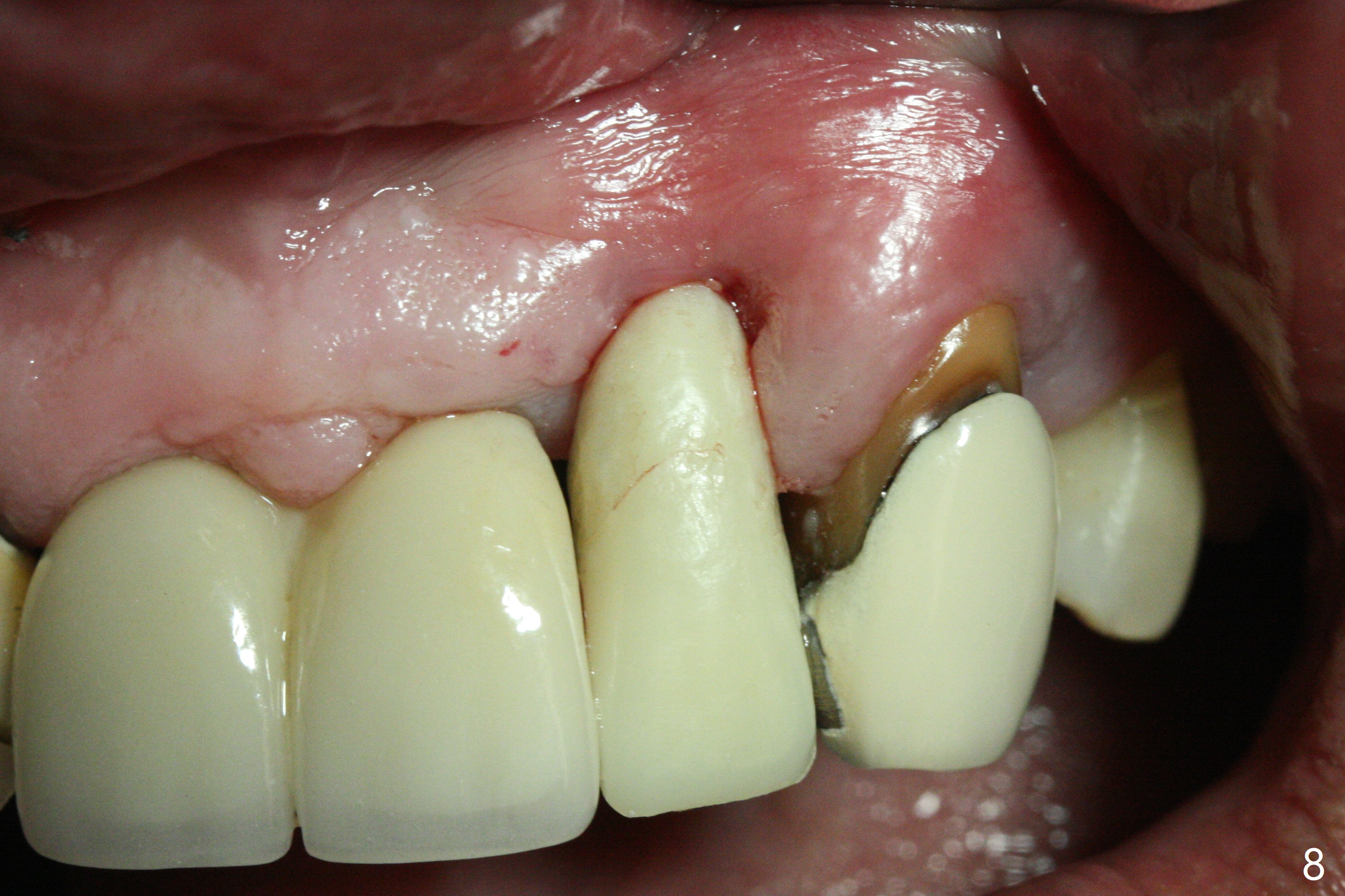
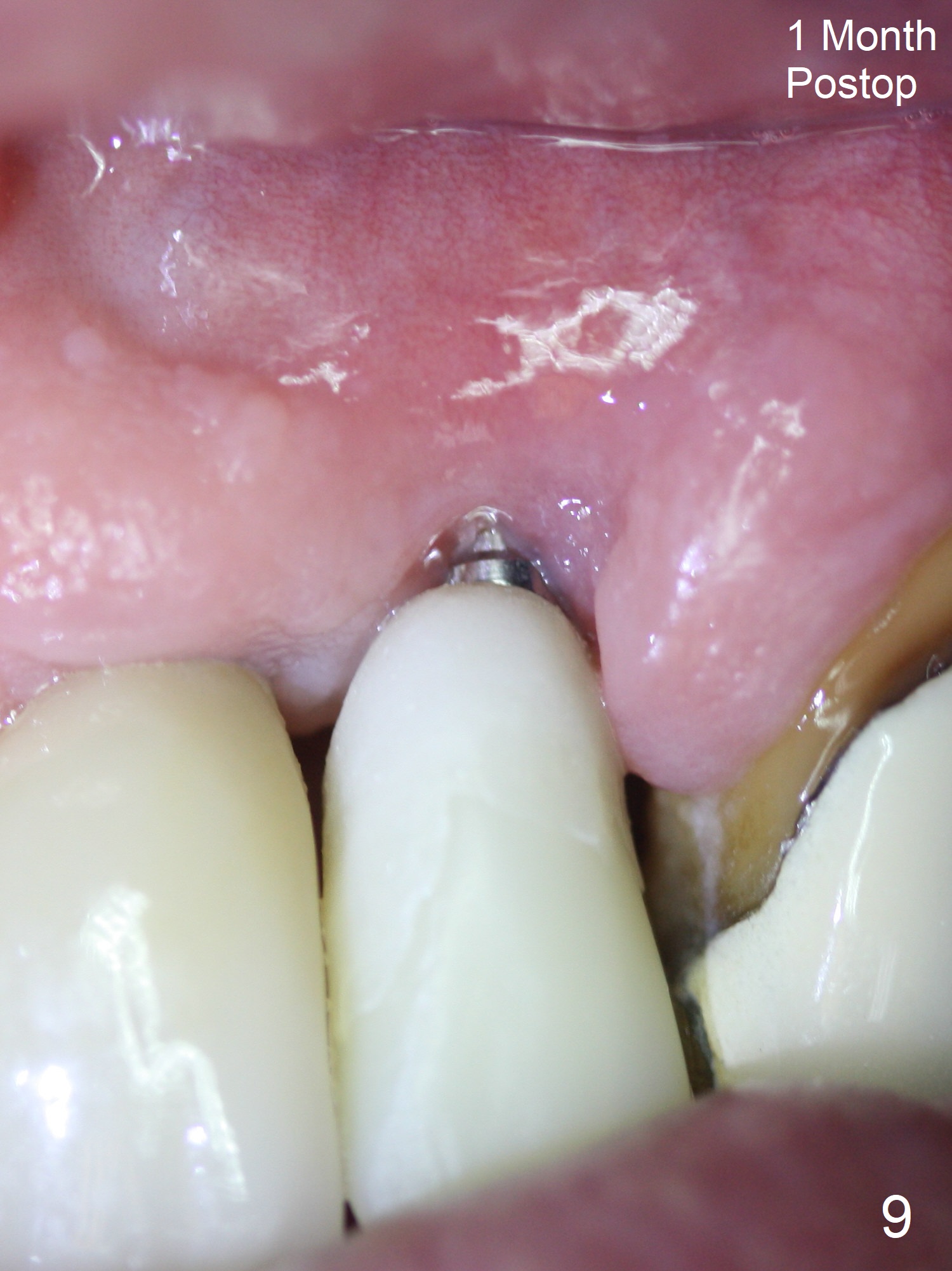
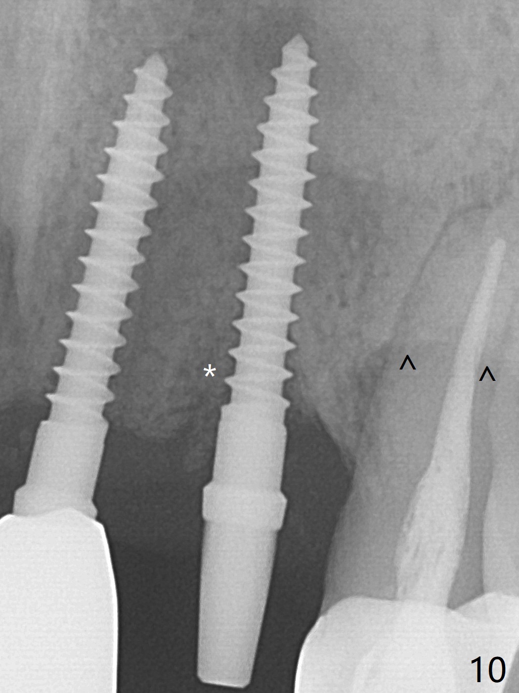
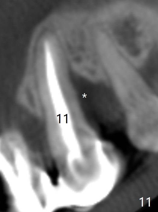
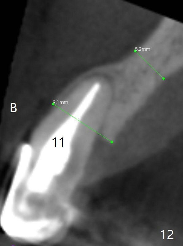
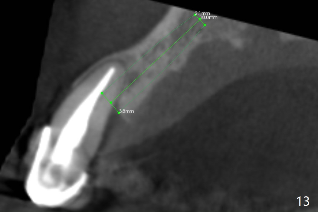
The tooth #10 has severe gingival recession (Fig.1) with loss of the buccal plate (Fig.2 *). After the initial osteotomy depth (Fig.3) increases by 2 mm, a 2.5x14(2) mm 1-piece implant is placed with insertion torque >60 Ncm (Fig.4). Palpation indicates the native bone apparently wider than CT shows. There is no sign of buccal or palatal plate perforation by palpation during osteotomy or implant placement. Vera Graft is placed repeatedly around the coronal threads (Fig.5-7 *). An immediate provisional is fabricated to close the socket (Fig.8). The buccal plate appears to collapse 1 month postop (cortical plate graft apparently more appropriate in this case); the margin of the provisional is trimmed so that the gingiva may grow incisally (Fig.9). The provisional dislodges several times postop due to short abutment. By nearly 4 months postop, the coronal bony defect seems to have been repaired (Fig.10 *). The tooth #11 has tenderness with bone loss (Fig.10 ^), corroborated by CT (distal bone loss, Fig.11 *). Since the apical bone is narrow (Fig.12), a narrow long implant is expected (Fig.13). Use an implant (3.5x13 mm) consistent with those at #14 and 15.
Upper Incisor, Arch Immediate Implant, Armaments 24/25 19/21/30 Shield Xin Wei, DDS, PhD, MS 1st Version 03/19/2018, Last Update 02/20/2021