.jpg)
,%20also%20bone%20graft.jpg)
|
|
|
|
|
|
|
|
|
|
||
|
|
|
|
||
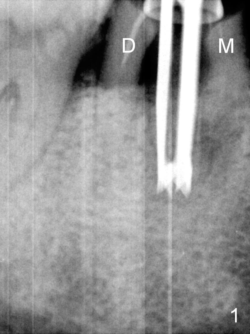
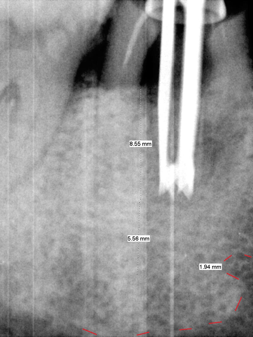
Osteotomy Via Roots
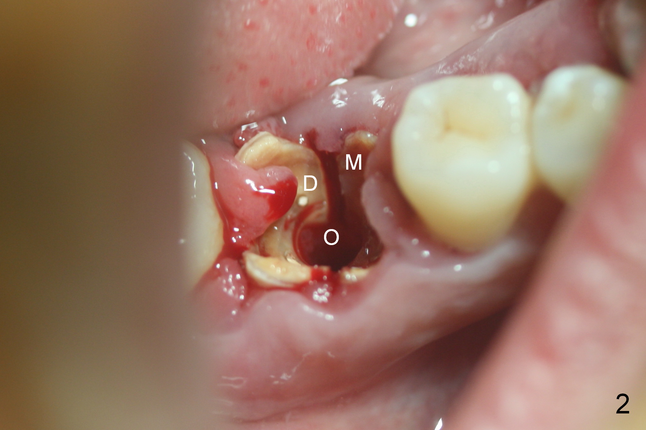
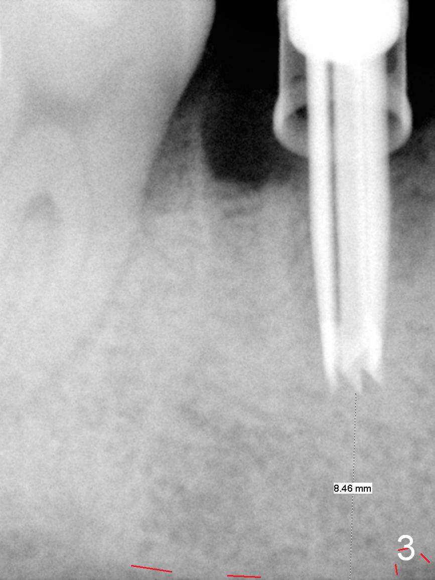
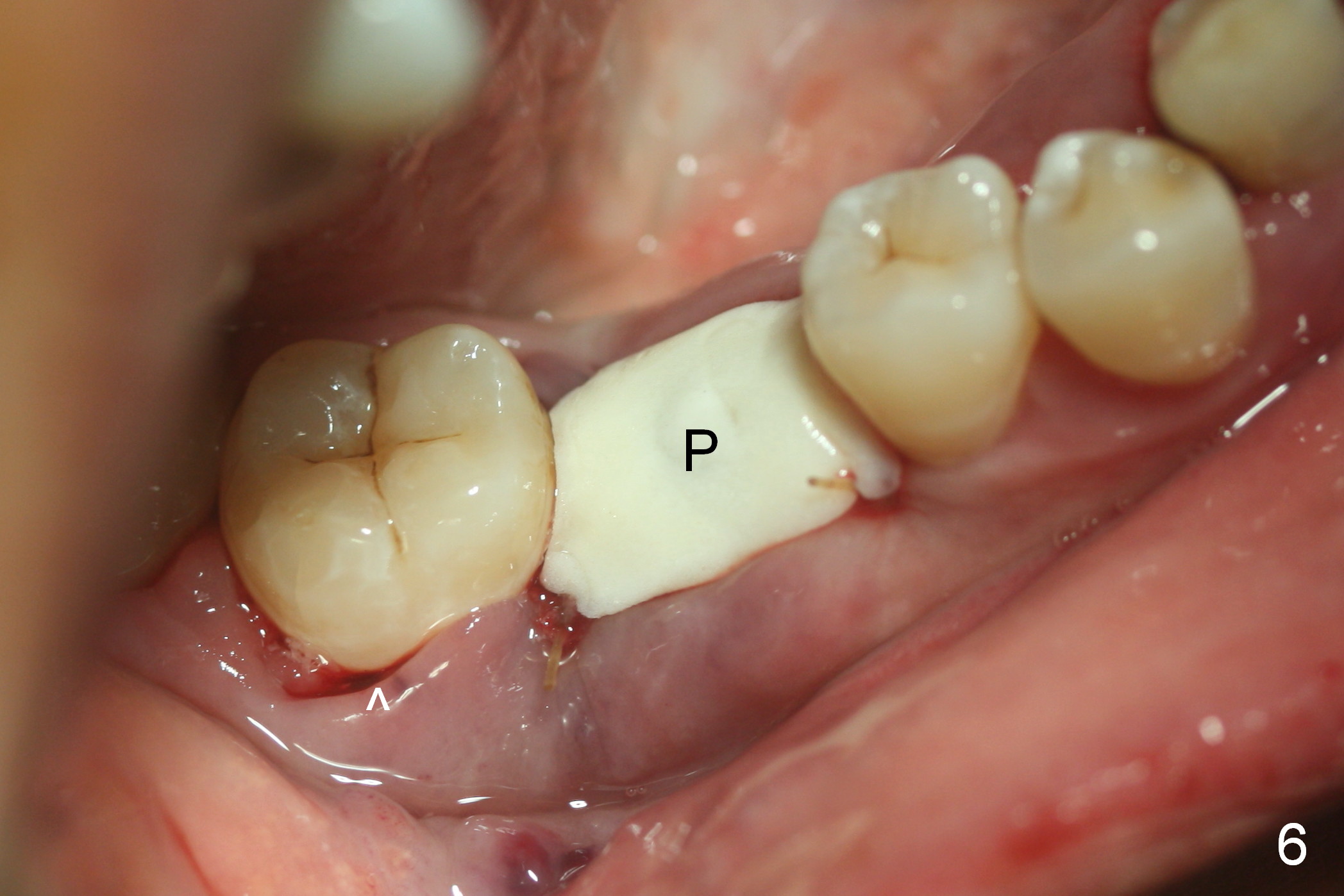
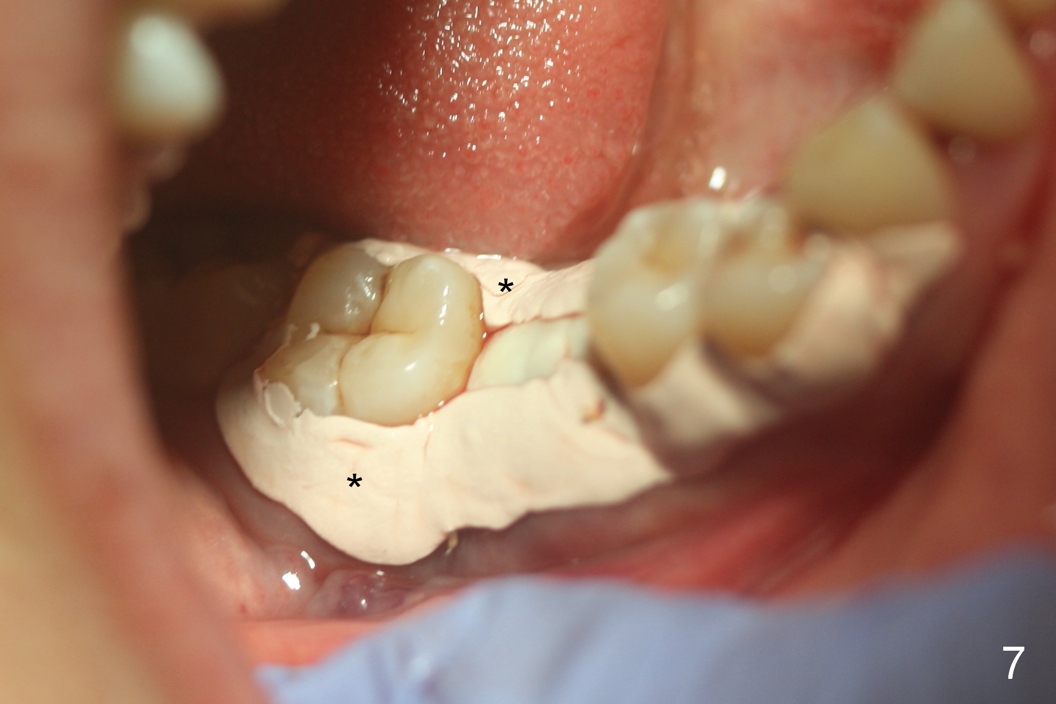
After cleaning the root surface with a surgical curette, a 1.6 mm pilot drill is used to start osteotomy (O) between the mesial (M) and distal (D) roots, followed by a marking bur and 3.8 mm Magic drill 13 mm deep (Fig.1,2). Following root removal, the 3.8 mm drill is reused for 11 mm (Fig.3). After deepening the osteotomy for another 2 mm, a 4.5x13 mm implant is placed with insertion torque of 50 Ncm, followed by allograft (*, .5-2 mm) and a 5x4(2) mm abutment (Fig.4,5). An immediate provisional is fabricated to close the socket (Fig.6 P (lock in)). The detached buccal gingiva (Fig.6 *) is kept in place by applying periodontal dressing (Fig.7 *). Red dashed lines in Fig.1, 3, 5 represents the superior border of the Inferior Alveolar Canal and Mental Foramen.
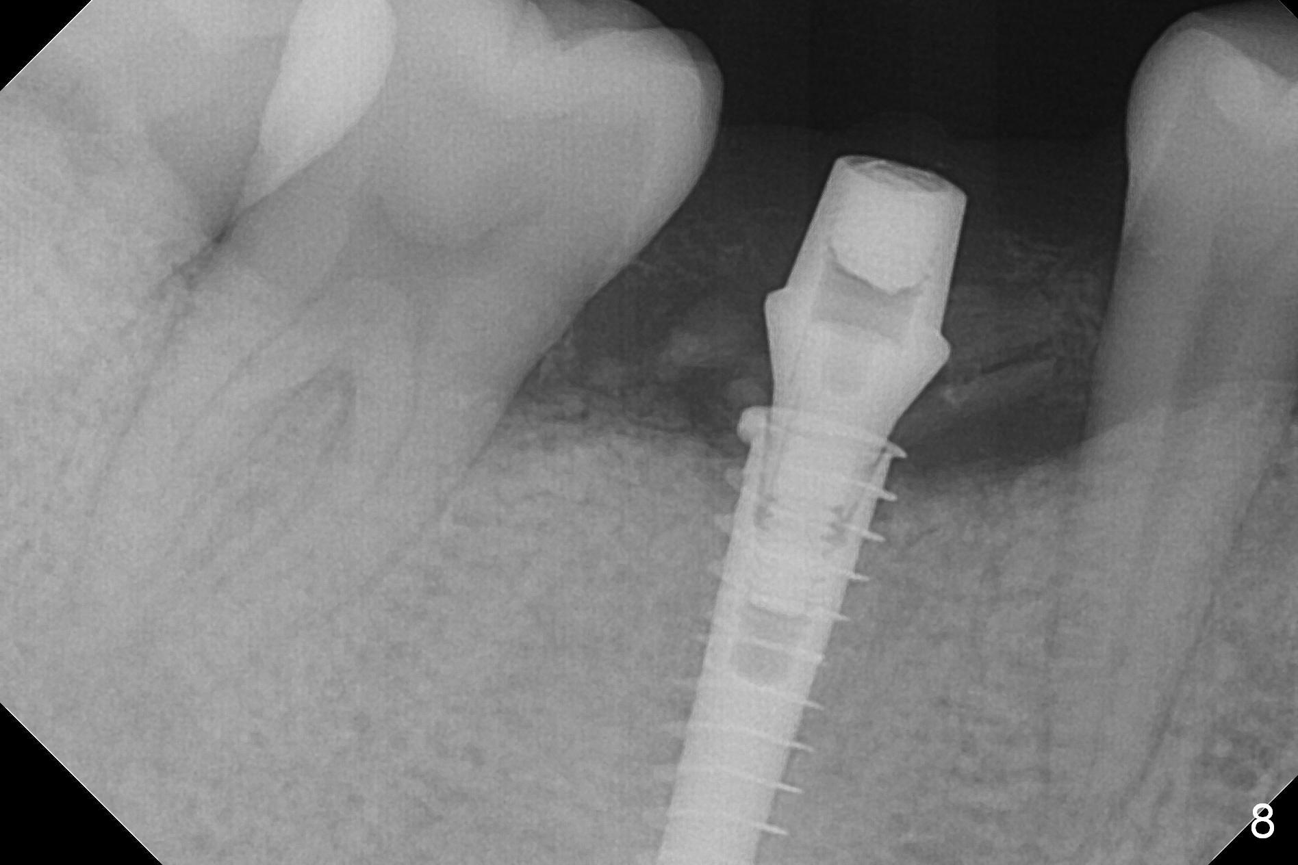
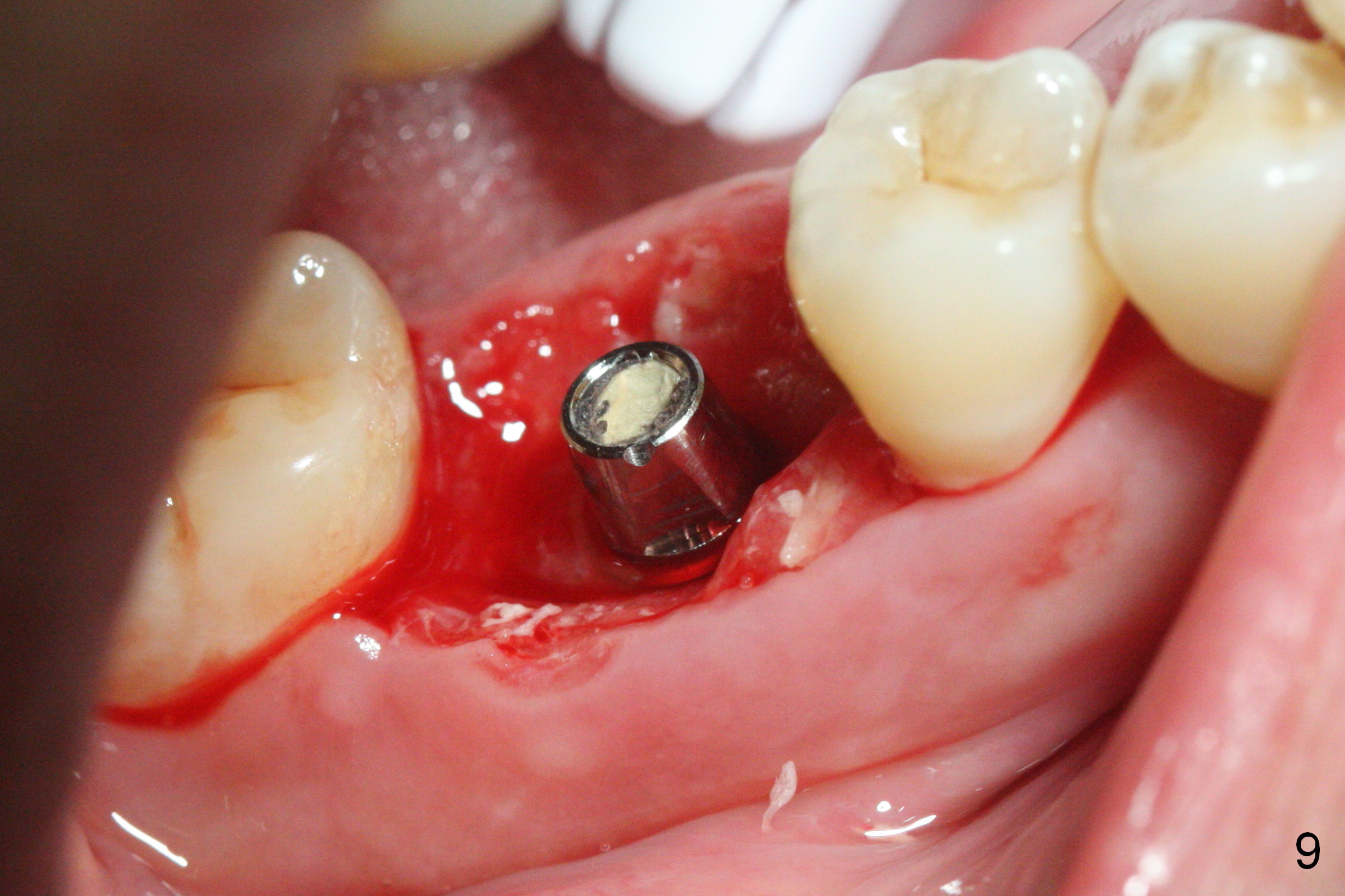
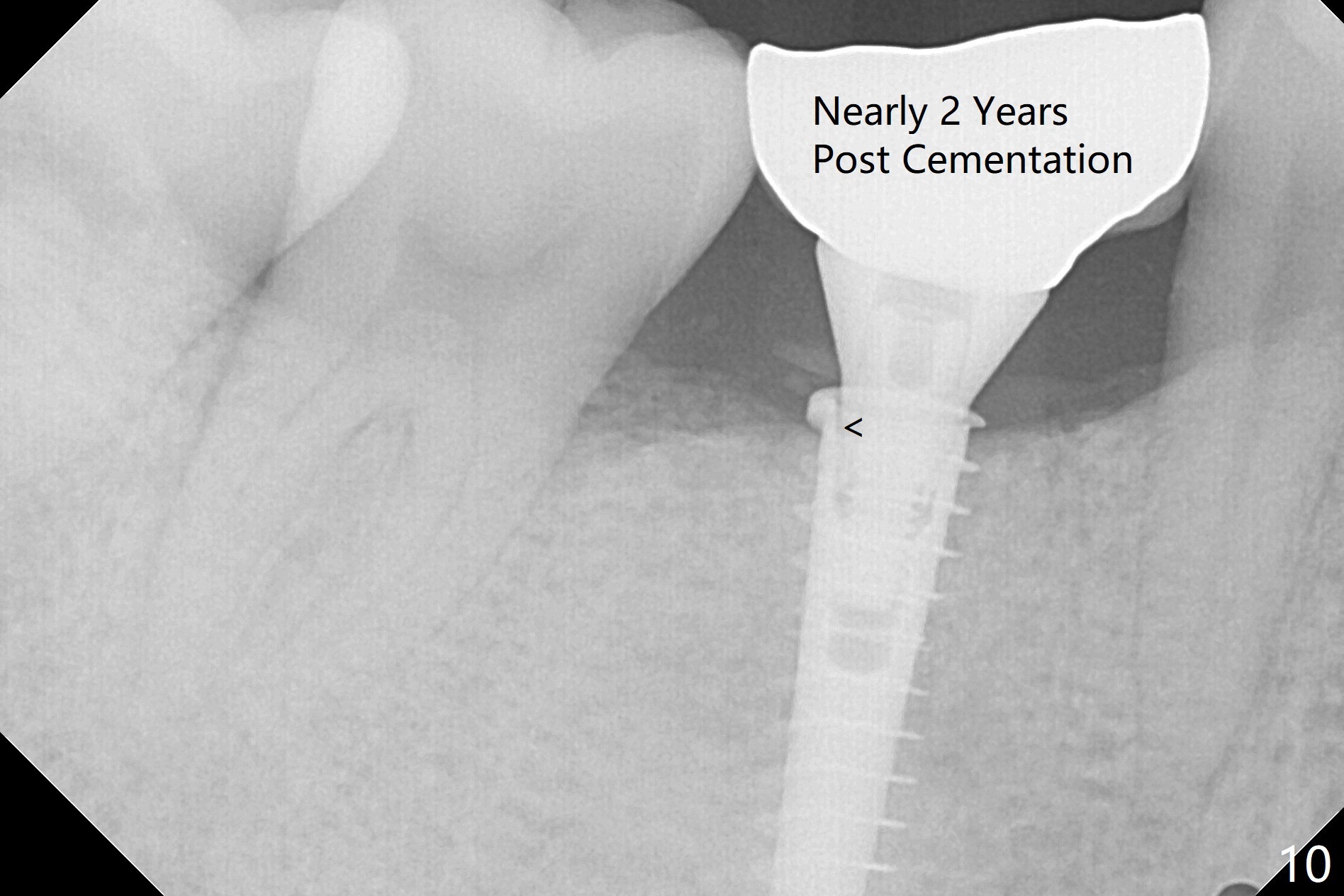
The patient returns 5.5 months postop for final restoration. The implant appears to have osteointegrated (Fig.8). When the locked in provisional is removed, the gingiva bleeds (Fig.9). She will return in 2 weeks. The abutment remains incompletely seated nearly 2 years post cementation (Fig.10).
Return to Lower Molar Immediate Implant, Prevent Molar Periimplantitis (Protocols, Table), IBS Xin Wei, DDS, PhD, MS 1st edition 08/08/2016, last revision 03/29/2021