|
|
|
|
|
|
|
|
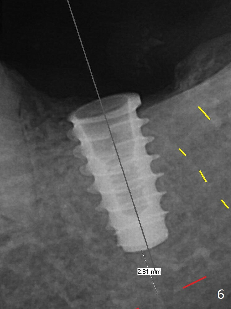
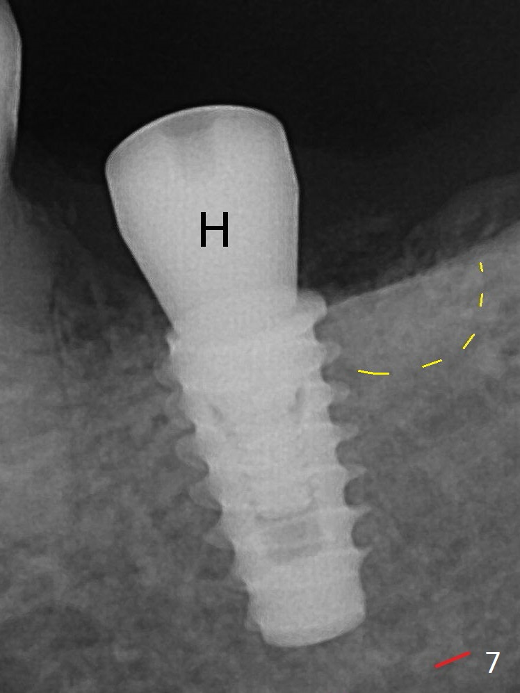
After Marking Bur and 4.3 mm Magic Drill, a 5x9 mm IBS implant is placed with 2.8 mm clearance from the Inferior Alveolar Canal (Fig.6). Following deepening the osteotomy with Final Drill, the implant is placed deeper (Fig.7). The osteotomy happens to be established in the mesial socket, since the distal socket has not completely healed (Fig.6 yellow dashed line). Granulation tissue is removed. Since the lingual crest is lower than the buccal one (Fig.1 B), there is lingual thread exposure after implant placement (Fig.5). The exposed thread is covered by bone graft (autogenous bone, allograft and Osteogen, Fig.5 pink circles). Some of the graft is apparently pushed into the distal socket (Fig.7 yellow dashed line) post GBR and suture. As the implant is placed twice, insertion torque is <10 Ncm (although the implant is stable). A 5x3 mm healing abutment is placed (Fig.7 H).
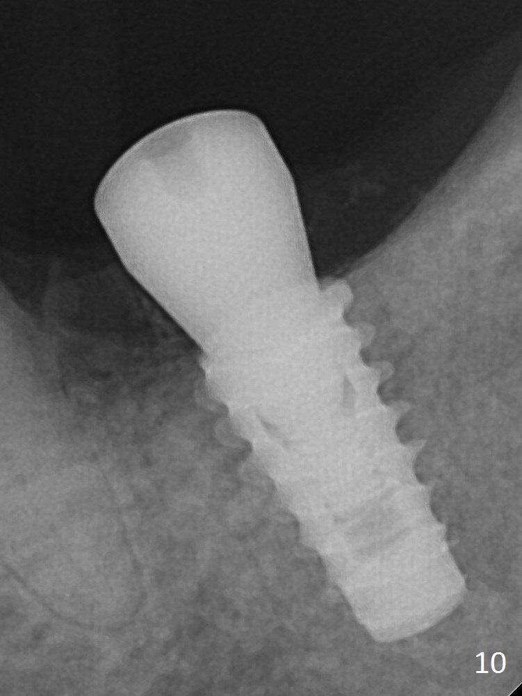
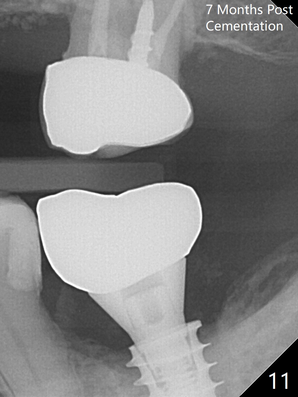
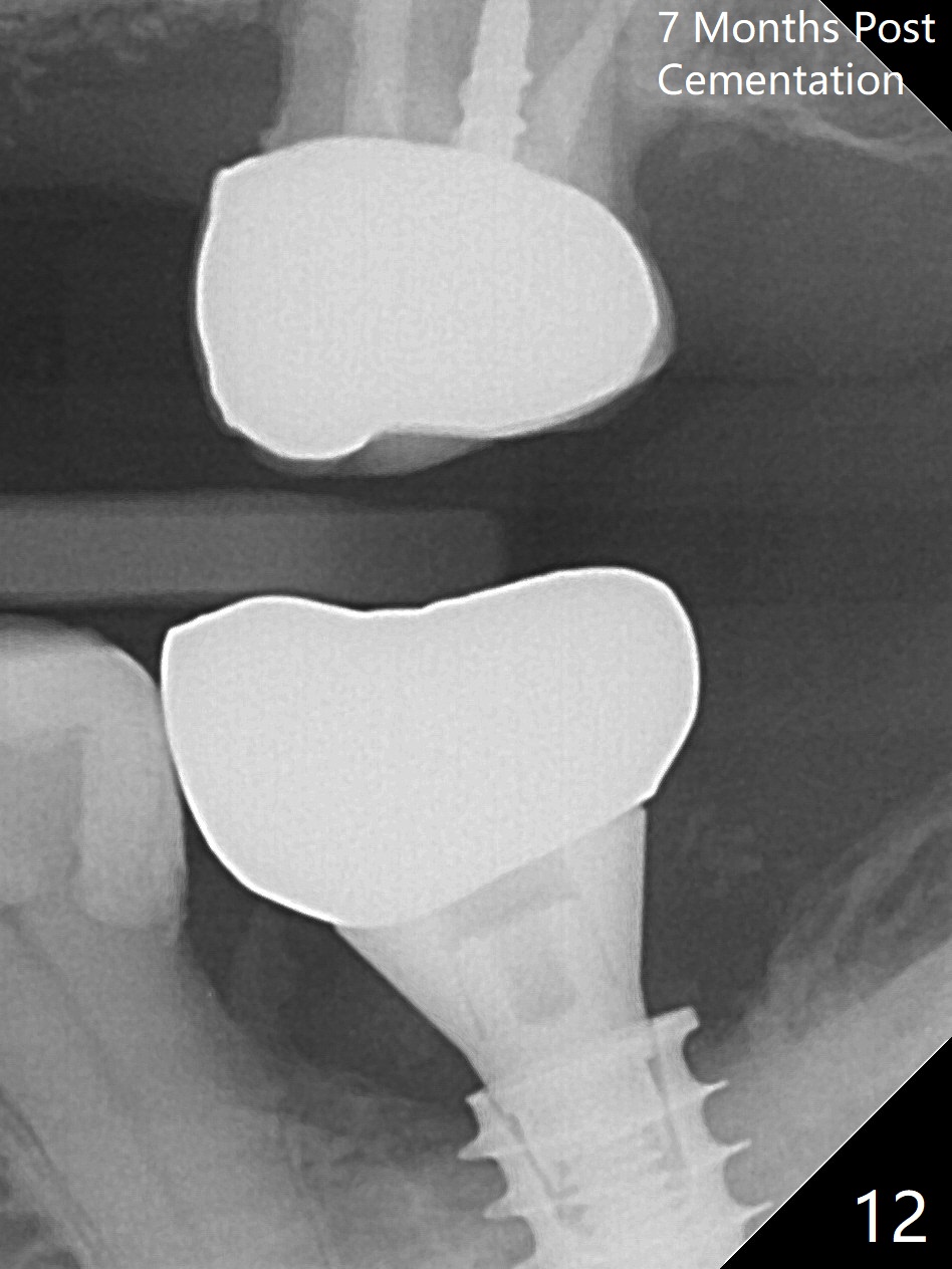
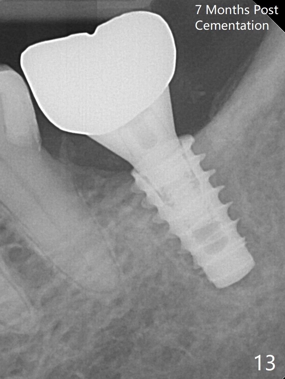
There is no apparent bone loss 4 months postop (Fig.10). There is chewing pain in spite of previous occlusal adjustment 7 months post cementation (Fig.11-13: increased radiolucency around the implant). The crown/abutment is removed and a healing abutment is placed (6x3 mm); the implant is found to be stable. The patient will return for re-evaluation in 3 months. Check proximal contact after crown reseating. Occlusion should be light, since #18 is the only molar with a natural opposing tooth (heavy occlusion).
Mesial Socket Placement Last Next
Xin Wei, DDS, PhD, MS 1st edition 01/20/2017, last revision 08/30/2018