|
|
|
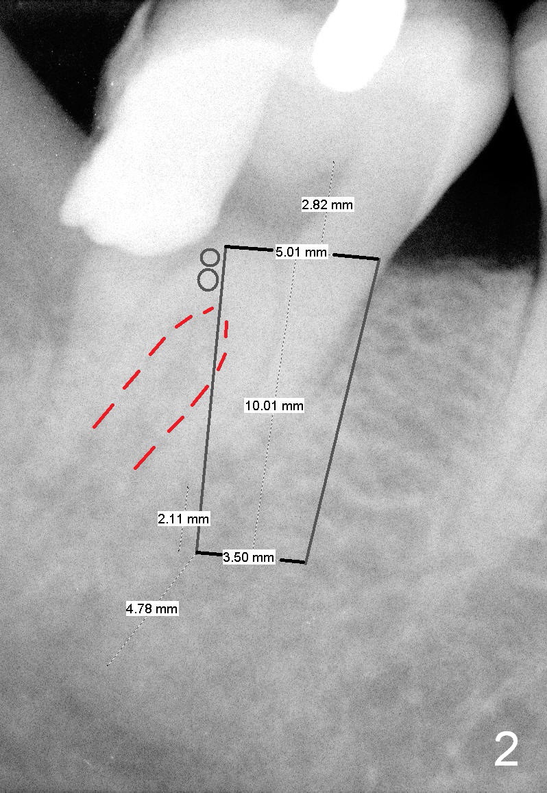
As planned, the septum (Fig.2 red dashed line) provides implant (black outline) with stability. Black circles: bone graft placed coronal to the septal crest.
When the implant (5x10 mm) is placed in the mesial socket, it has more bone contact and is farther away from the Inferior Alveolar Nerve (4-5 mm) than placed along the axis of the socket.
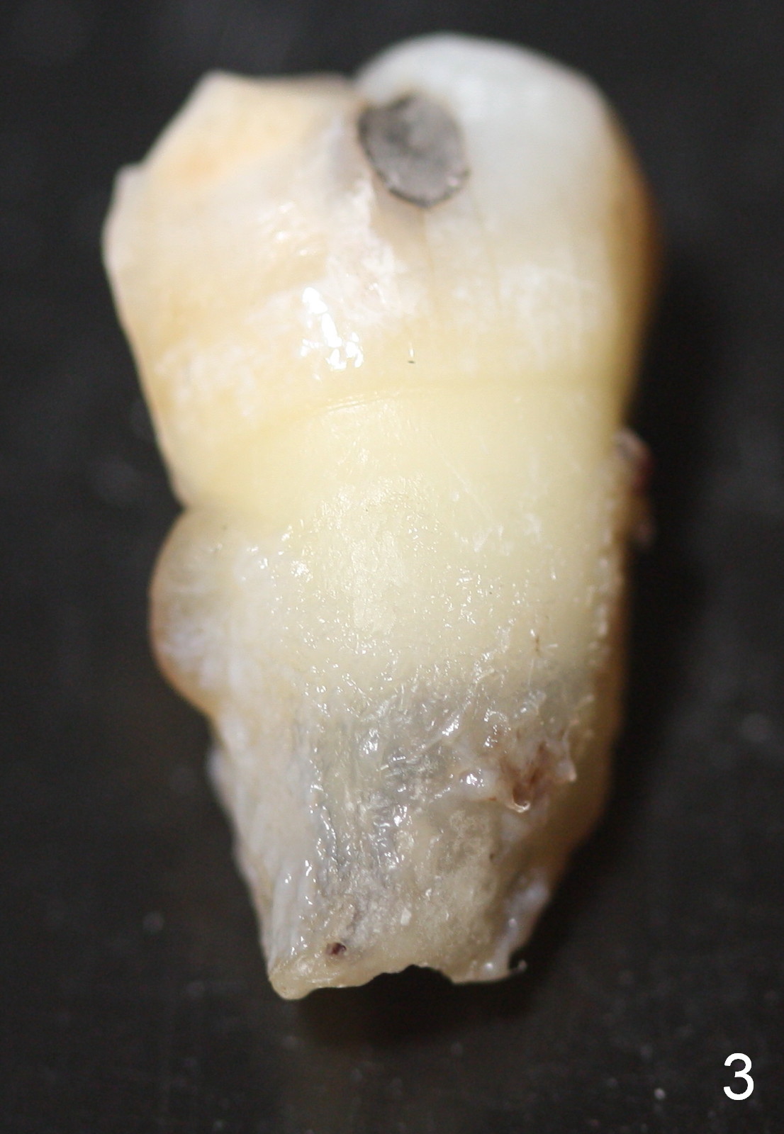
Although there appear two roots of the 2nd molar (Fig.2), the extracted tooth looks to have a single root (Fig.3, buccal view). In fact the mesial and distal roots fuse buccally, whereas have a little separation lingually. In fact the septum between the roots is not as big as it looks and fractures while the tooth is extracted.
When the septum is fractured, the implant size has to be increased, counting more on the contact with the buccal and lingual plates of the socket.
Xin Wei, DDS, PhD, MS 1st edition 06/25/2015, last revision 06/25/2015