.jpg)

|
|
|
|
|
|
|
|
|
|
|
|
|
|
|
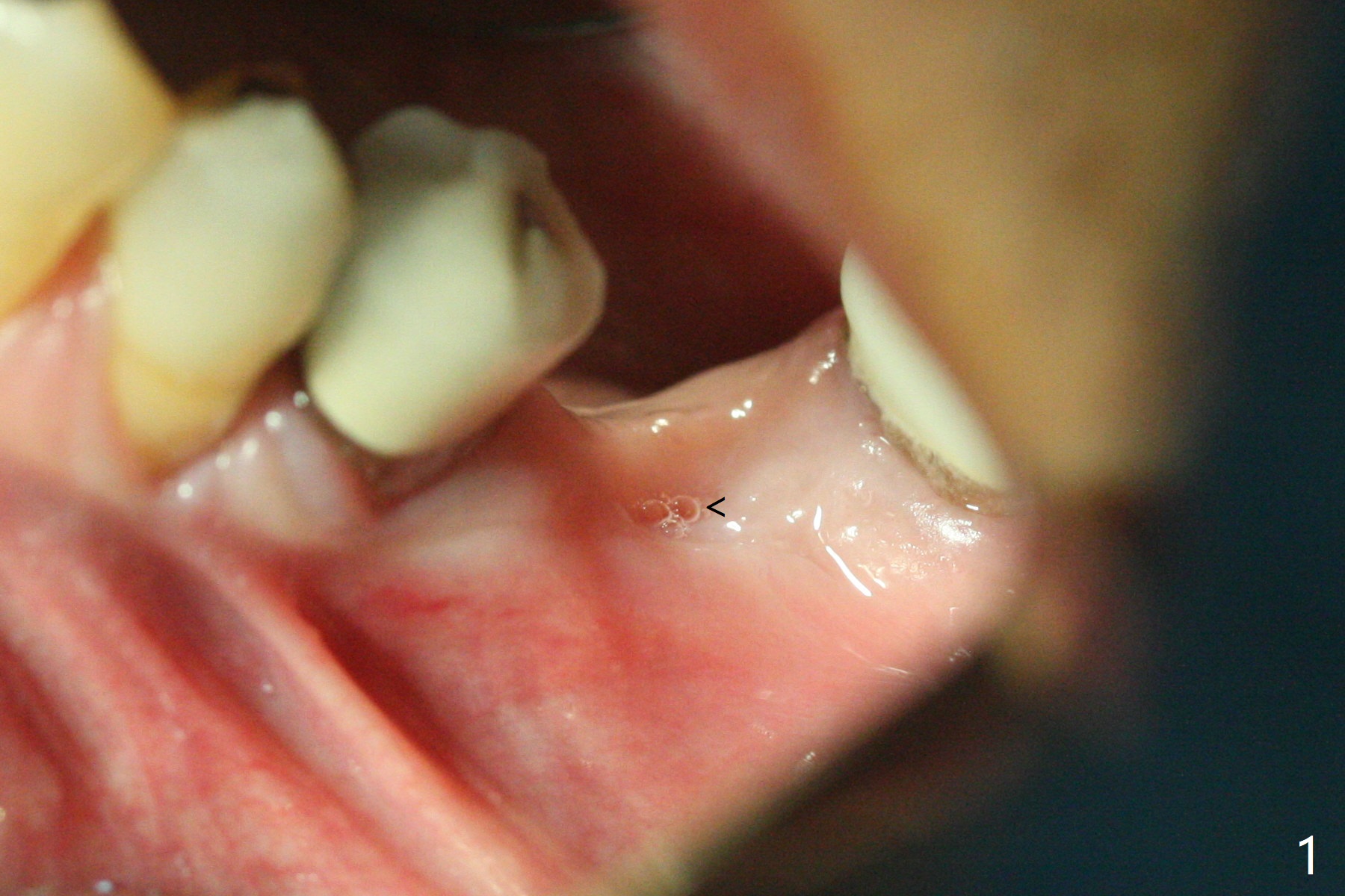
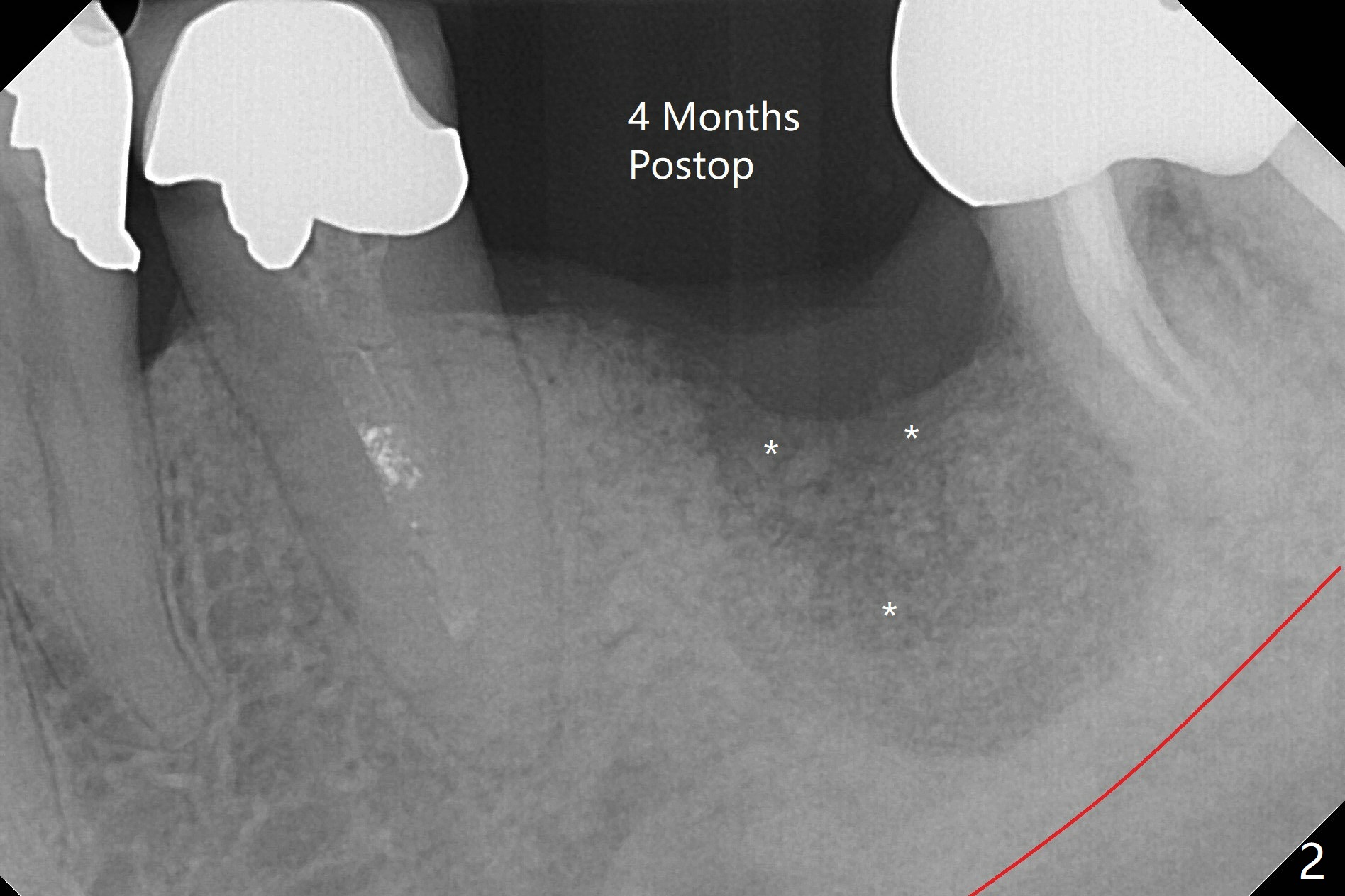
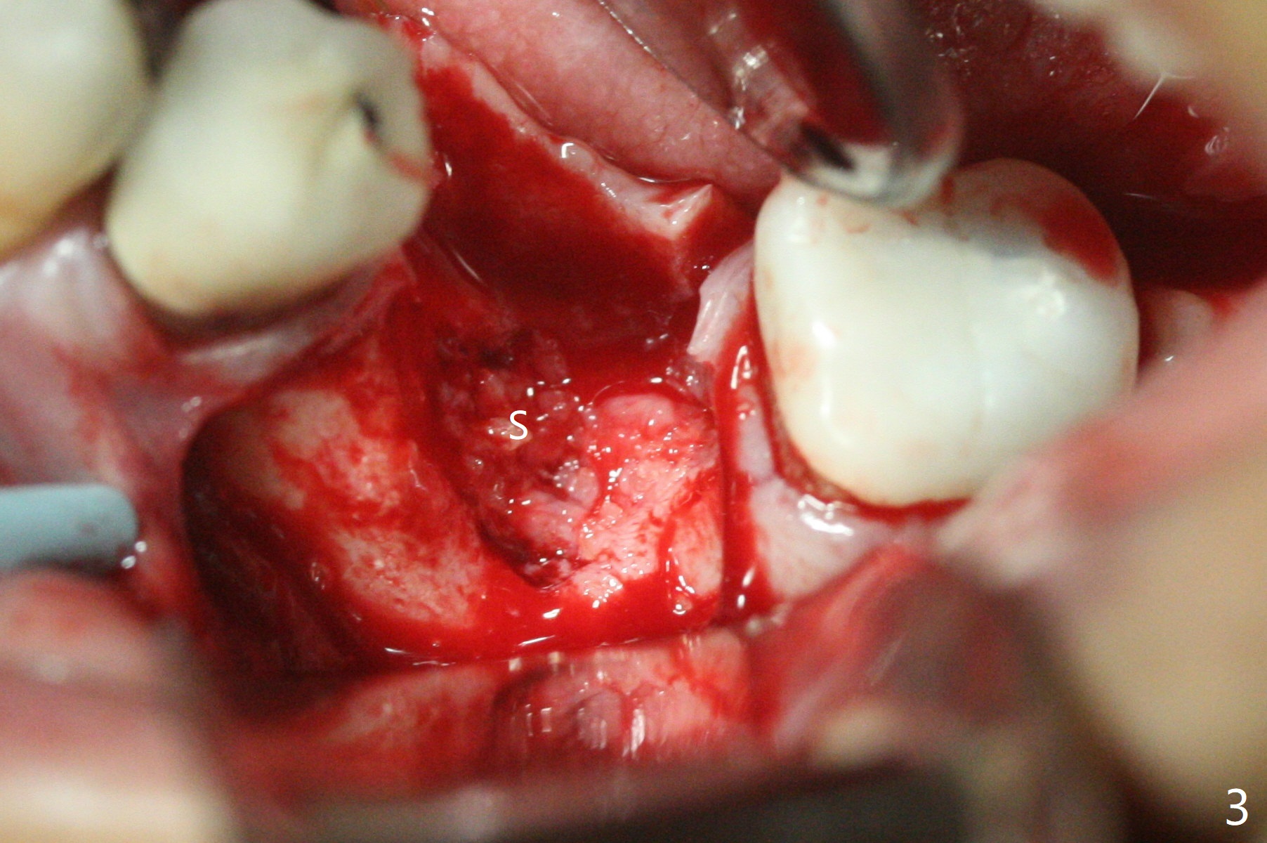
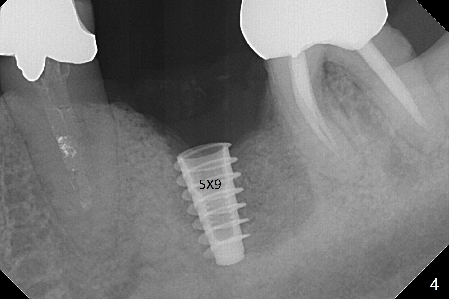
Incompletely Healed Bone Graft
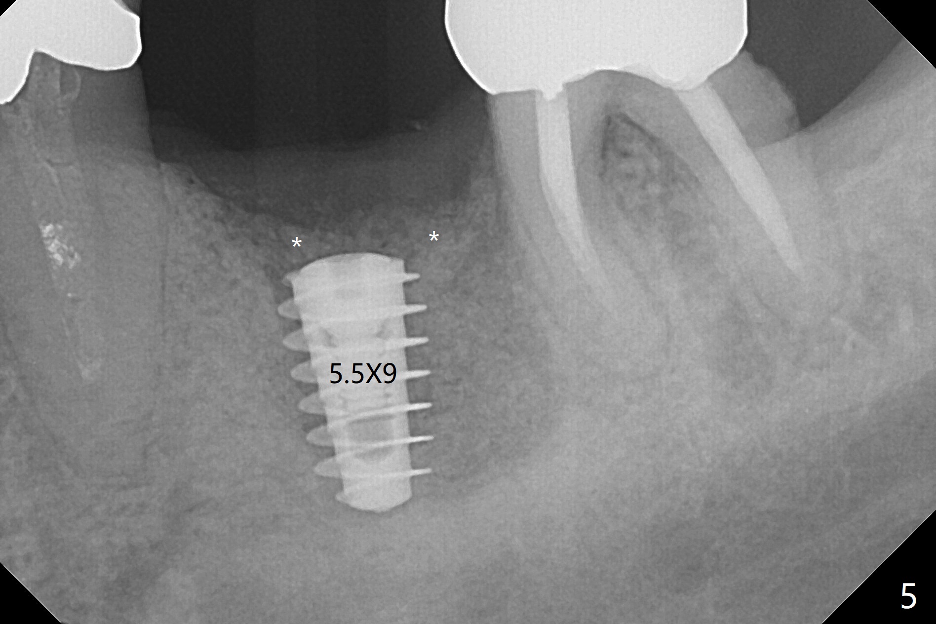
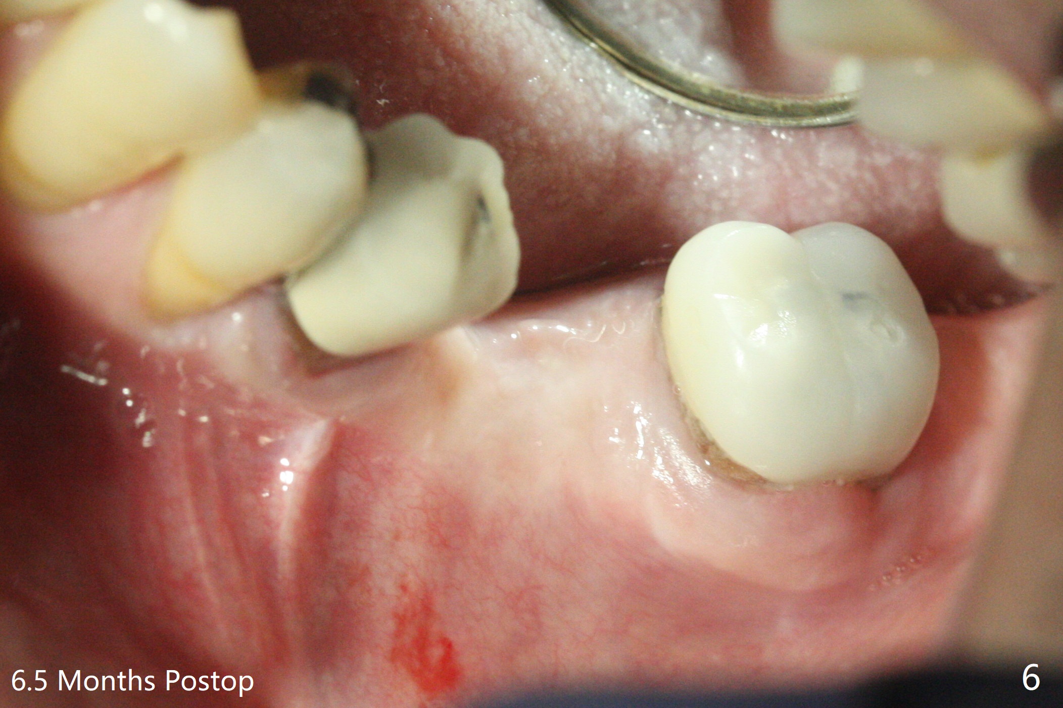
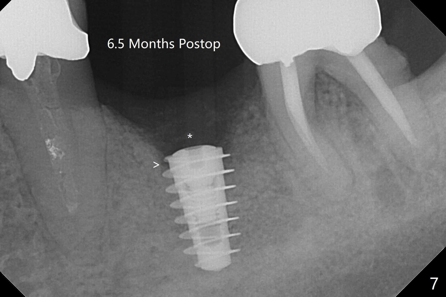
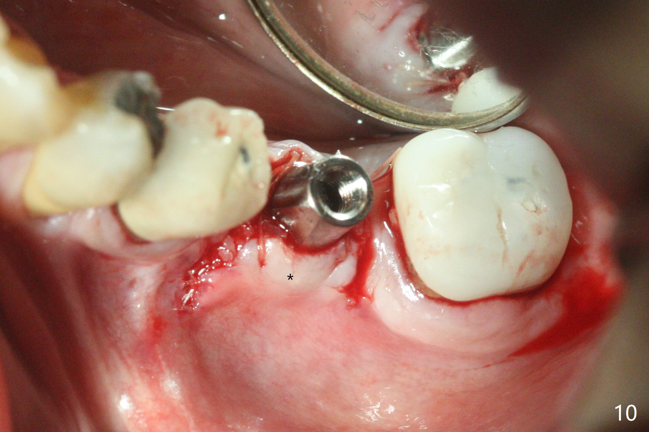
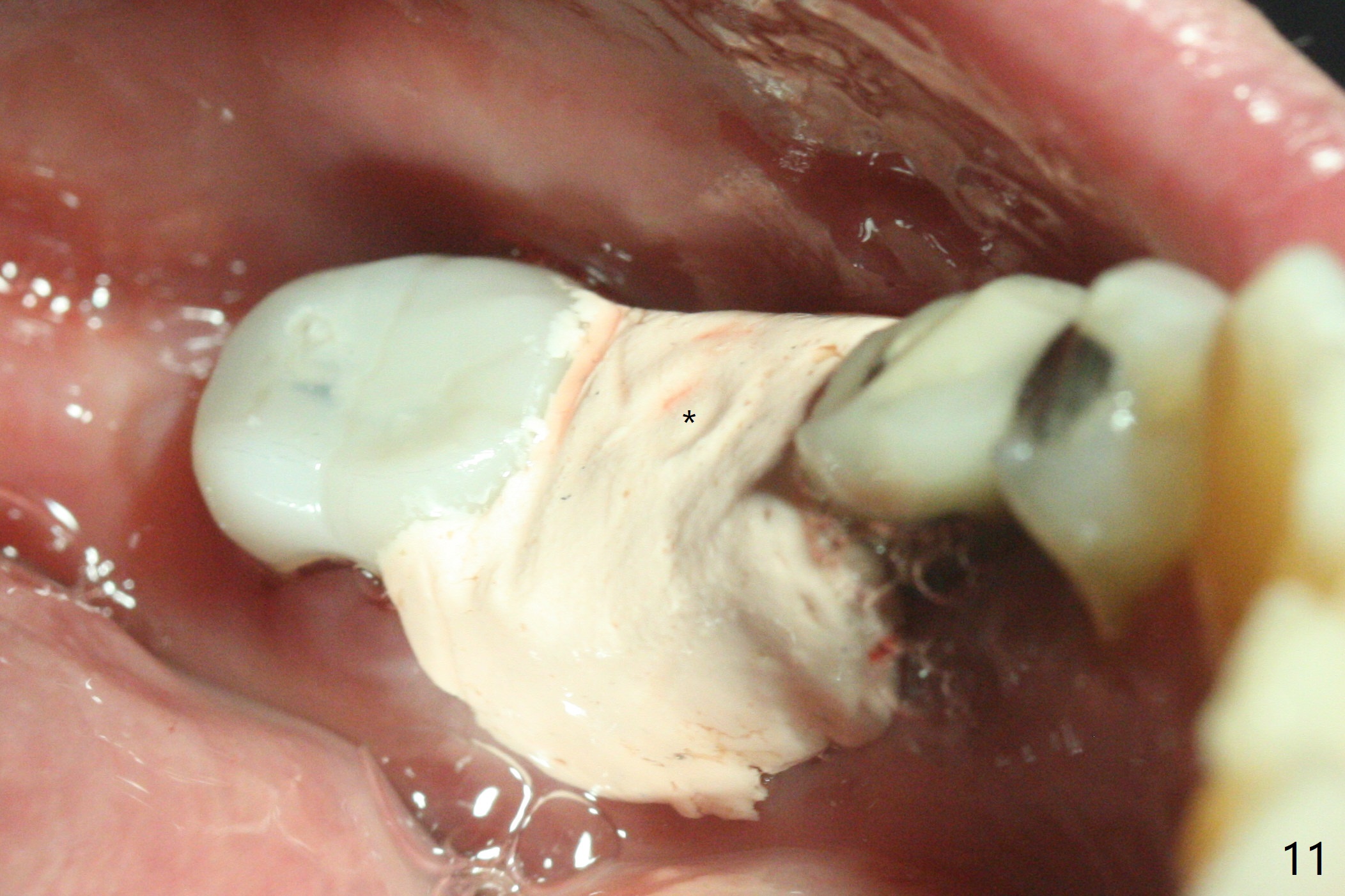
When the patient returns 4 months post implant removal and bone graft, the ridge at #19 is concave with a small hole which communicates with the underlying bone (Fig.1 <). The low density bone in the middle of the socket is ignored (Fig.2 *, 3 S (soft)). With under drilling, primary stability of tissue-level taps is lower. A bone-level dummy implant is placed with low torque (Fig.4). After removal of granulation tissue distal to the osteotomy, a larger implant is inserted with ~15 Ncm; mixture of autogenous bone and allograft is packed (Fig.5 *). The latter is covered by 12x12 mm BioXclude and sutured with 4/0 Chromic Gut tension free. 植牙后6.5月牙槽嵴饱满,角化龈宽(图六(刚开始浸润麻醉))。术后6.5月切开证实植体上面没有骨质覆盖(如图七:*),第一螺纹可能暴露(>)。由于骨质吸收,大号基台置入没阻挡,或者困难(图八),第一螺纹暴露(>)需要再次植骨(图九:*)。放置大号基台(6x6(4)毫米)和植骨使萎缩牙槽嵴部分得到修复(图十,与Uncover前(图六)对比)。Uncover时即刻置入修复性基台好处是利用基台牙龈外部分强化牙周敷料固定,基台中央放置一个棉球后,敷料可以插入基台中央进一步增加固位(图十一(舌侧观):*)。
Return to
No Deviation
开场白
Xin Wei,
DDS, PhD, MS 1st edition 07/17/2020, last revision
03/07/2021