|
|
|
|
|
|
|
|
|
|
|
|
Pain Control
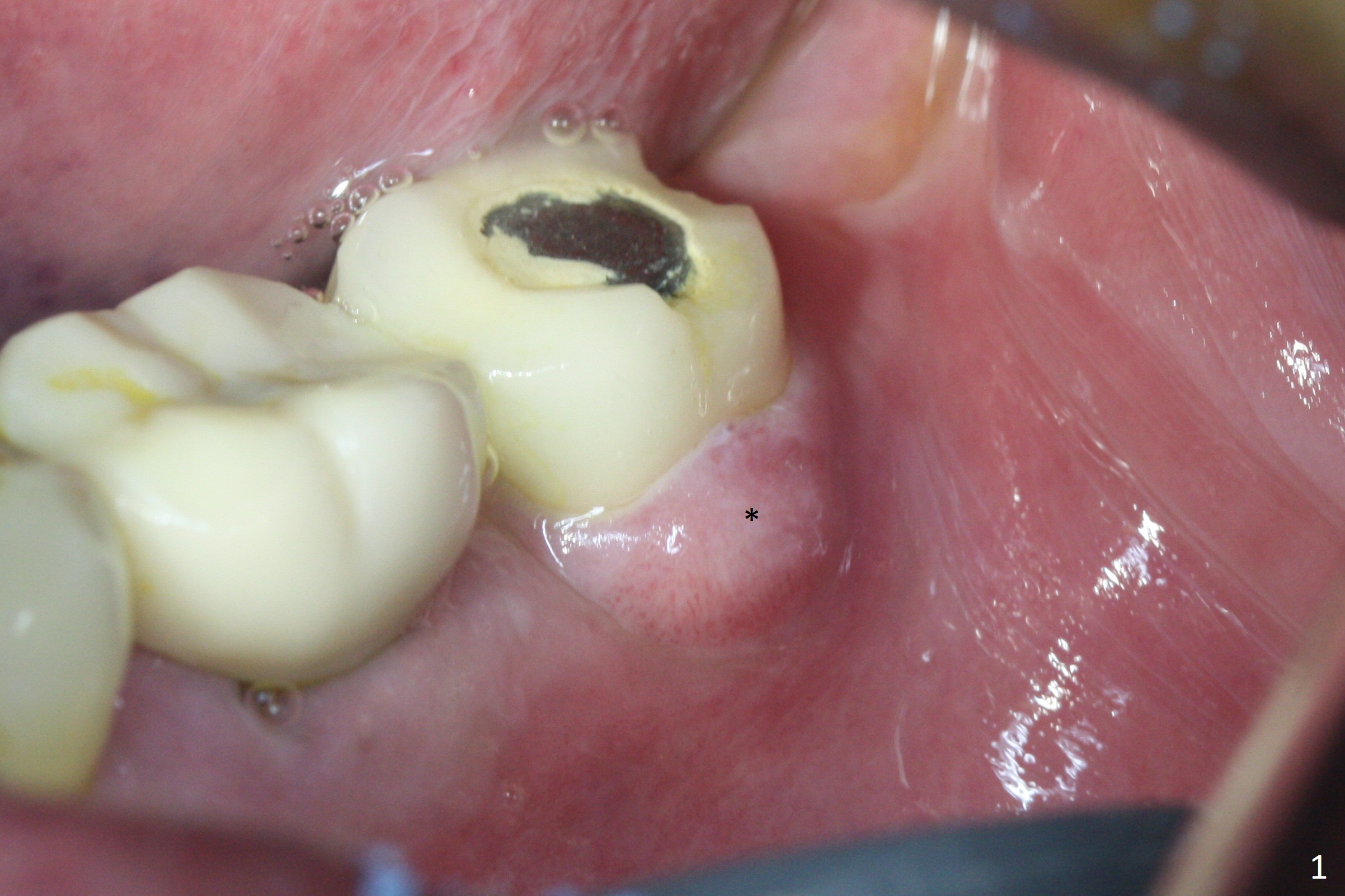
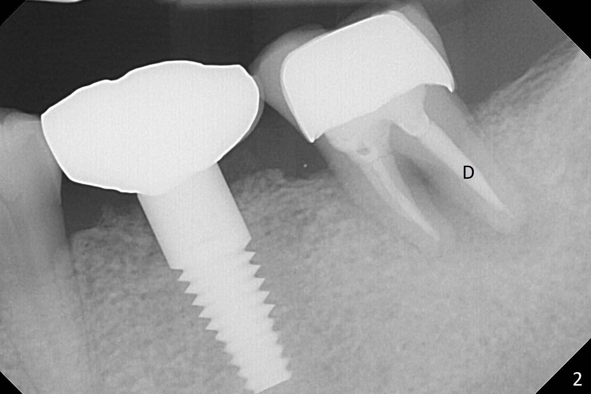
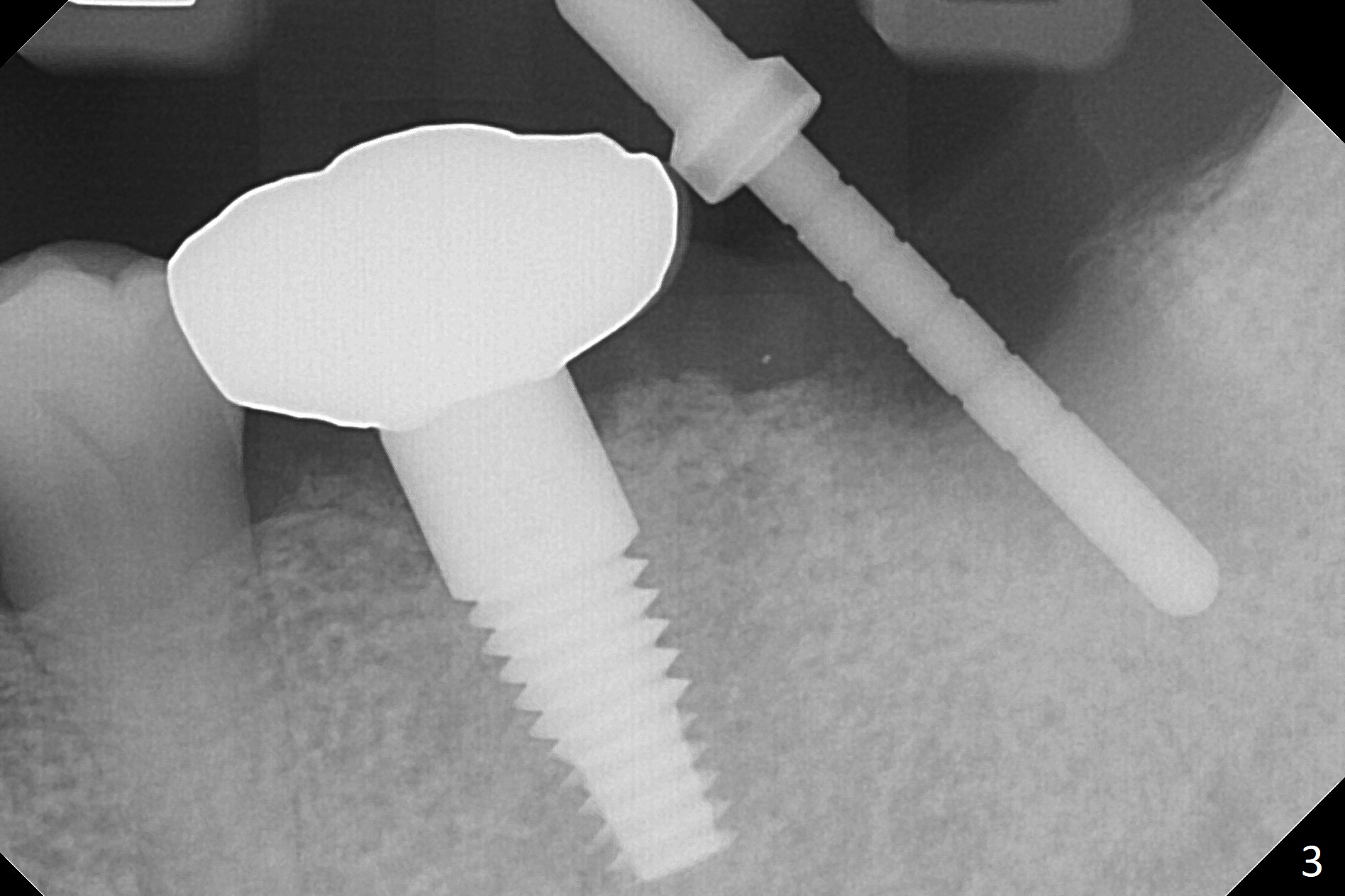
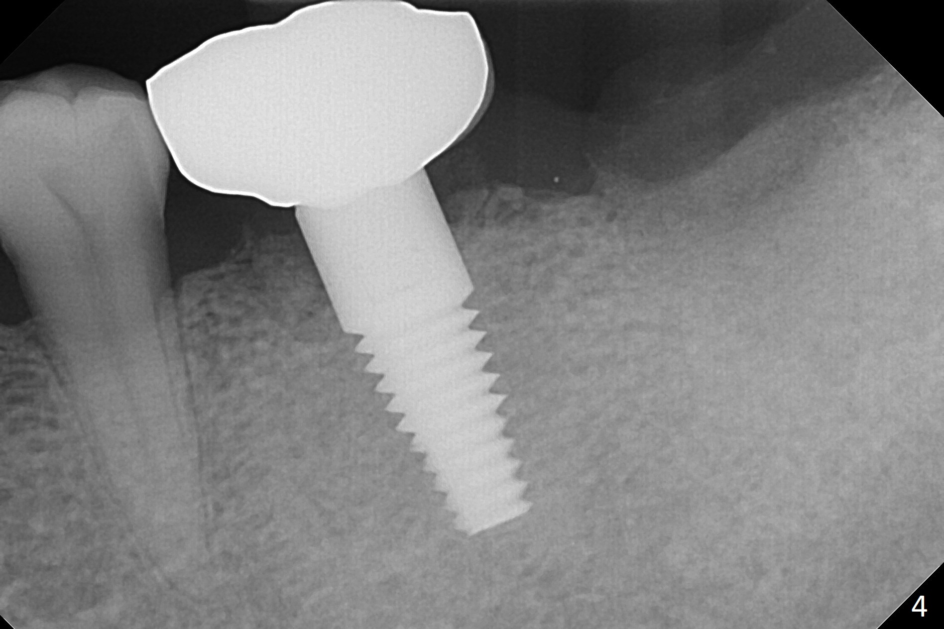
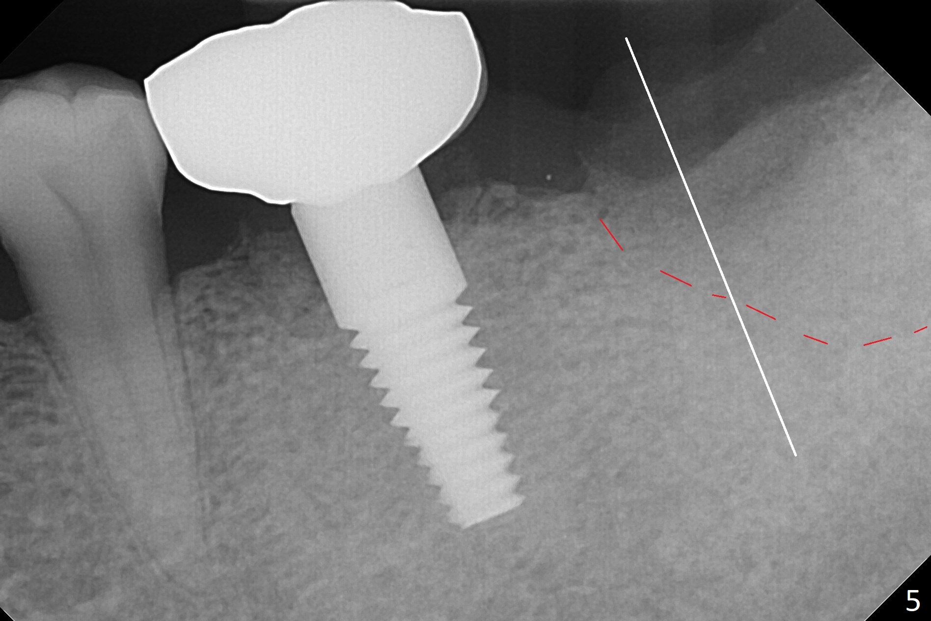
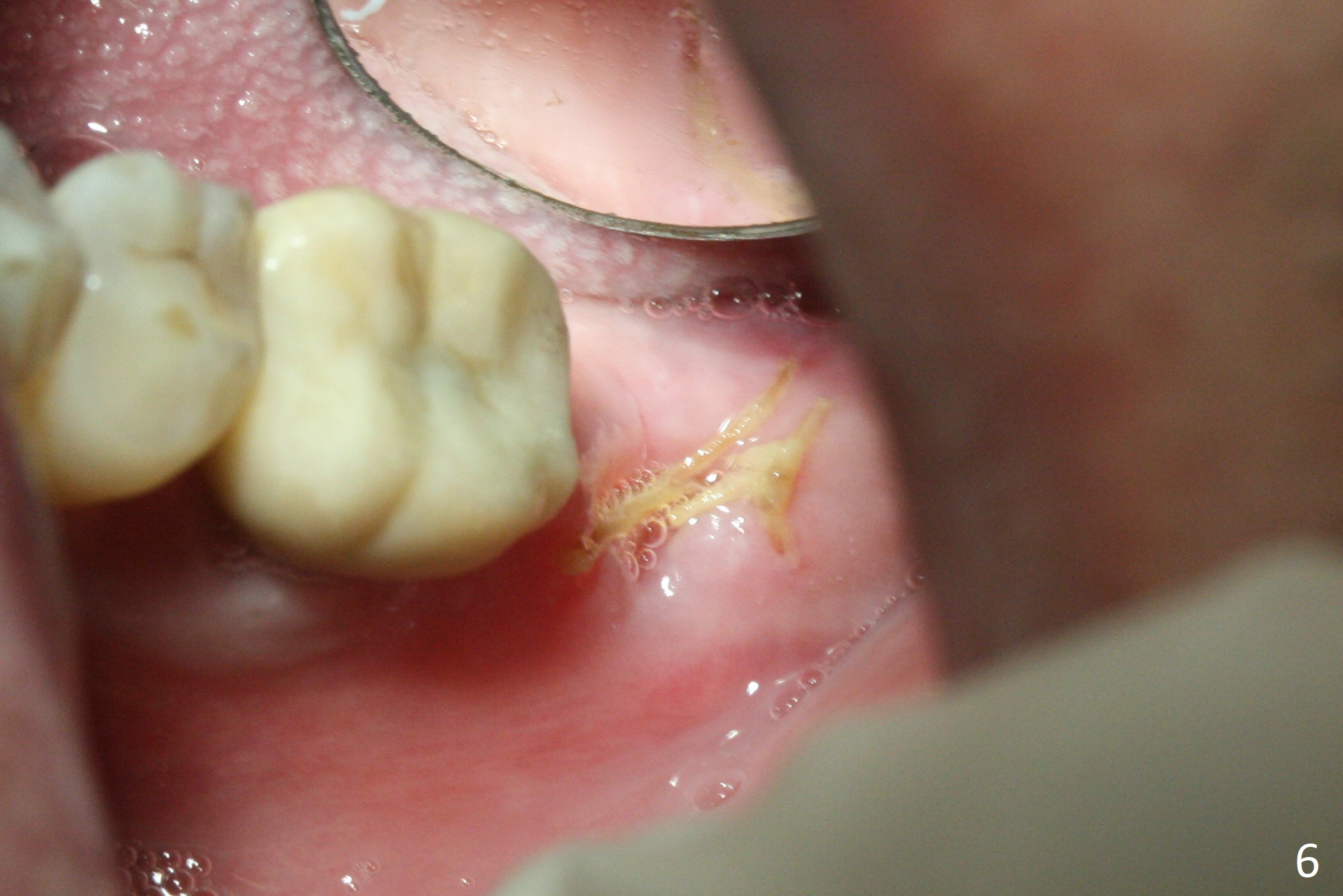
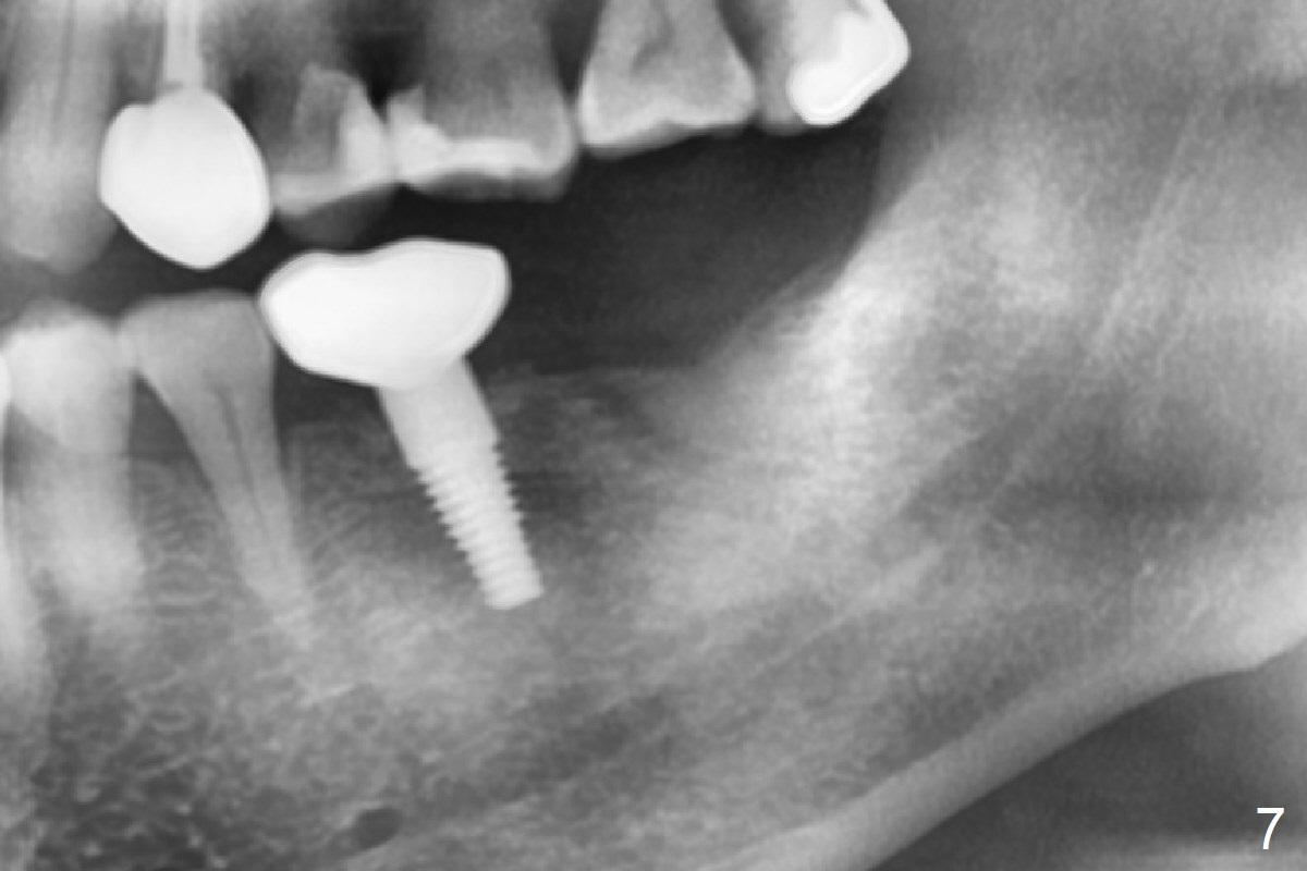
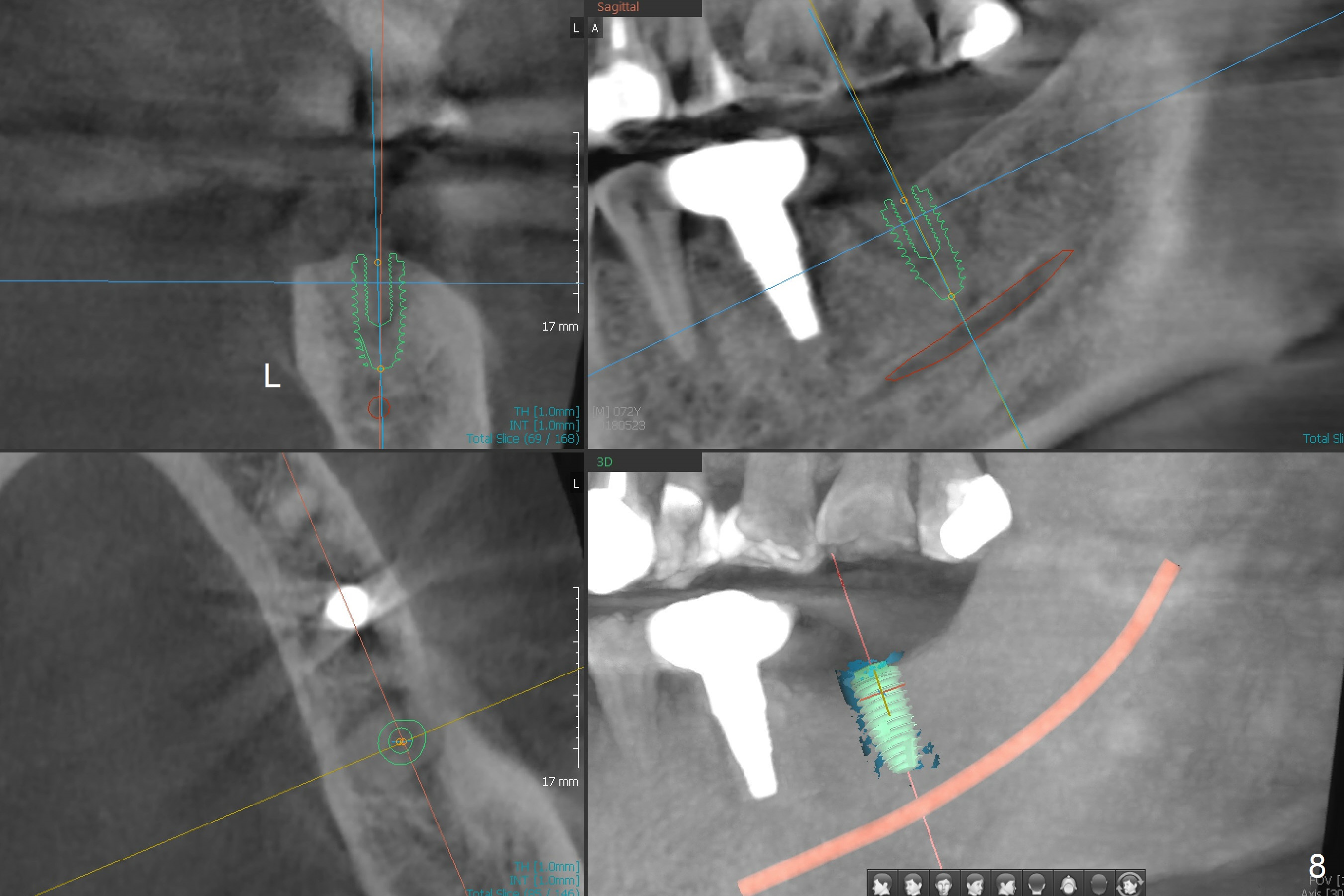
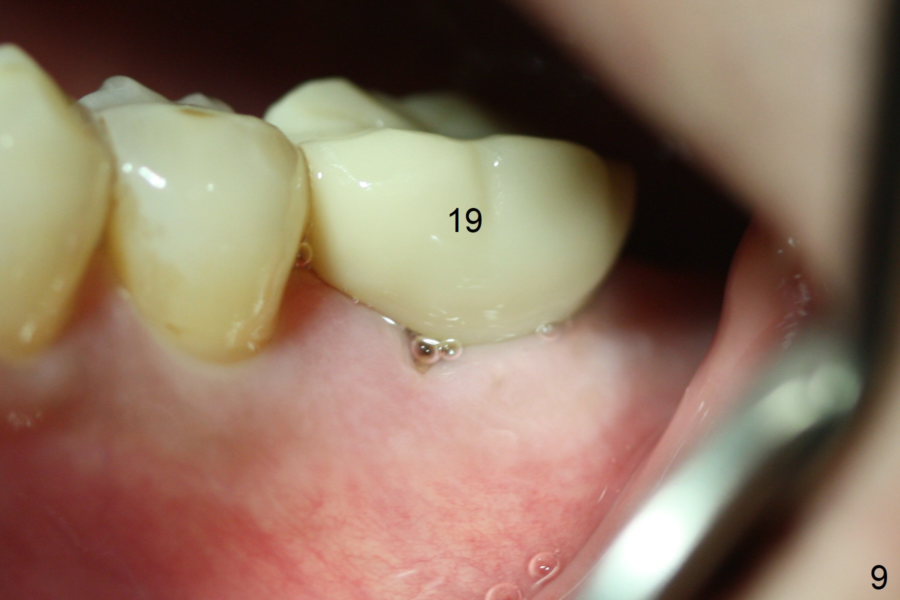
Due to severe furca (Fig.1 *) and distal root (Fig.2 D) infection at #31, there is pain when osteotomy is initiated in the apical native bone for 2 mm after extraction (Fig.3). The distal root is found to have vertical fracture. Socket preservation is performed with Vera Graft, collagen plug and 6-month membrane (Fig.4). In fact it would be possible to initiate osteotomy (Fig.5 white line) in the mesial slope (red dashed line) for implant placement (less infection (far from the lesion), less pain). There is postop pain and swelling, but the symptoms are less 7 days postop (Fig.6). The patient returns 6 months post socket preservation (Fig.7). The bone density at the healed socket is high (>1000 units); a 5x10 mm implant will be placed with guide (Fig.8). After implant placement at #18, make a buccal incision to explore the buccal gingival defect at #19 (Fig.9, 1), most likely due to extra bone graft.
Return to
Lower
Molar Immediate Implant, Prevent
Molar Periimplantitis (Protocols,
Table),
Armaments
Xin Wei, DDS, PhD, MS 1st edition 11/14/2017, last revision 04/14/2019