|
|
|
|
|
|
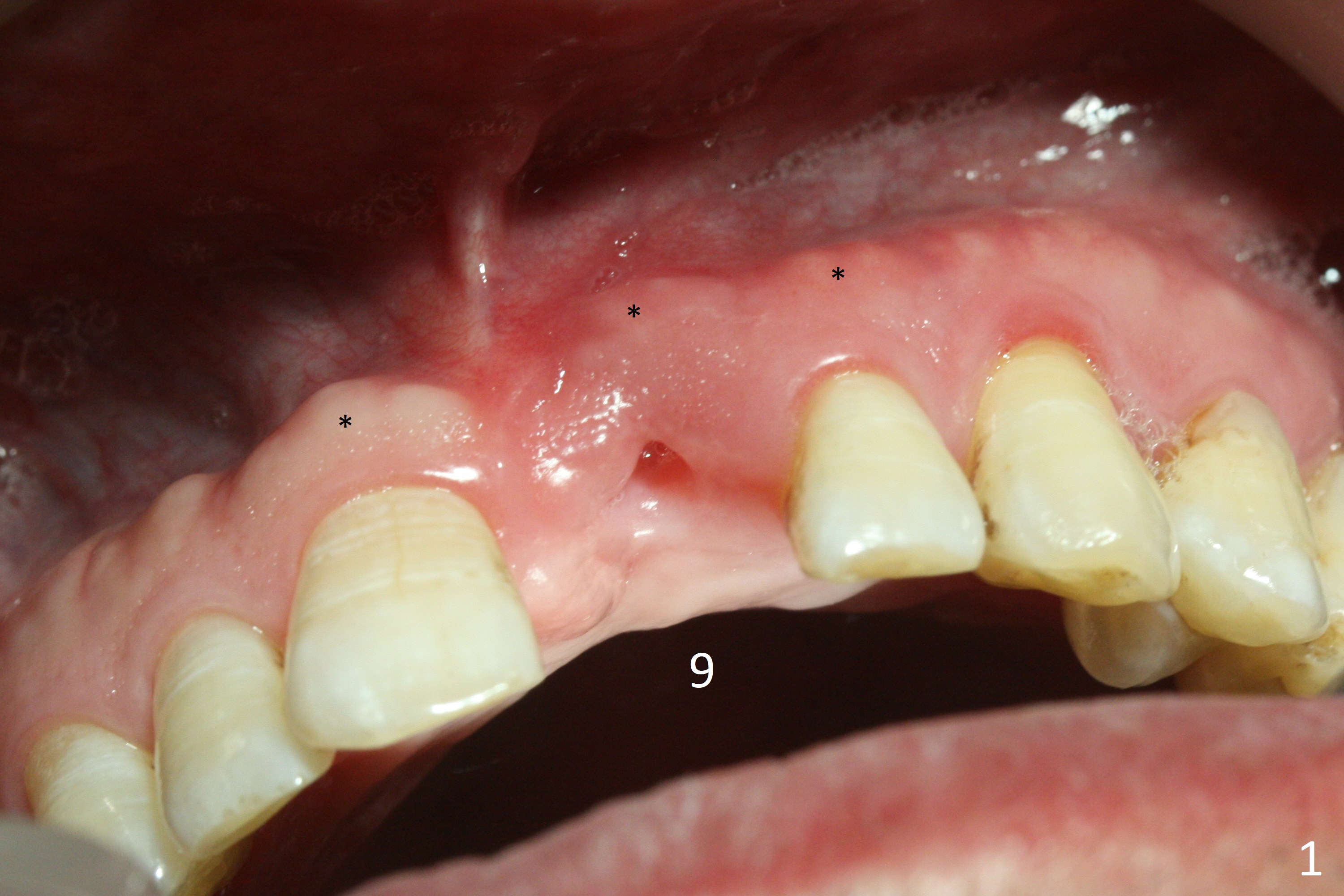
Preop examination shows mobility I of the teeth #8 and 10 and apparent occlusal trauma from #22-26 implant bridge. After occlusal equilibrium, incision reveals low, but moderate ridge at #9 (Fig.1). * coronal portion of the Incisive Canal. Once it is discerned, osteotomy should be distal.
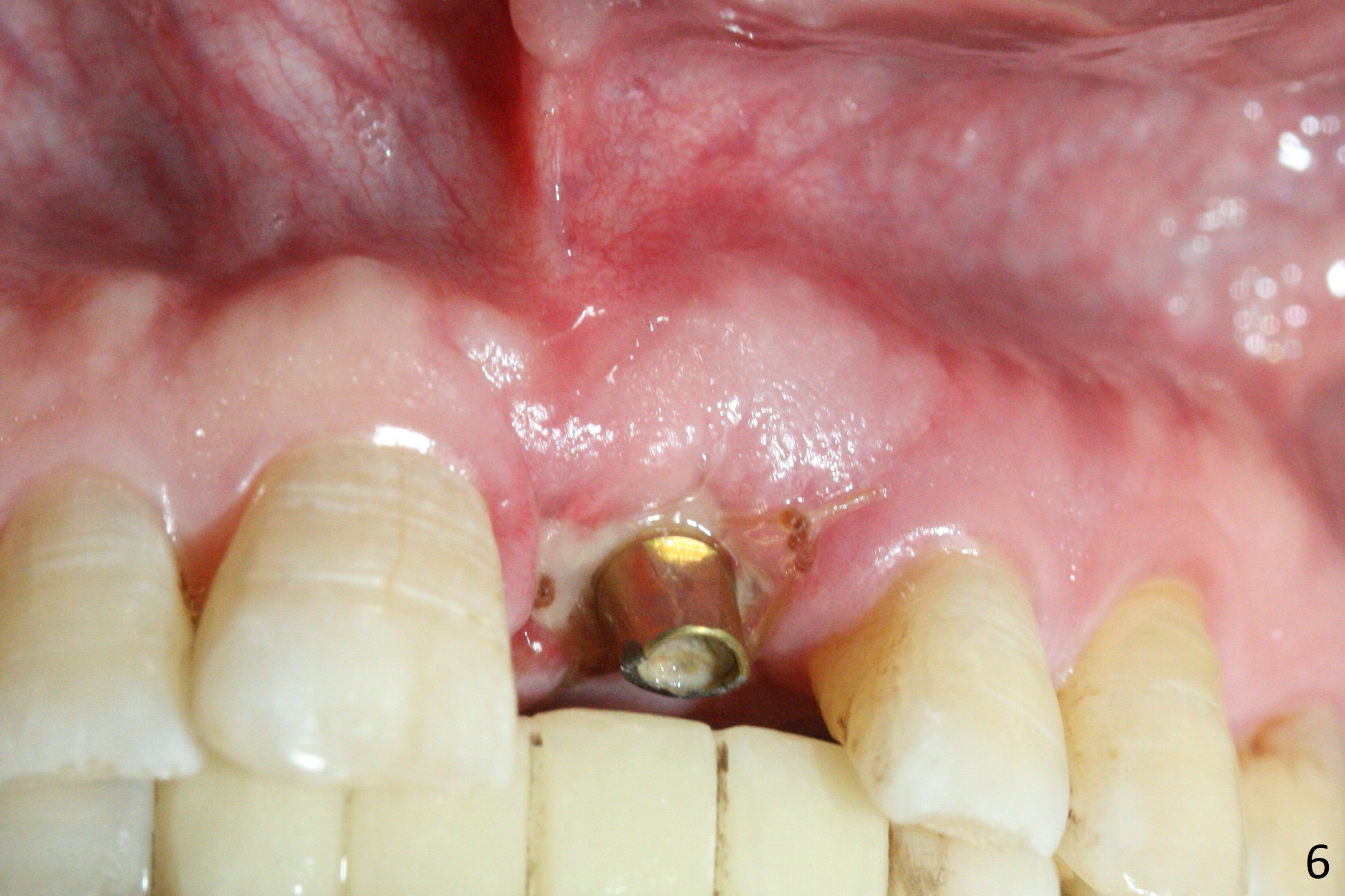
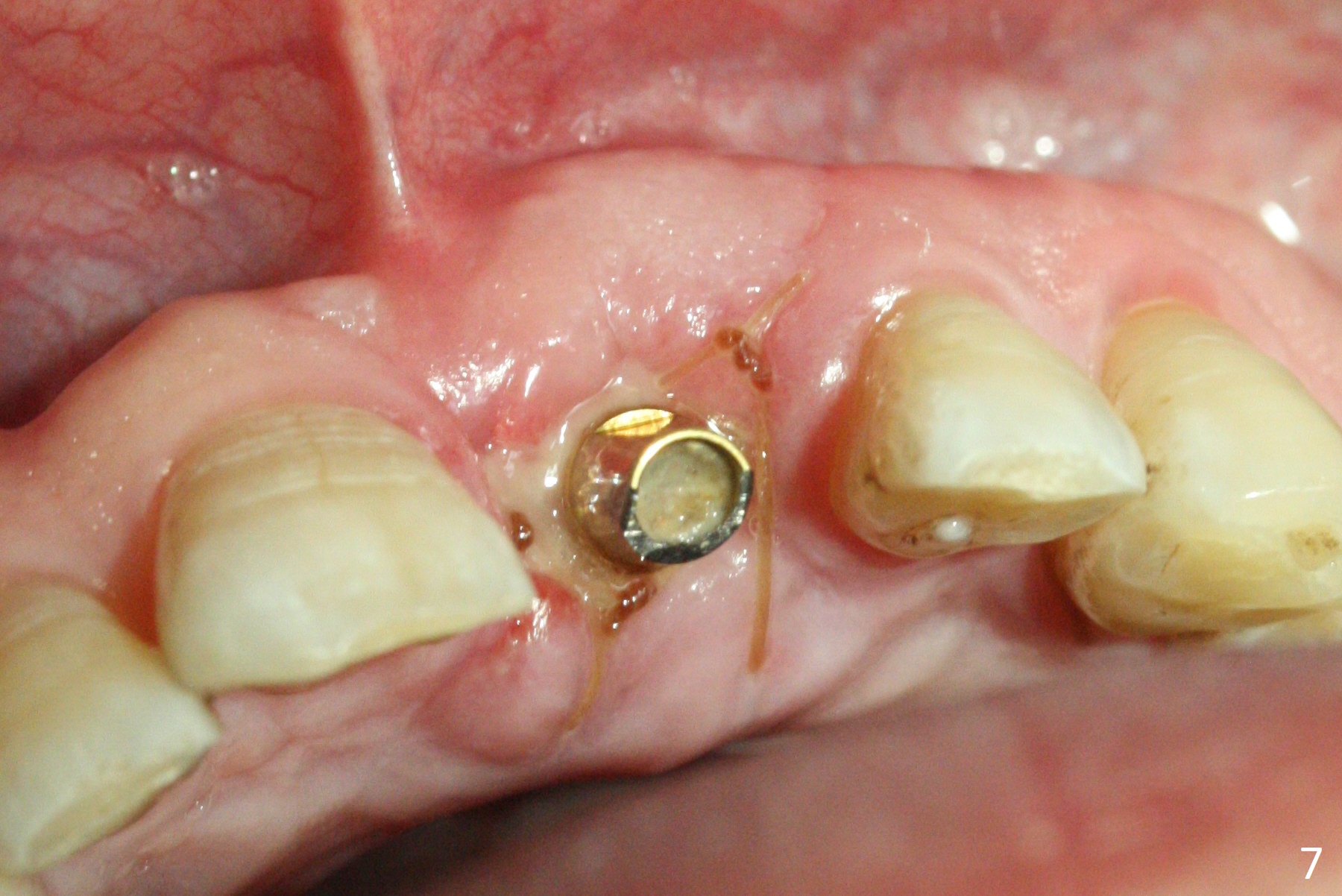
The buccal plate looks bulky with sufficient keratinized gingiva due to placement of the abutment and bone graft 1 week postop (Fig.6,7 (crown dislodgement)). The abutment seems to be short and palatal.
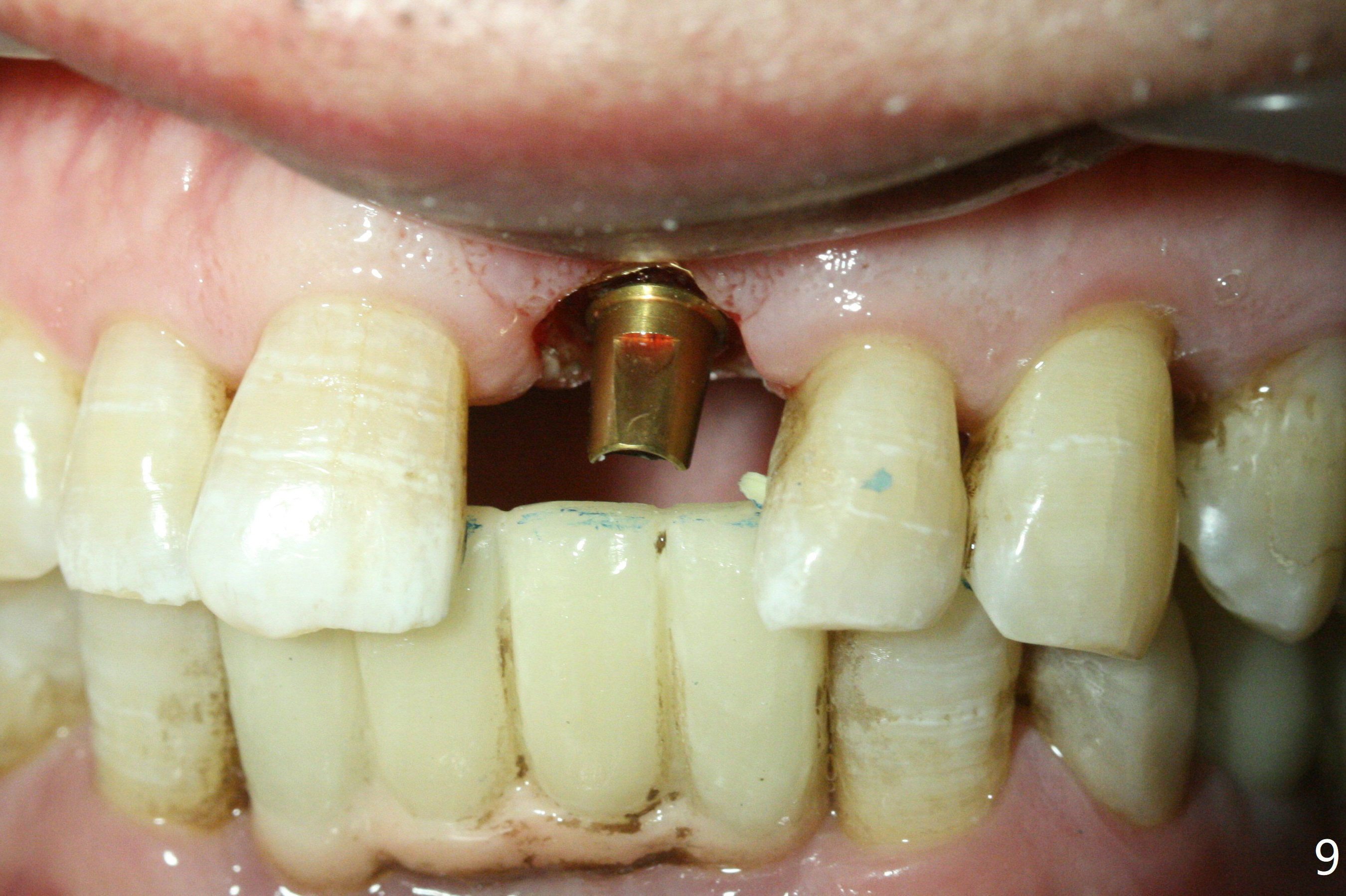
Impression is taken 2 months postop because of instability of the immediate provisional (Fig.9 after Laser gingivectomy).
Incisive Canal Last Next Xin Wei, DDS, PhD, MS 1st edition 12/04/2017, last revision 08/13/2018