|
|
|
|
|
|
|
|
|
|
|
|
|
|
|
|
|
|
|
|
|
|
|
1st Molar Will Have Premolar Width
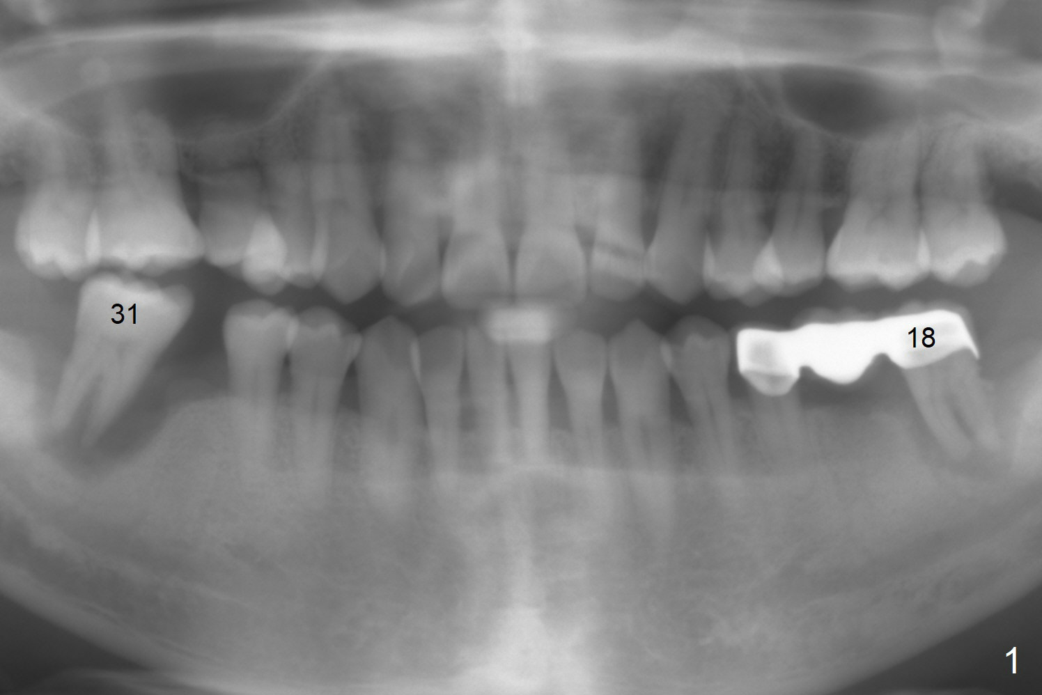
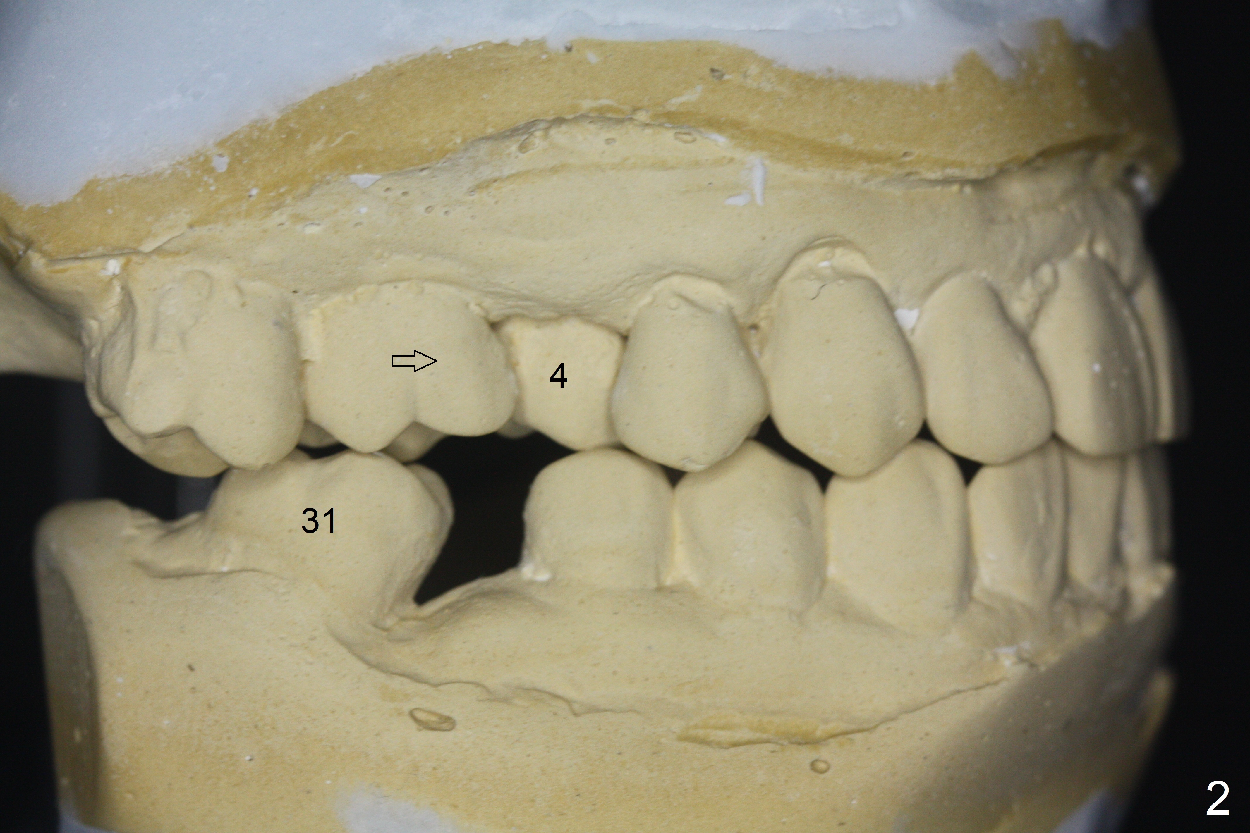
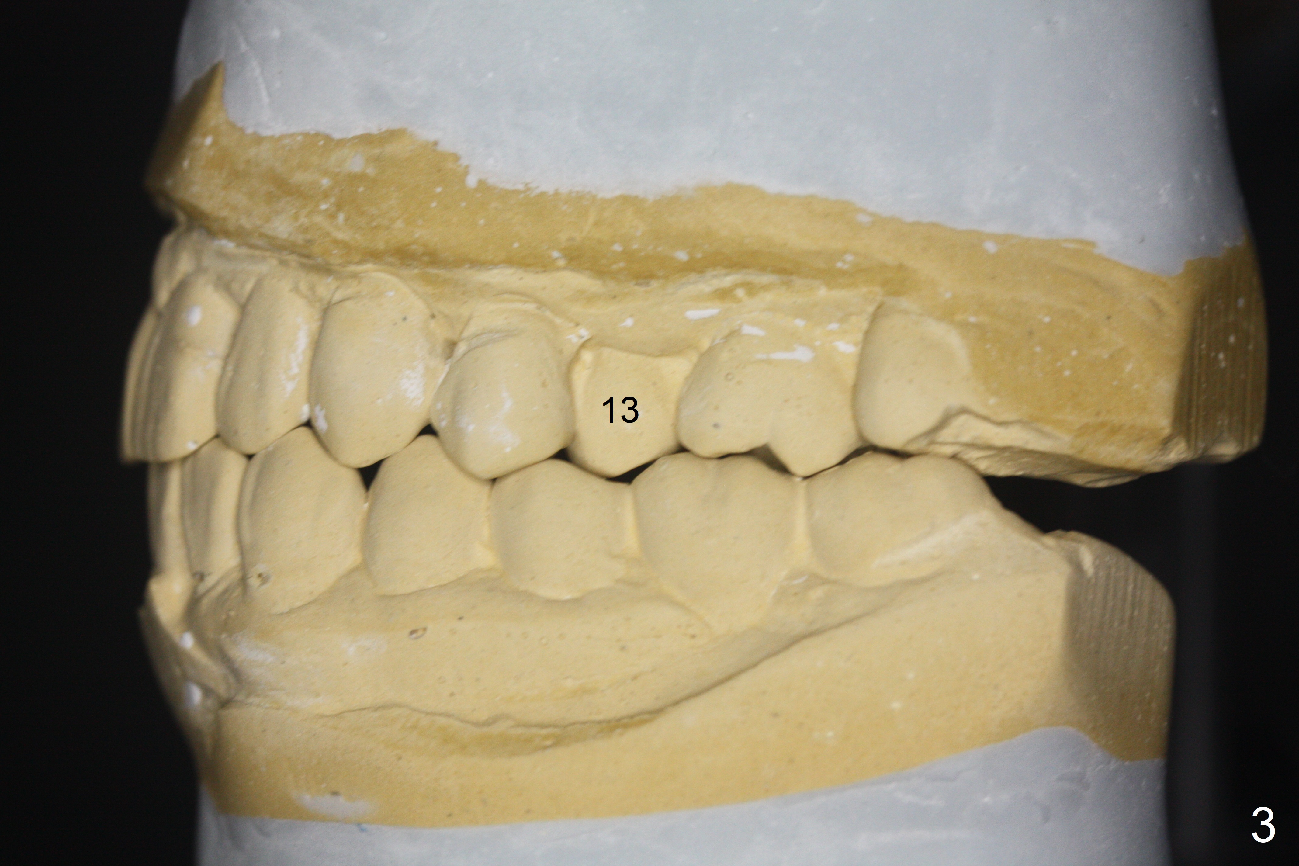
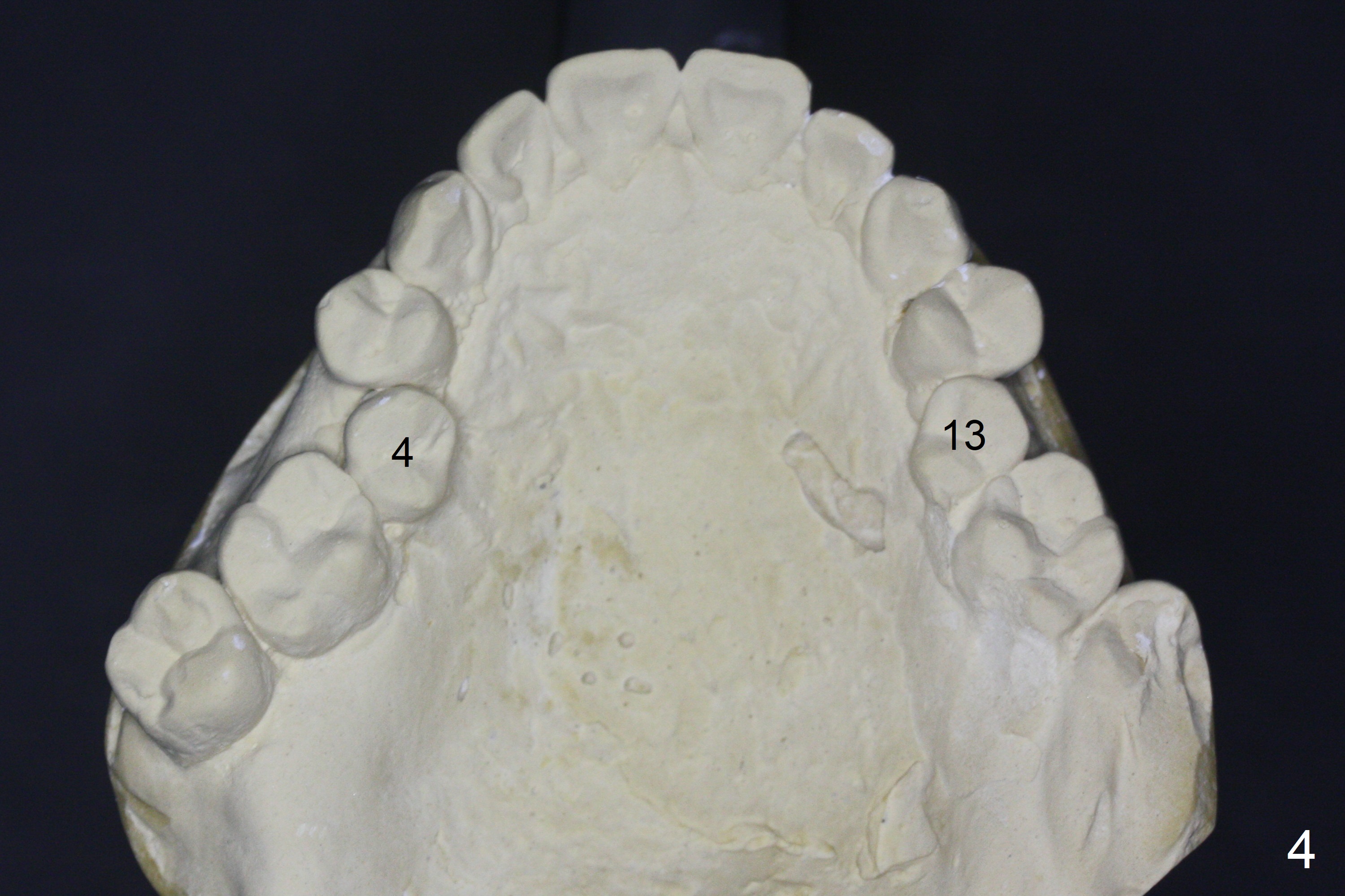
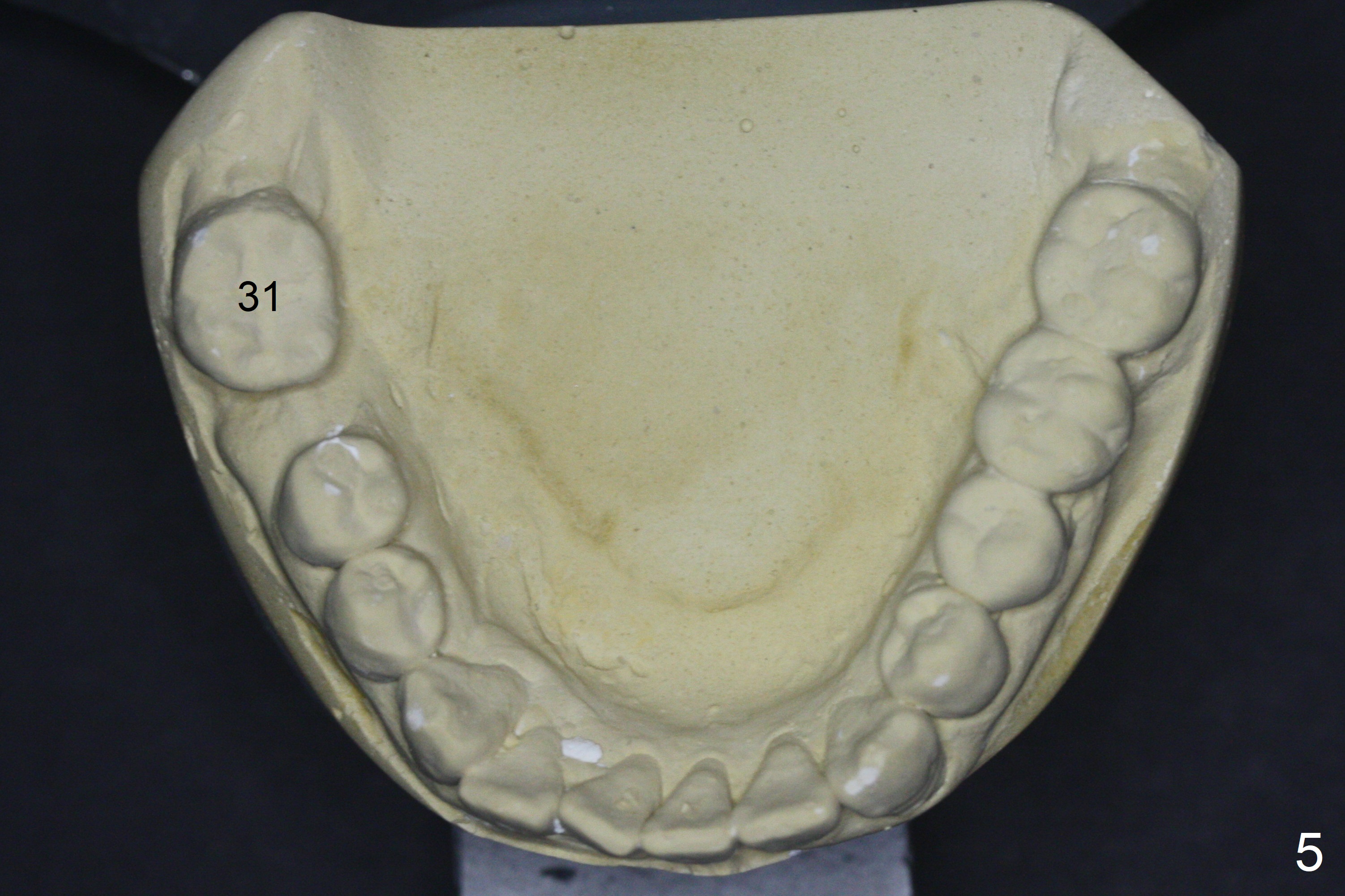
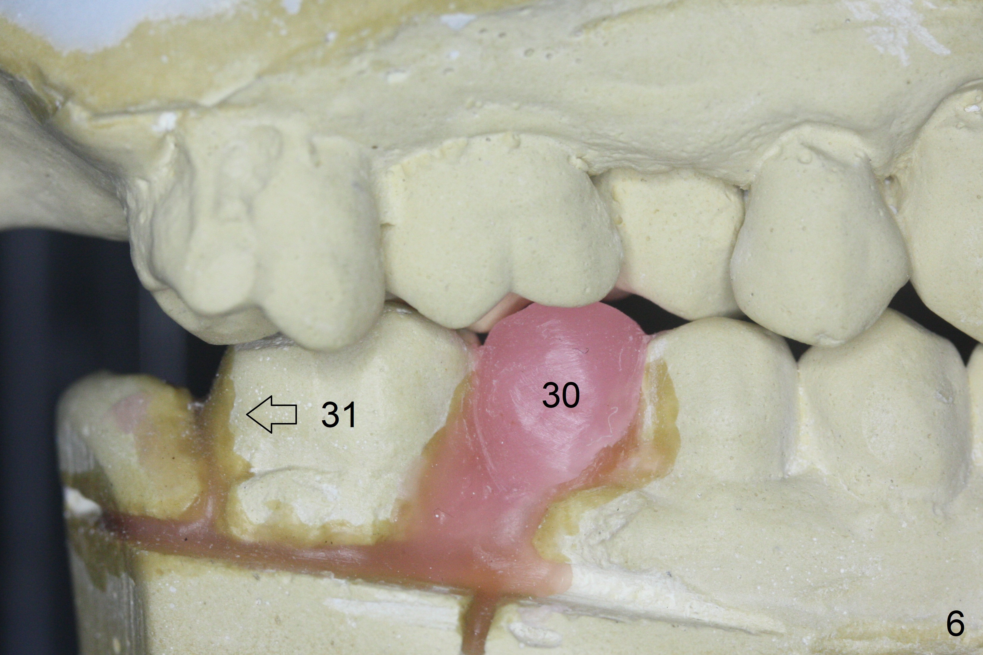
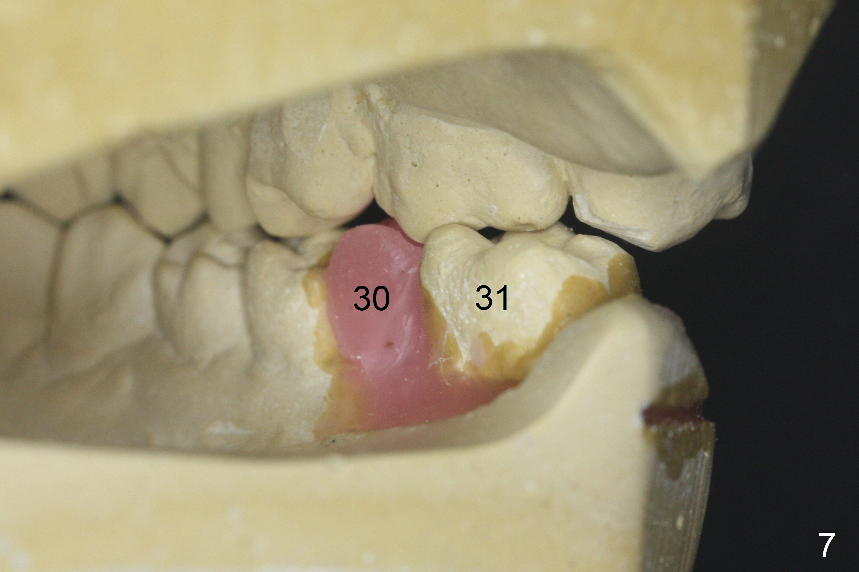
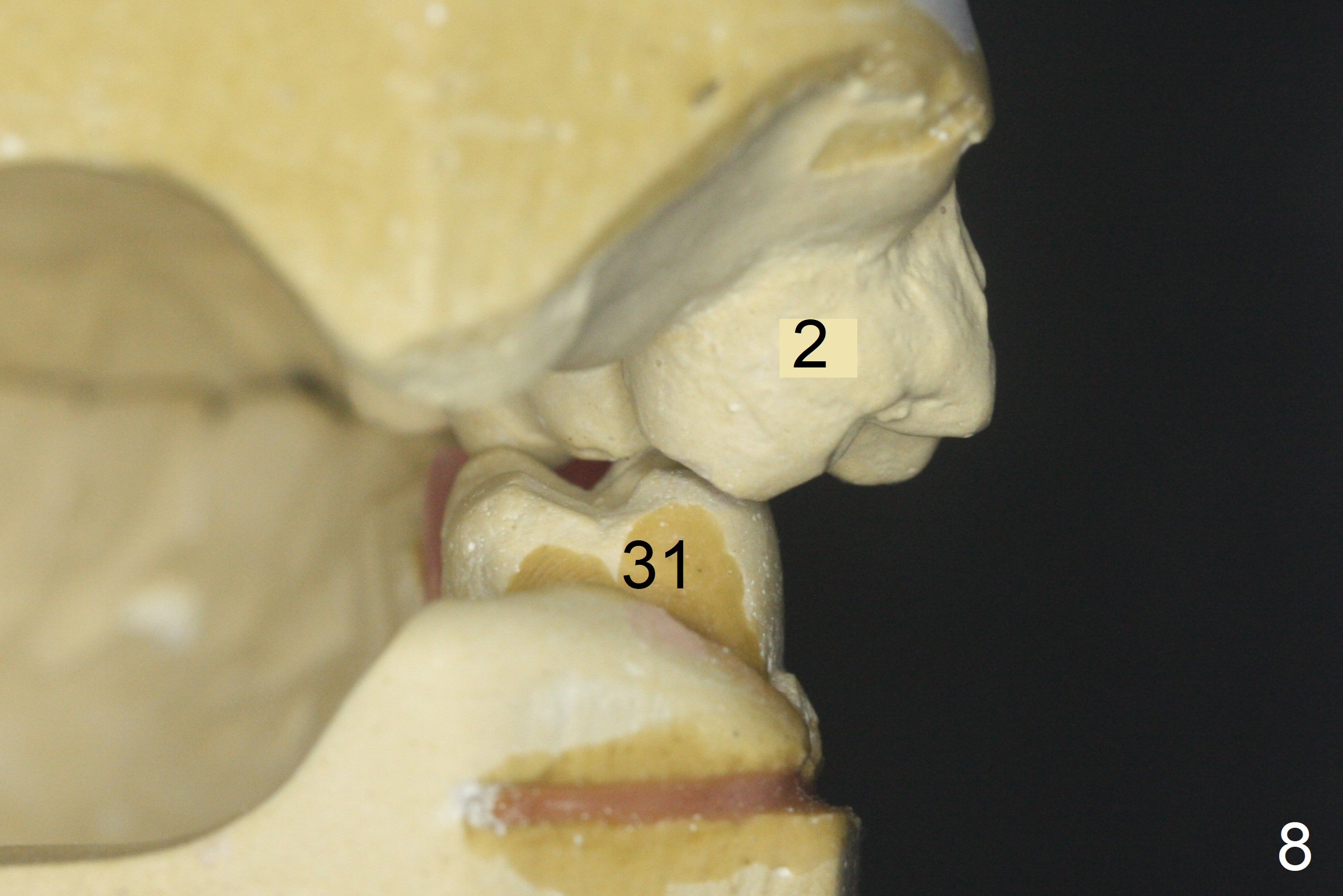
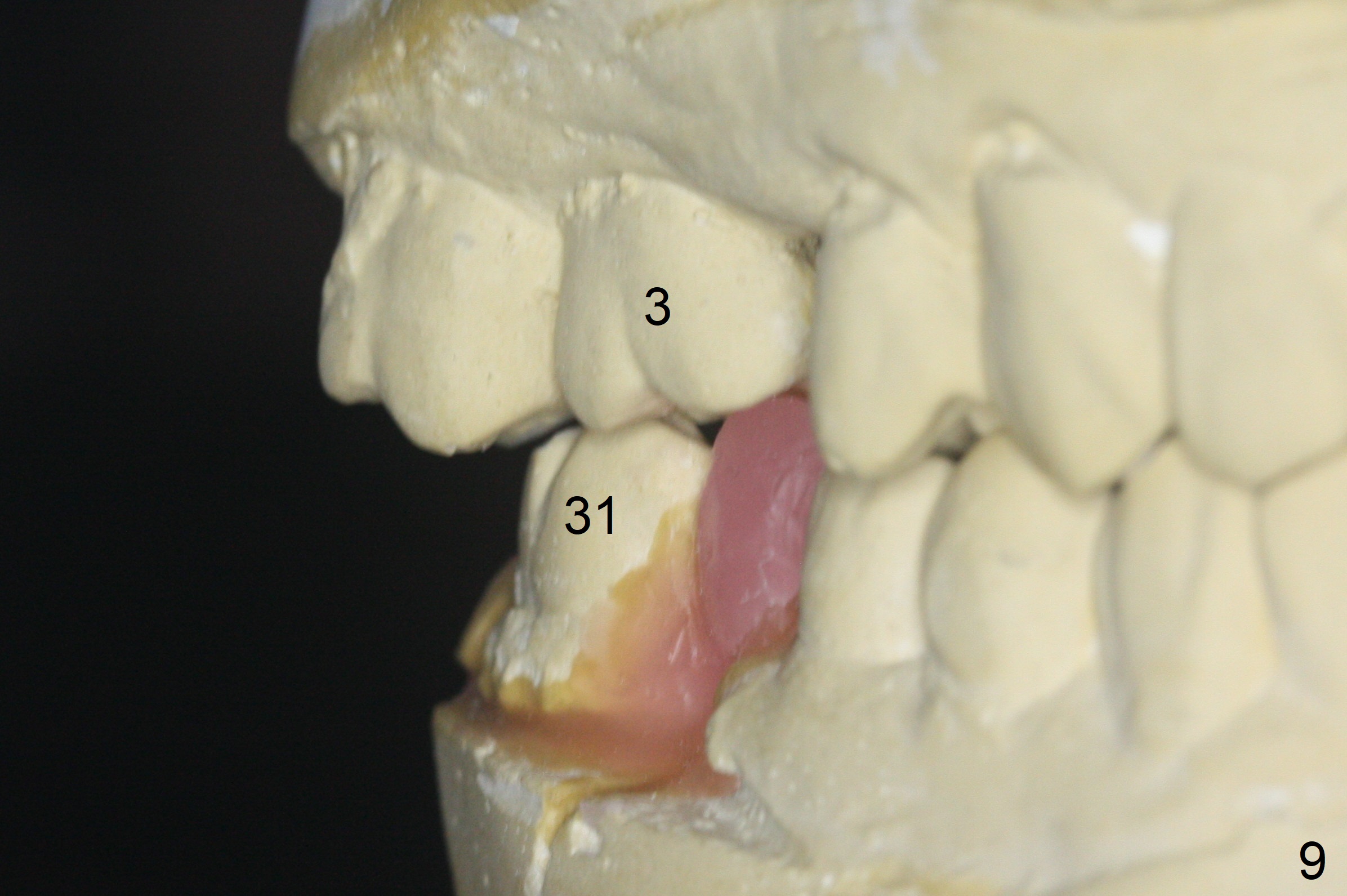
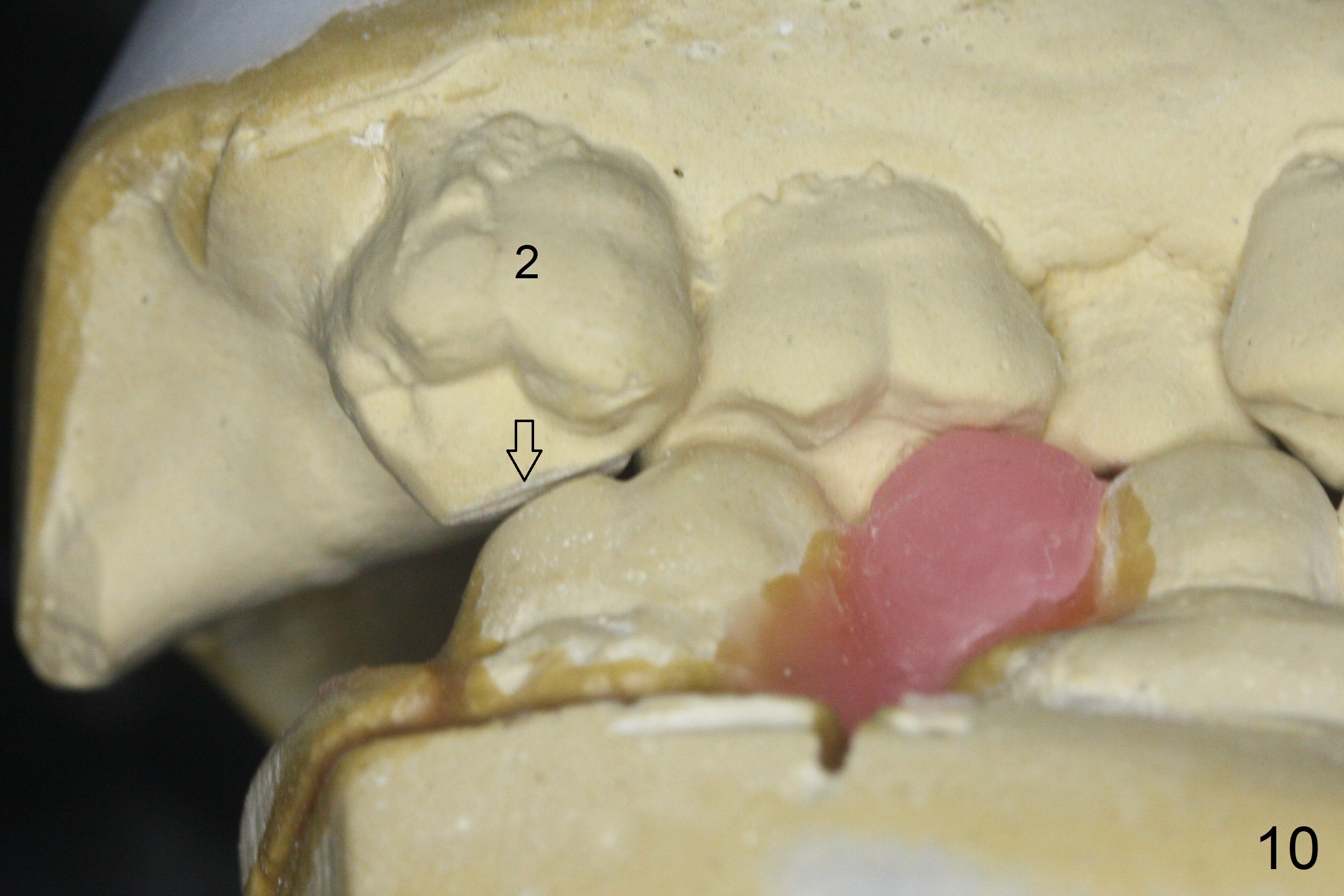
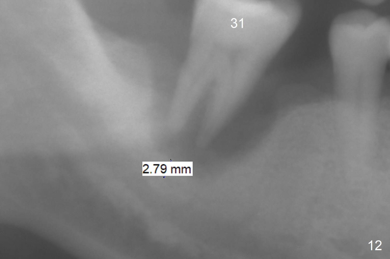
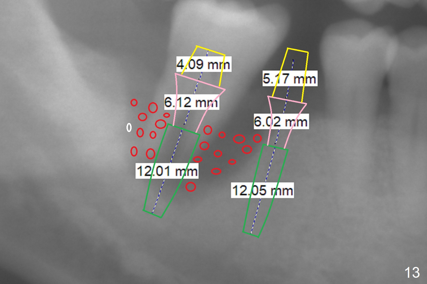
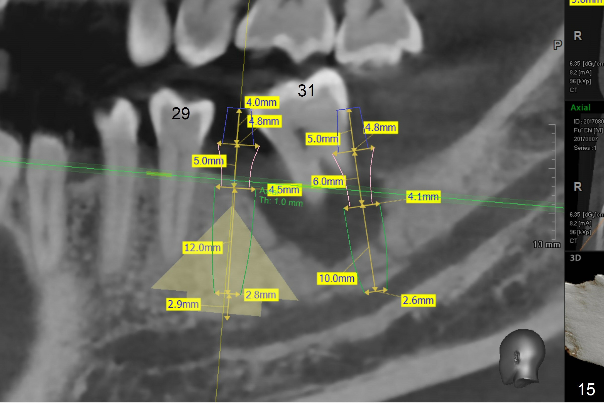
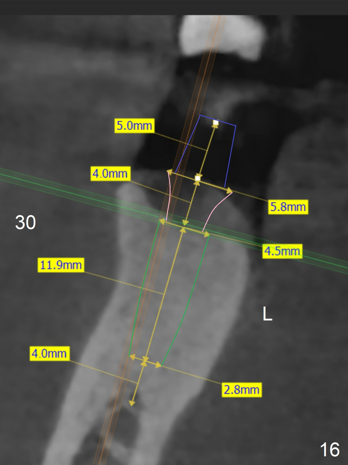
A 64-year-old man has discomfort with the lower left bridge and a loose tooth on the left (Fig.1). It appears that 4 implants are to be placed at #18,19,30 and 31. Since the tooth #4 and 13 are palatalized (Fig.2-4), the teeth #2 and 3 are mesialized (Fig.2 arrow). Besides, the ridge at #30 must be atrophic due to long termed edentulism. A small-diameter implant will be placed at #30 with premolar width (Fig.6-13), while the implant at #31 will be placed distal (arrow, as compared to Fig.2). To establish harmonious occlusion at provisional and final stages, the palatal slope of the mesiopalatal cusp of the tooth #2 will be adjusted (Fig.10).
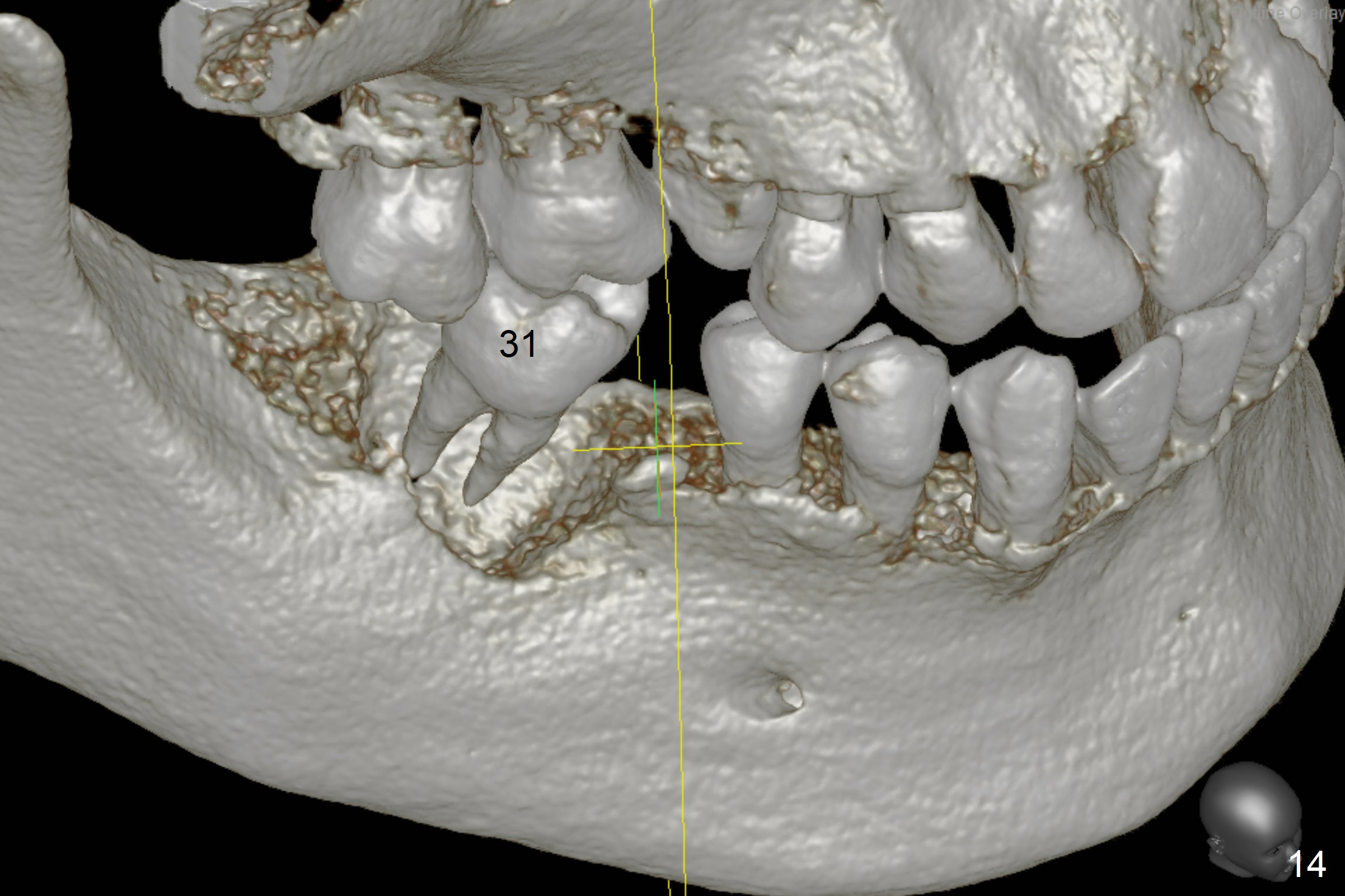
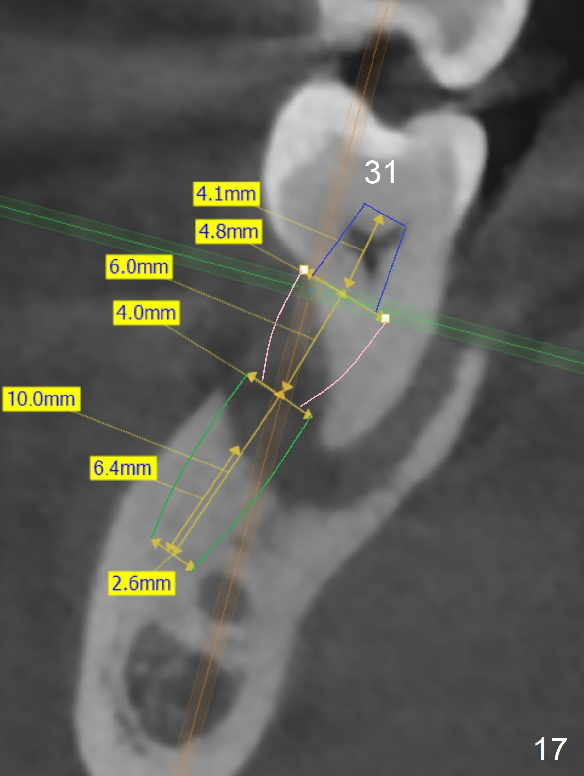
Prepare 3 large pieces of gauze with the vasoconstrictor. If one of them is still too small for the socket, insert another piece of gauze without the vasoconstrictor for pressure hemostasis. Prepare PRF (x4) and Sinus Master Kit in case short stoppers are needed (Fig.12). CBCT taken immediately preop shows relatively wide ridge at #30 (Fig.14,16). The implant at #31 should be place in the distobuccal aspect of the socket (Fig.15,17): 2 mm drill with 7 mm stopper, 2.8 mm round drill at 5 mm (50 RPM) and 3.6 mm at 3 mm.
Return to
Lower
Molar Immediate Implant, Prevent Molar Periimplantitis (Protocols,
Table),
IBS,
No Antibiotic
Xin Wei, DDS, PhD, MS 1st edition 07/29/2017, last revision 04/15/2020