,%206.8x5.5(2),%207.8x4(3).jpg)
|
|
|
|
|
|
|
|
|
|
|
|
|
|
|
|
|
|
||
Short Implants in Mandibular Posteriors
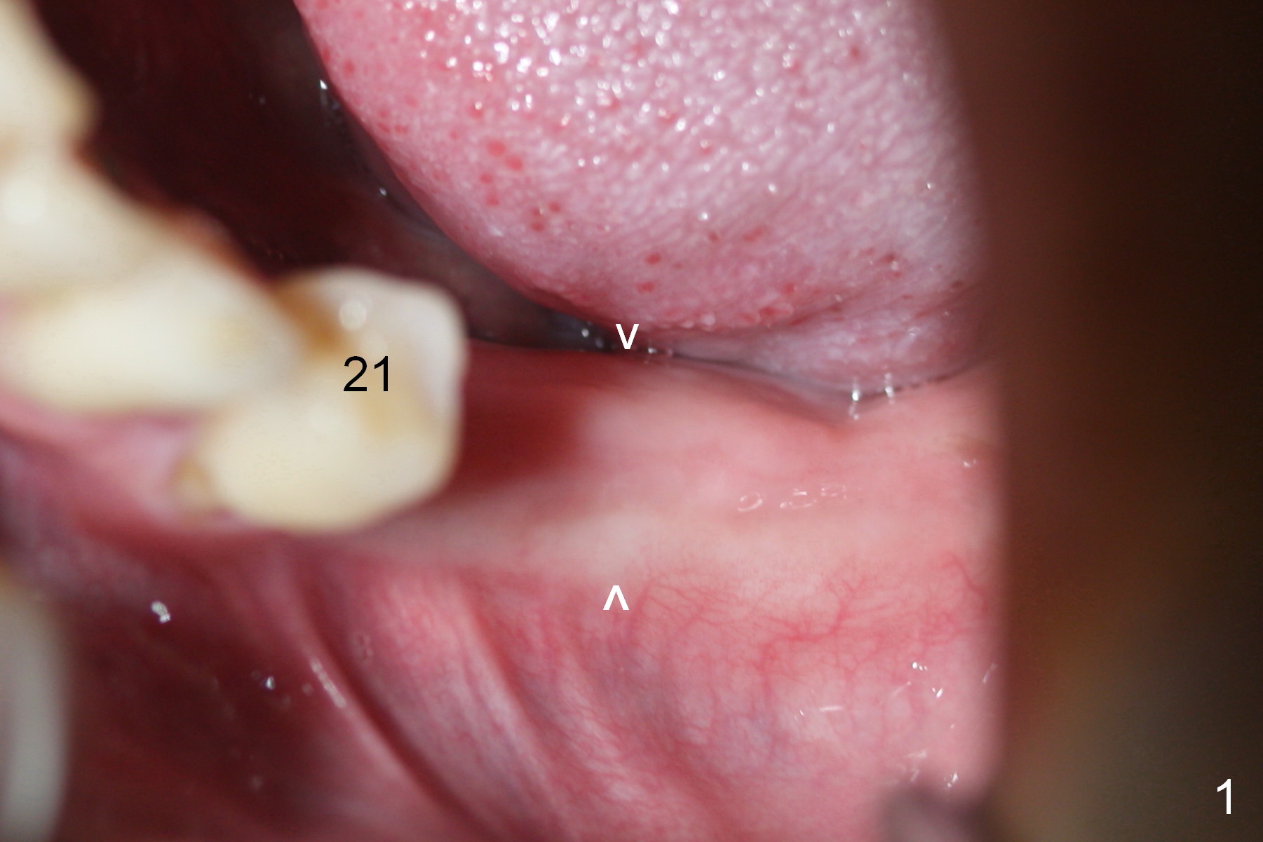
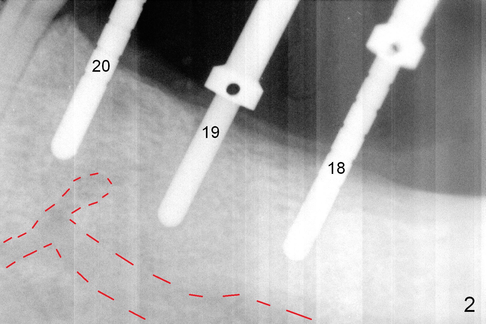
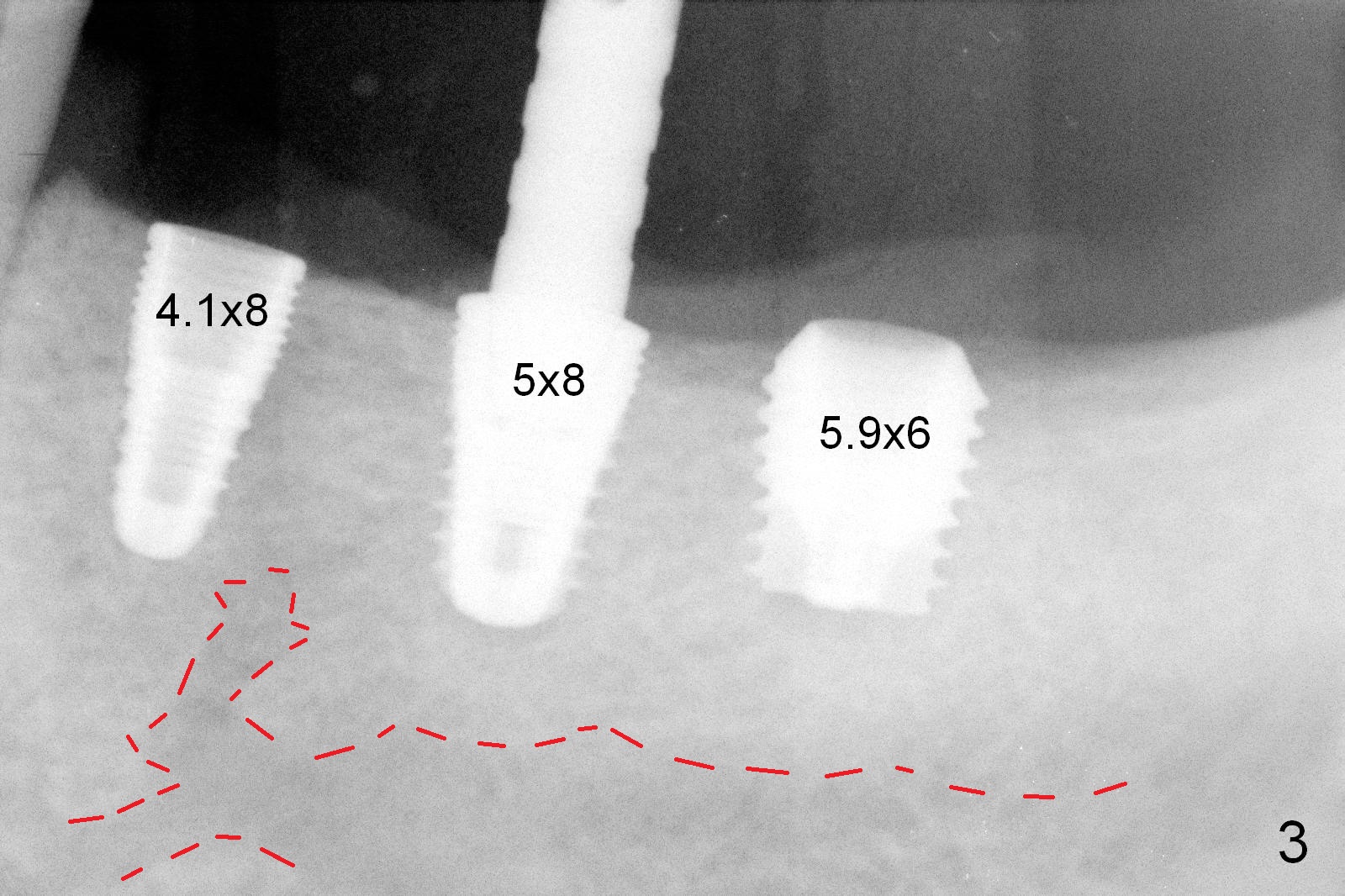
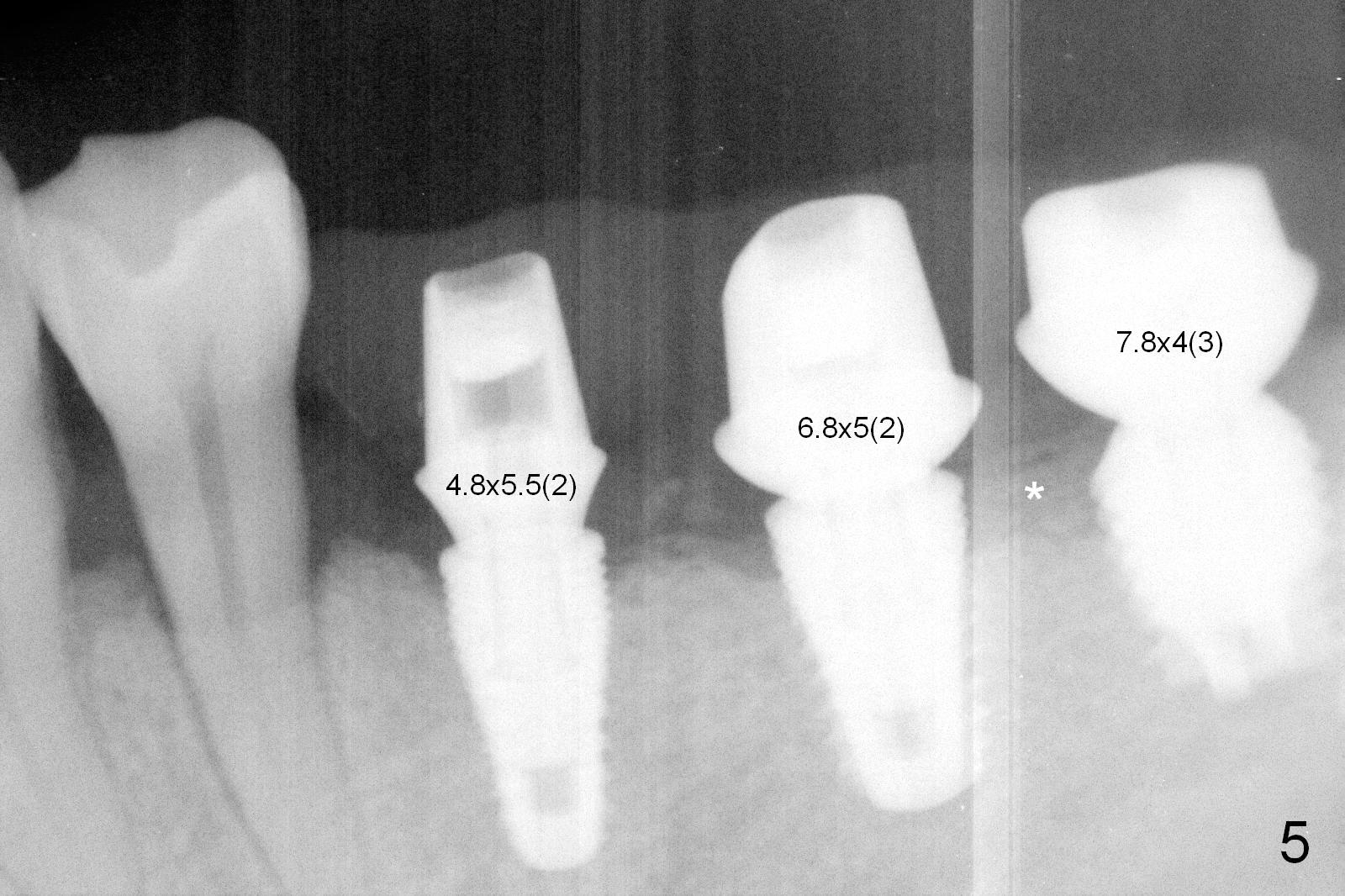
Although bone height is limited, the crest of the lower left area appears to be wide (Fig.1 arrowheads). Initial osteotomy depth is 6 mm (at #18) and 8 mm (19,20) with sufficient clearance from the Inferior Alveolar Canal (Fig.2 red dashed line). The size of the implants at the sites of #18-20 is shown in Fig.3 in millimeters. The insertion torque is around 50 Ncm. When abutments are immediately placed (the diameter and size shown in mm in Fig.4,5) and autogenous bone is packed around the implants/abutments, the ridge looks particularly wide (arrowheads). A splinted immediate provisional is fabricated.
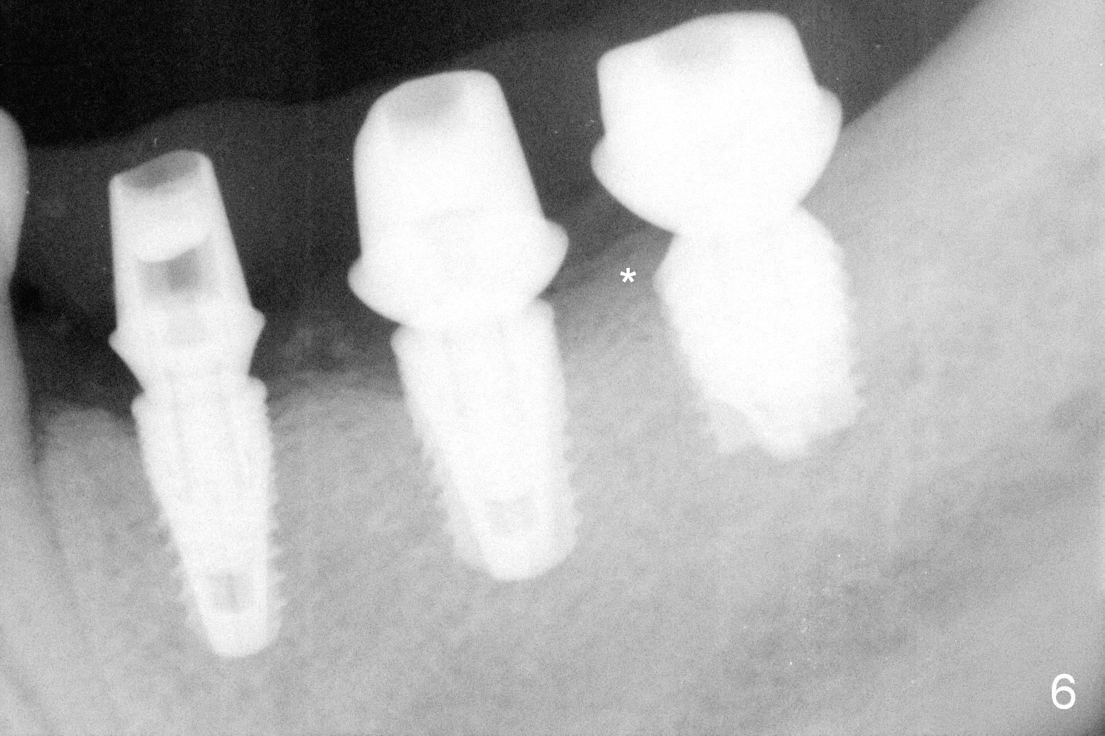
The patient returns for final restoration 4 months postop. He is a smoker with poor oral hygiene. The provisional is fractured at #19 (distobuccal). There is a gap mesial to #18 abutment. It appears that there is mild bone loss, particularly mesial to the implant at #18 (Fig.5,6 *). The implants should have been placed deeper to prevent periimplantitis. When the abutment at #18 (7.8x4(3) mm) is removed, there is food debris mesial, although the gingiva remains healthy. A smaller abutment with shorter cuff is placed (6.8x4(2) mm) before impression.
When the provisional is removed for final restoration, #18 mesial gingiva is recessive and does not attach to the implant well. There is light bleeding on Piezo. All suggest early sign of periimplantitis.
Five months post cementation, the patient reports that #18 implant is loosening. It appears that the implant has to be redone, placed deeper (Fig.3). There is still bone apically. Encourage smoke cessation, better oral hygiene and implant placement at #2. He is a bruxer.
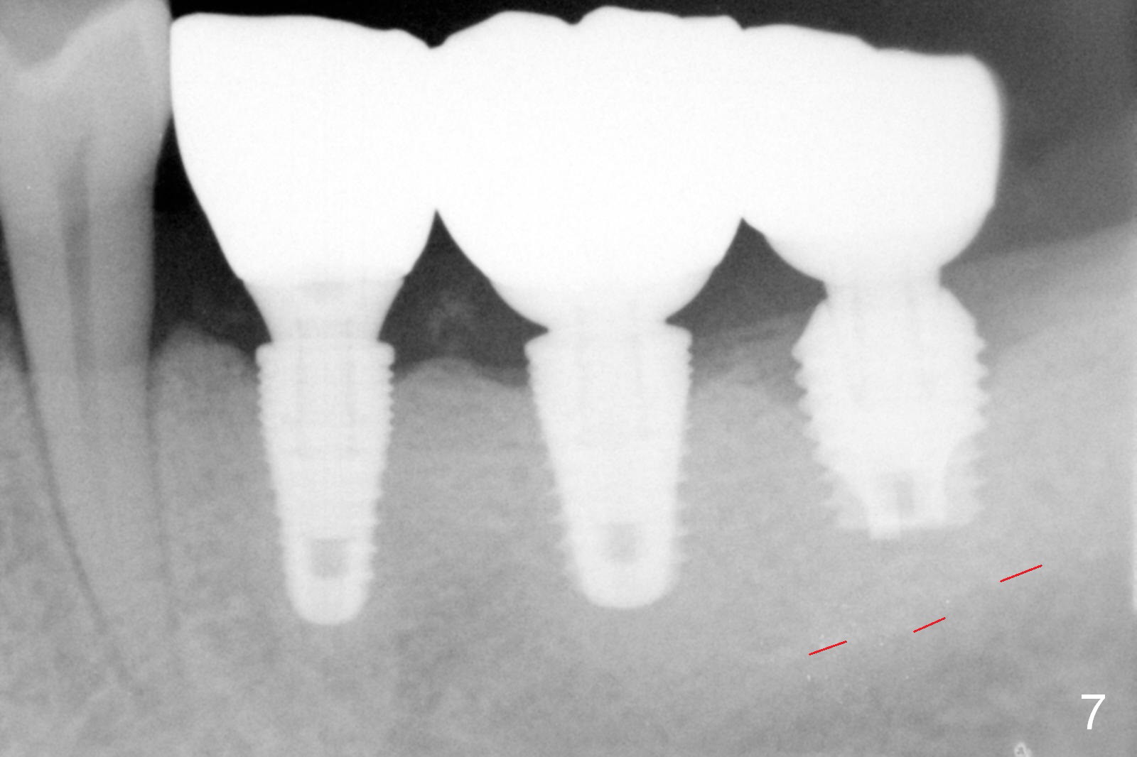
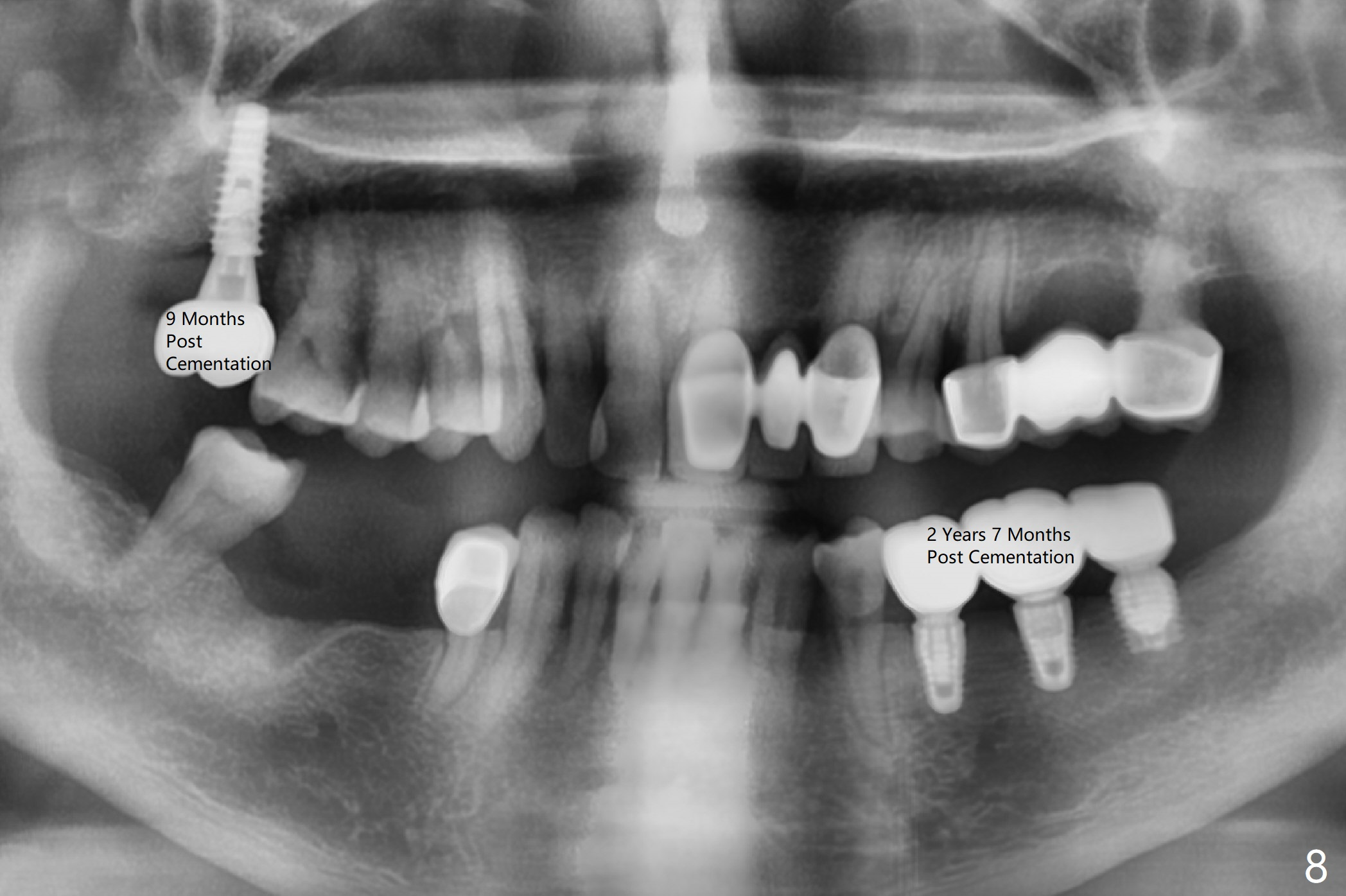
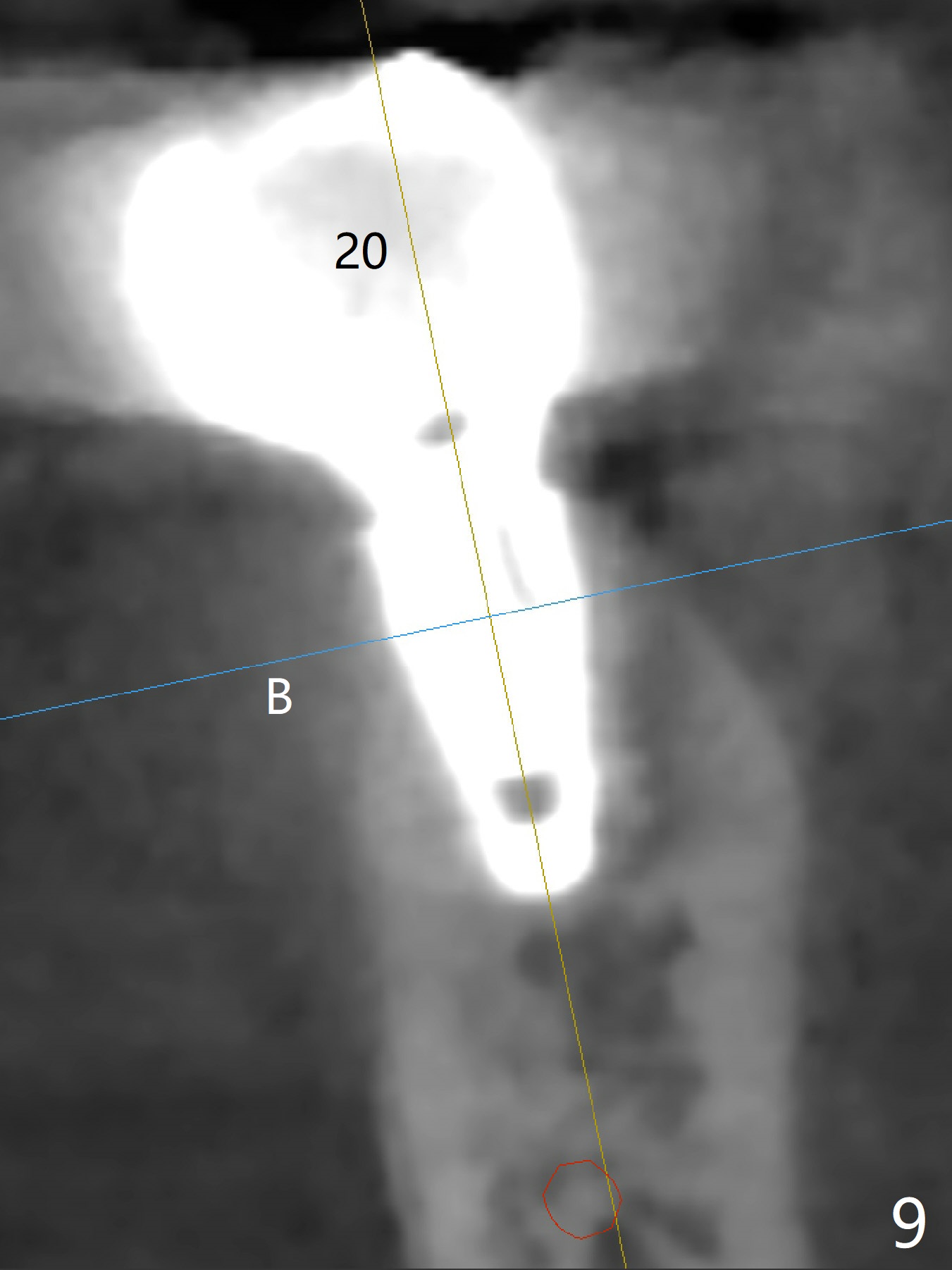
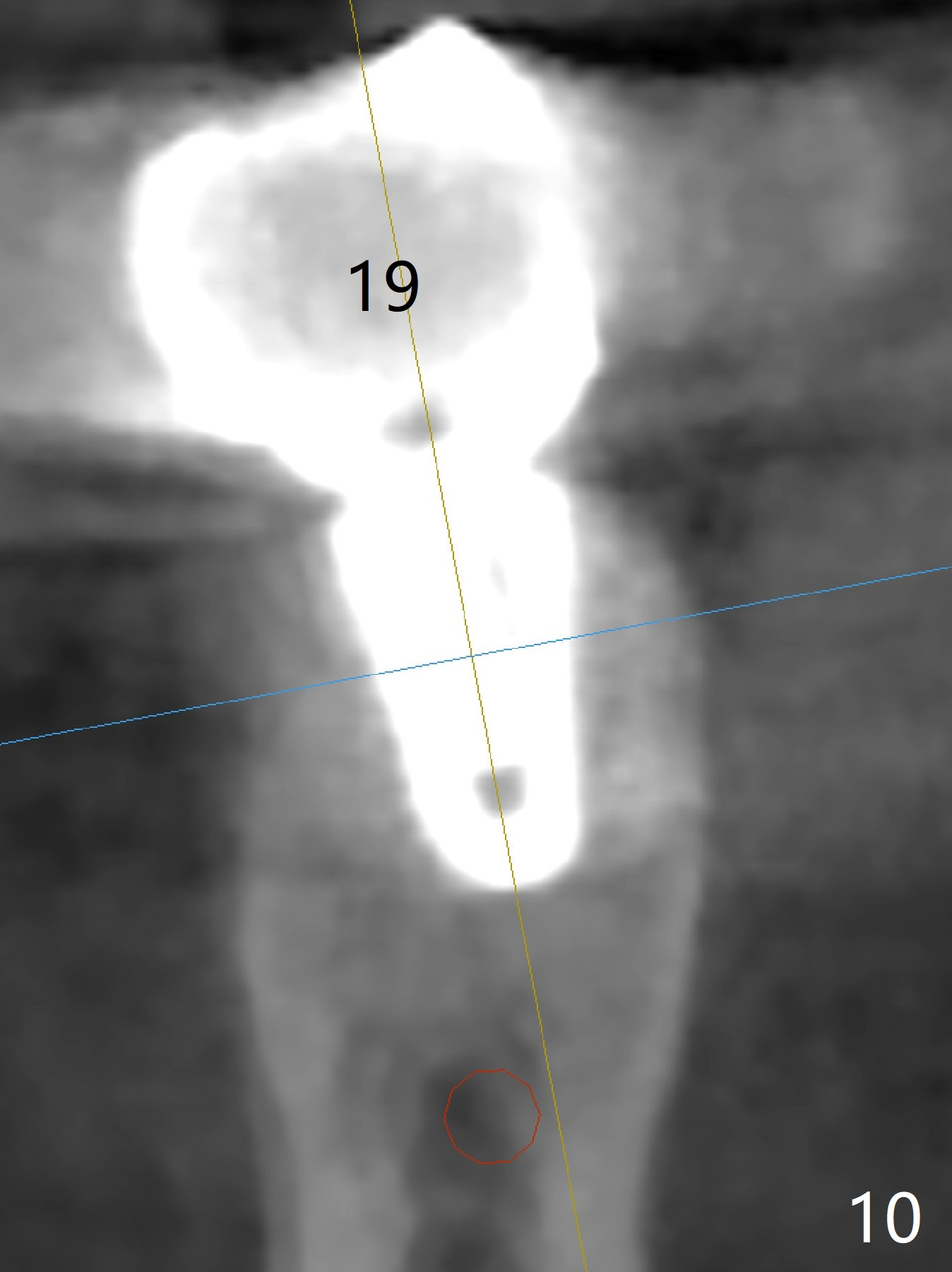
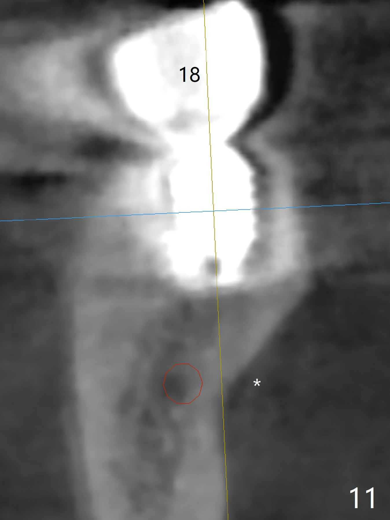
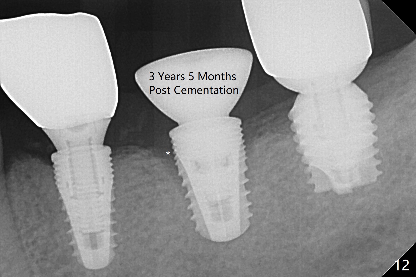
Five months post cementation, the patient returns because of loose abutment screw at #18. Bone resorption is noted (Fig.7 arrowheads). His oral hygiene is fair. He refuses implant at #2, saying that he dares not to chew on the left. When the screw became loose again 3 months later, he accepted the treatment. While he was returning to his home country, the lower right bridge was sectioned and the tooth #30 was removed. He is thinking of 2 implants at #29 and 30 (Fig.8). Bone loss appears not to get worse at #18-20 (Fig.9-11 (CBCT coronal sections, 2 years 7 months post cementation)). The implant at #19 is loose with a gap between the implant and the bone 3 years 5 months post cementation (Fig.12 *). SM implant does not last under stress (bruxism) and rigid implant/abutment connection.
Return to
Lower Arch Reconstruction
with Implants,
Posterior Immediate Provisional,
IBS,
29,30,
2,
Trajectory II
Xin Wei, DDS, PhD, MS 1st edition 10/14/2015, last revision 08/15/2021