|
|
|
|
|
|
|
|
|
|
|
|
|
|
|
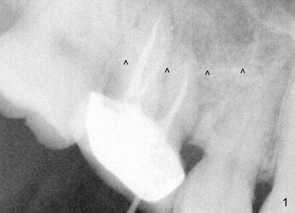
How to Gain Bone Over the Sinus Floor
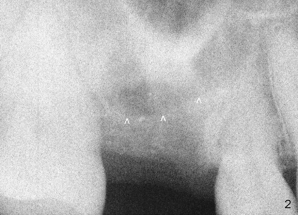
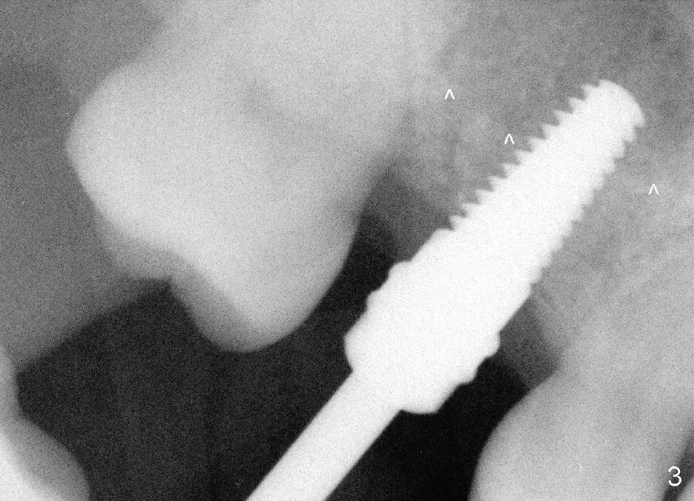
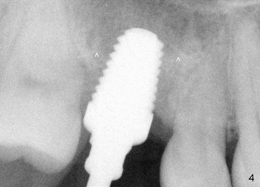
The tooth #3 of a 45-year-old man has endo failure (Fig.1), which shows that the sinus floor (^) is lower than the root tips. Fig.2 is taken a few days post extraction. Note low bone density in the socket, which is the basis for bone condensation. Five months later the patient returns for implantation. A ~12 mm incision is place in the edentulous ridge. The osteotomy forms by using osteotomes (rounded tapered 2-4 mm), followed by insertion of a 4.5x14 mm tap at the depth of 11 mm (Fig.3). It appears that the tip of the tap is in the sinus, but the top of the osteotomy has solid bone when the tap is removed. The bone is being expanded until 7x14 mm tap at the depth of 11 mm (Fig.4). The roof of the osteotomy is still intact. It appears that bone expansion intentionally lifts the sinus floor.
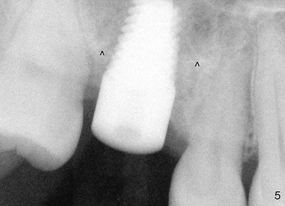
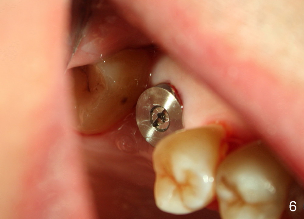
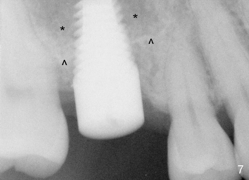
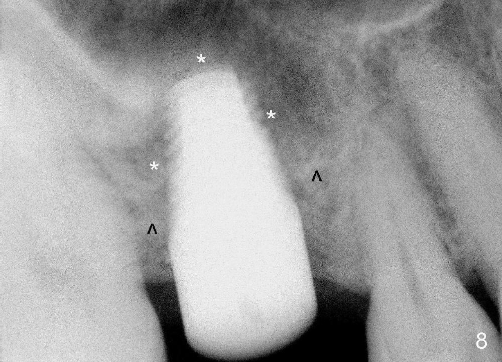
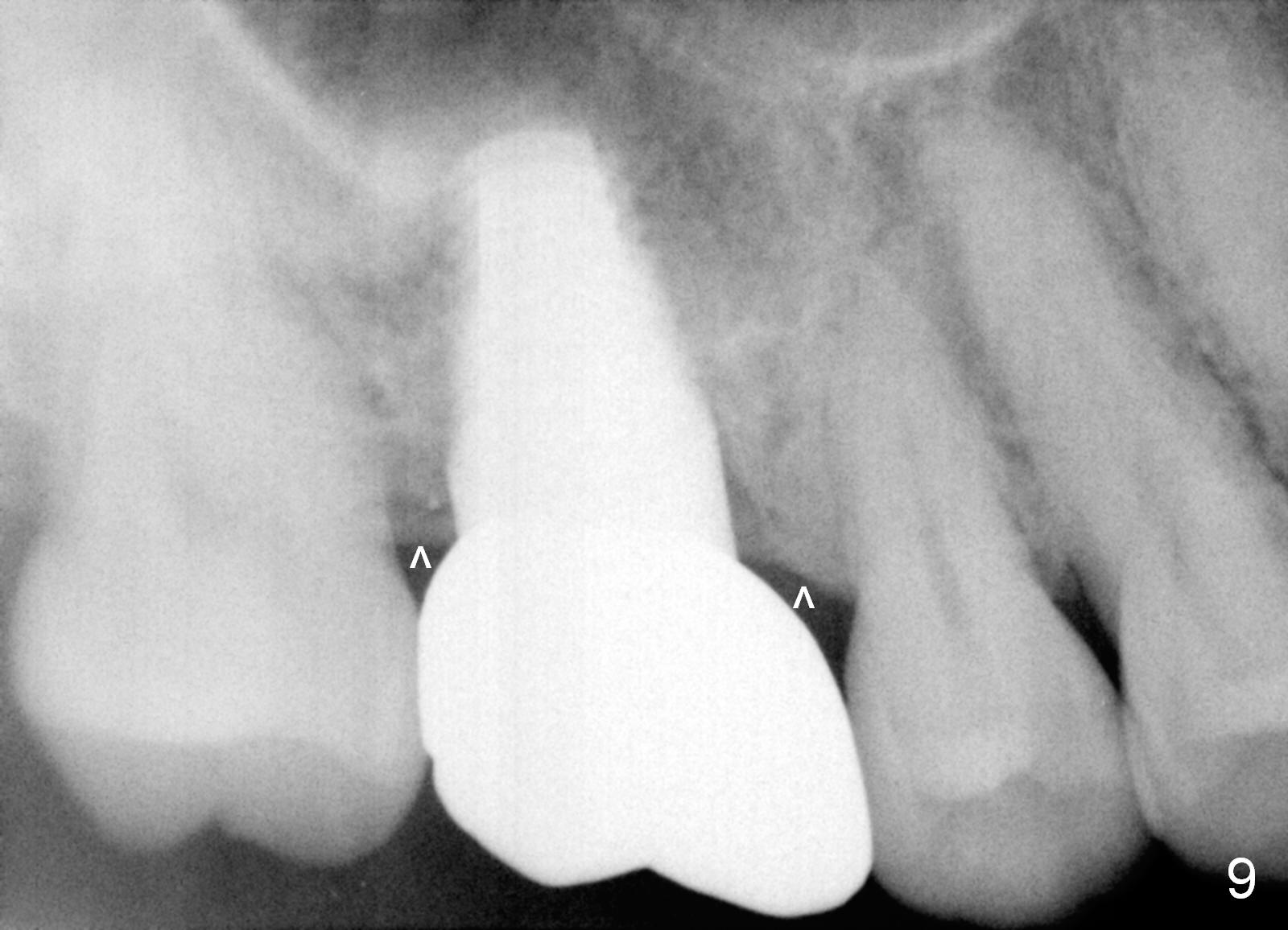
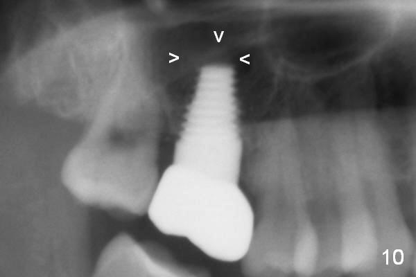
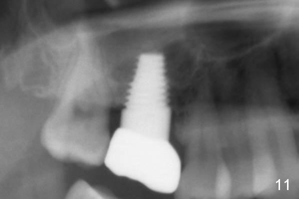
Our experience demonstrates higher failure rate associated with shorter implant in this situation. The depth of the osteotomy is then increased to 14 mm using the same series of osteotomes and taps. The bone at the top of the osteotomy finally wears off when the last tap is applied (7x17 mm). But the sinus membrane is intact. Osteogen is pushed into the osteotomy before placing a 7x14 mm implant with insertion torque > 60 Ncm (Fig.5). The implant contacts the gingiva tightly; no suture is necessary. There is no intra- or postop nasal hemorrhage. The wound heals 8 days postop (Fig.6). There is bone surrounding the apical portion of the implant in the sinus (Fig.7,8 *) 3.5 months postop. The implant is stable. There appears no crestal bone resorption 7.5 months post cementation (Fig.9 ^). Sinus lift is visible 16 months post cementation (Fig.10 (trimmed panoramic X-ray) arrowheads). There is discomfort between #2 and 3, probably due to poor oral hygiene. There is no deep pocket or bone loss (Fig.11, 26 months post cementation).
Return to Sinus Lift
Xin Wei, DDS, PhD, MS 1st edition 05/17/2014, last revision 11/17/2016